Glaucoma Diagnosis: First 90 Days

A glaucoma diagnosis can feel overwhelming, but the first 90 days are crucial for understanding your condition, starting treatment, and establishing a plan to protect your vision long term. Early follow-up, regular eye pressure monitoring, and clear communication with your glaucoma specialist can make a significant difference in preserving sight.

Your First 90 Days With Glaucoma: A Step-by-Step Action Plan

Many patients ask me: I have been diagnosed with glaucoma. What do I do now. Here is what I tell them: A glaucoma diagnosis does not mean you are going blind. It means you now have information most people get too late. The next 90 days are the most important window — not because the disease moves fast, but because the habits you build now protect your vision for the next 30 years.

This guide, written by Dr Shibal Bhartiya, tells you exactly what to do, in order.

Dr Shibal Bhartiya is a fellowship-trained glaucoma specialist and Mayo Clinic Research Collaborator with over 25 years of experience. Her approach focuses on identifying risk before damage is irreversible, simplifying treatment decisions, and protecting vision long-term. Emphasis on early detection, risk assessment, and continuity of care. She is rated 5 stars across 1,500+ patient reviews on Google.


Day 1–7 of Glaucoma Diagnosis: Get the Basics Right

Learn to put in your eye drops correctly

This is the single most important skill you will learn. Studies show that over 60% of patients use eye drops incorrectly — and incorrect technique means the drop misses the eye, or drains immediately into the tear duct and does nothing.

Do this:

Wash your hands. Tilt your head back. Pull your lower eyelid gently down to form a pocket. Hold the bottle above the eye without touching it. Squeeze one drop into the pocket — not onto the eyeball directly. Close your eye gently. Press the inner corner of your eye (near the nose) firmly with one finger for 60 seconds. This blocks the tear duct and keeps the drug in the eye where it belongs. Do not blink vigorously. Do not wipe.

If you use more than one drop type, wait five minutes between them. The first drop dilutes and flushes out the second if you use them together.

Ask your doctor or optometrist to watch you do it once. Ask for a correction if your technique needs adjustment.

Here’s a video demostration:

Set your alarms — and take them seriously

Glaucoma drops work only when taken on time, every day, for life. A single missed day matters less than a pattern of casual delays.

Most drops are once daily, ideally at night. Set a recurring alarm on your phone with a label — “Left eye drop, right eye drop, press corner.” Place the bottle next to your toothbrush. The habit links to the existing habit.

If you use drops twice daily, set both alarms. Never rely on memory alone.

File your papers before they disappear

You walked out of the clinic with reports. Photograph or scan every one of them today — the visual field test, the OCT nerve scan, the IOP readings, the prescription. Put them in a dedicated folder on your phone or email them to yourself with the subject line “Glaucoma Records — [your name].”

You will need these at your next visit, at any second opinion, and if you travel and need emergency eye care. Doctors cannot make good decisions without your baseline.


Week 2–4 of Glaucoma Diagnosis: Build the Follow-Up Structure

Your 30-day appointment is not optional

Glaucoma drops take four to six weeks to show their full pressure-lowering effect. Your doctor needs to see you at 30 days to measure whether the drop is working — and to catch side effects early. Do not skip this.

At this visit, your doctor will check:

  • Your intraocular pressure (IOP) against your baseline
  • Whether the drop is causing redness, allergy, or discomfort
  • Whether you need a dose adjustment or a switch to a different medication

Set a calendar reminder for this appointment the day you are diagnosed. If the appointment was not scheduled, call the clinic and schedule it yourself before the week is over.

Know what side effects to watch for

Most glaucoma drops are well-tolerated. But some cause changes you should know about.

Prostaglandin analogues (bimatoprost, travoprost, latanoprost) can darken the iris over time in some patients, and may cause eyelash growth or mild redness. These are cosmetic and not dangerous — but tell your doctor.

Beta-blockers (timolol) can slow your heart rate and cause breathlessness in patients with asthma or heart disease. If you feel unusually short of breath or very tired after starting drops, contact your doctor the same day.

Alpha agonists (brimonidine) sometimes cause an allergic reaction with marked redness and discharge, usually within weeks of starting. Stop the drop and call your doctor if this happens.

None of these mean you must stop treatment. They mean the treatment may need adjustment.


Month 1–2 of Glaucoma Diagnosis: Tell Your Family

Your siblings and children need an eye check — now

Glaucoma has a strong genetic component. First-degree relatives of a glaucoma patient have a four to nine times higher risk of developing the disease. Most of them will have no symptoms at all until damage is advanced.

Tell your siblings and adult children this week. Ask them to see an ophthalmologist for a baseline pressure check, optic nerve assessment, and field test. This is not alarmist. It is the most useful thing your diagnosis can do for your family.


Month 1–3: Address the Controllable Risk Factors

Stop smoking — this one is not negotiable

Smoking constricts blood vessels and reduces blood flow to the optic nerve. It worsens the vascular risk that many glaucoma patients already carry. The damage from smoking adds to the damage from pressure — and your nerve cannot absorb both.

If you smoke, speak to your doctor about cessation support. This is as important as the drops.

Get your metabolic parameters checked

High blood pressure, diabetes, thyroid disease, and sleep apnoea all affect glaucoma progression through vascular and metabolic pathways. If these are uncontrolled, your optic nerve faces risk from two directions simultaneously.

Ask your physician to check your blood pressure, fasting glucose, HbA1c, and thyroid function if these have not been done recently. If you snore heavily or feel exhausted in the mornings, mention it — untreated sleep apnoea is a recognised glaucoma risk factor that is almost always missed.

Exercise — the right kind

Moderate aerobic exercise (brisk walking 30 minutes, five days a week) lowers intraocular pressure by a clinically meaningful amount in most patients. Avoid high-resistance head-down exercises like heavy weightlifting or inverted yoga poses — these transiently spike IOP.


Month 2–3: Ask About Laser Treatment

SLT — Selective Laser Trabeculoplasty

If your glaucoma is open-angle type, your doctor may recommend SLT as a first-line treatment or as a supplement to drops. SLT uses a laser to improve fluid drainage from the eye. It is done in the clinic in five to ten minutes, is painless, and works in approximately 75 to 80% of patients.

The effect lasts three to five years and can be repeated. SLT does not burn tissue — it sends a gentle energy pulse that stimulates the drainage cells to work better.

Ask your doctor at the 30-day or 90-day visit: “Am I a candidate for SLT?”

LPI — Laser Peripheral Iridotomy

If your glaucoma is narrow-angle or angle-closure type, LPI is a preventive procedure that creates a small opening in the iris to prevent a sudden pressure spike (acute angle-closure attack). LPI is typically recommended before an attack happens — it takes three to four minutes per eye and prevents one of the most painful ophthalmic emergencies.

If your doctor mentioned narrow angles at any point, ask specifically whether you need LPI. Do not wait.


Throughout: Keep Your Perspective

Do not search the internet at 2am

Glaucoma outcomes in treated patients are overwhelmingly good. The disease moves slowly in the vast majority of cases. Patients who take their drops, attend follow-ups, and manage their risk factors maintain useful vision for life in most cases.

The stories of severe vision loss you will find online mostly involve patients who were never diagnosed, or who stopped treatment. You are neither.

Reach out if you need support

A new diagnosis changes how you think about your body. Some patients find this unsettling, and that is entirely normal. Several Indian and international glaucoma patient forums, and online communities run by ophthalmologists offer peer support from people at every stage of the same journey.

You do not have to figure this out alone.


Your 90-Day Checklist

  • Eye drop technique confirmed by a doctor or technician
  • Alarm set — every day, same time
  • All reports photographed and filed digitally
  • 30-day follow-up appointment booked
  • Side effects list saved on your phone
  • Siblings and adult children informed and booked for screening
  • Smoking cessation initiated if applicable
  • Blood pressure, glucose, HbA1c, thyroid checked
  • SLT or LPI discussion had with your doctor
  • One support resource bookmarked

Frequently Asked Questions

Do I have to take eye drops for life?

In most cases, yes. Glaucoma is a chronic condition and eye drops control pressure — they do not cure the disease. Stopping drops allows pressure to rise again and damage to resume. Some patients reduce or stop drops after successful laser treatment (SLT), but this is a decision made with your doctor based on your pressure readings, not independently.

What if I forget a drop one day?

Take it as soon as you remember, unless it is almost time for the next dose. Do not double up. One missed dose will not cause a crisis. A habit of casual misses will. Reset the alarm and continue.

Can I drive after putting in my eye drops?

Most glaucoma drops do not affect vision significantly. Some patients notice mild blurring for a few minutes immediately after instillation — wait for this to clear before driving. If your doctor has dilated your pupils at a clinic visit, do not drive until dilation wears off, typically three to four hours.

My pressure was normal at diagnosis. Do I still have glaucoma?

Yes — this is called normal-tension glaucoma (NTG). Roughly 30 to 40% of glaucoma patients in India have pressures within the statistical normal range. The diagnosis is made on optic nerve appearance and visual field changes, not pressure alone. NTG is treated the same way — the target is to lower pressure further from your individual baseline.

Is glaucoma hereditary? Do I need to tell my family?

Yes, and yes. First-degree relatives — parents, siblings, children — have a four to nine times higher risk. Most will have no symptoms. Tell them this week and ask them to see an ophthalmologist for a baseline check that includes pressure, nerve assessment, and a visual field test.

Will I go blind?

Treated glaucoma in a compliant patient who attends follow-up carries a very low risk of blindness. The risk is real only when the disease is undiagnosed, undertreated, or ignored. You have been diagnosed. That is the most important step already taken.

What is SLT and should I ask about it?

Selective Laser Trabeculoplasty (SLT) is a five-minute clinic procedure that improves fluid drainage from the eye. It works in approximately 75 to 80% of open-angle glaucoma patients and can reduce or eliminate the need for drops for three to five years. Ask your doctor at the 30-day visit whether you are a candidate.

Can I exercise with glaucoma?

Yes — moderate aerobic exercise is actively beneficial and lowers IOP. Brisk walking, cycling, and swimming are all good. Avoid heavy resistance training with breath-holding (Valsalva manoeuvre) and inverted positions, both of which spike pressure transiently. If exercise is a regular part of your routine, tell your doctor so they can factor it into your pressure readings.

My drops are making my eyes red. Should I stop?

Do not stop without speaking to your doctor first. Redness is common with several drop classes and is often manageable — a preservative-free formulation or a switch in medication resolves it in most cases. Stopping drops independently allows pressure to rise. Call the clinic and describe the symptom.

How often will I need follow-up forever?

Once stable on treatment, most patients are reviewed every three to six months. This includes a pressure check and, once yearly or more often if needed, a repeat visual field test and OCT nerve scan to confirm the disease is not progressing. Glaucoma never becomes self-managing — the follow-up rhythm continues for life, but it is not onerous once the initial titration phase is complete.


This page is part of the Glaucoma Hub hub. Read about our full approach to glaucoma care and monitoring. Please also read our guide to Understanding Your Visual Field Test. You may want to read a patient’s experience with glaucoma eye drops, and of one with SLT.


About the Author

This article was written by Dr Shibal Bhartiya, fellowship-trained glaucoma specialist and Mayo Clinic Research Collaborator, Clinical Director at Marengo Asia Hospitals, Gurugram, known for ethical, patient-centred glaucoma care and independent glaucoma second opinions. She is also the Program Director for Community Outreach & Wellness; and for the Marengo Asia International Institute of Neuro and Spine.

She has published peer-reviewed research on glaucoma management, examining how treatment decisions should balance medical evidence, patient preferences, and long-term vision outcomes.

As Editor-in-Chief of Clinical and Experimental Vision and Eye Research and Executive Editor of the Journal of Current Glaucoma Practice (Pubmed Indexed, official journal of the International Society of Glaucoma Surgery), Dr Shibal Bhartiya brings editorial and research depth to every clinical decision. Her 200+ publications, including 90+ PubMed-indexed publications and 28 edited textbooks span glaucoma biology, surgical outcomes, health equity, and emerging diagnostics.

1500+ Five Star Patient Reviews Google Business Profile

If you are unable to come to Dr Bhartiya’s clinic: Read more about teleconsultation

Read her research on PubMed | Google Scholar | ResearchGate | ORCID

Upload your reports for a structured review.| www.drshibalbhartiya.com | +91 88826 38735

Leave a review on Google
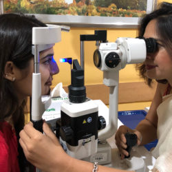

HOW TO DO VISUAL FIELD

A visual field test checks your side (peripheral) vision and helps detect or monitor glaucoma and other optic nerve conditions. During the test, you look straight ahead and press a button whenever you notice lights appearing in different parts of your vision.

Automated static perimetry is the clinical gold standard for tracking glaucoma progression. Yet it is notoriously anxiety-inducing. High fixation losses and false positives corrupt diagnostic data when a patient is stressed. Active coaching before and during the test stabilises fixation, yields clean reproducible data, and transforms a feared exam into a collaborative clinical tool.

Dr Shibal Bhartiya is a fellowship-trained glaucoma specialist and Mayo Clinic Research Collaborator with over 25 years of experience. Her approach focuses on identifying risk before damage is irreversible, simplifying treatment decisions, and protecting vision long-term. Emphasis on early detection, risk assessment, and continuity of care. She is rated 5 stars across 1,500+ patient reviews on Google.


How Patient-First Coaching Transforms Glaucoma Perimetry

Ask any glaucoma patient what part of their routine checkup they dread most. Nine out of ten will say the visual field test.

Sitting alone in a dark room, staring at a central yellow light, clicking a button for faint flashes you think you might be missing — it feels less like a diagnostic test and more like a high-stakes exam you are destined to fail.

A patient recently left a review that captured exactly why we approach this differently. They noted how other clinics seat you in the machine and tell you to press the clicker. No explanation. No preparation. Just anxiety and confusion. They described how, in our clinic, the entire experience was different. We walked them through what the visual field map actually shows. We explained the rhythm of the test before they started. They felt like a partner in their own care — not a passive subject.

You can read their experience here on Google.

When a patient understands that missing some flashes is a normal part of the machine’s threshold calculation, their heart rate drops. Their blinking stabilises. Their anxiety disappears.

That extra ten minutes of human coaching does not just produce a more comfortable patient. It produces pristine, accurate diagnostic data — the data we rely on to protect their optic nerve for decades.

What Actually Happens During a Visual Field Test

You sit with one eye covered and rest your chin on the machine. Your job is simple: keep looking at the central target and press the button whenever you notice a light anywhere in your side vision.

You are not expected to see every flash.

In fact, the machine deliberately presents lights that become increasingly faint to identify the threshold where vision transitions from “seen” to “not seen.” Missing some lights is not failure — it is how the test works.

Blink normally. Take short pauses if needed. If your attention drifts for a moment, do not panic and start clicking rapidly to catch up. The best visual field tests are usually not the fastest tests. They are the calmest.


The Most Common Mistake Patients Make

Patients often believe this is an intelligence test or a reaction-time test.

It is neither.

Trying too hard can sometimes reduce accuracy. Clicking every time you think a light might have appeared creates false positives. Chasing missed flashes leads to fatigue and fixation loss.

The goal is not perfection. The goal is honest responses.


Why One Visual Field Rarely Tells the Whole Story

A visual field is not interpreted in isolation.

Sleep, dry eye, anxiety, distraction, cataract, learning the machine, and even understanding instructions can influence a result.

That is why glaucoma decisions are usually made by combining visual fields with optic nerve examination, eye pressure, imaging, and change over time.

Protecting vision is rarely about one dramatic test result. It is about recognising patterns early and responding before change becomes irreversible.


FAQs

How do I prepare for a visual field test?

No special preparation is usually needed. Wear your glasses if advised, stay relaxed, and try to rest your eyes before the test.

Is a visual field test painful?

No. A visual field test is non-contact, painless, and usually takes only a few minutes for each eye.

Why do visual field tests need to be repeated?

Visual field tests help monitor change over time. In glaucoma, repeated tests are often more useful than a single result because they help detect progression early.

Why is the visual field test for glaucoma so stressful?

The test is designed to find the absolute limit of your peripheral vision. It presents flashes that are intentionally very faint, so feeling like you are missing lights or guessing is completely normal. This design triggers anxiety when the process is not explained beforehand. Preparation changes the entire experience.

How does anxiety affect the accuracy of a glaucoma perimetry test?

High anxiety leads to irregular blinking, rapid head movements, and false-positive clicking. These introduce significant noise into the results. An ophthalmologist cannot reliably distinguish true disease progression from a stressful test day. A coached, relaxed patient produces far more clinically reliable data.

What if I think I did badly on my visual field test?

Many patients feel they performed poorly, especially during early tests. A difficult test does not automatically mean glaucoma has worsened. Ophthalmologists interpret reliability measures, compare previous results, and look for repeatable patterns over time.

Am I Doing My Visual Field Test Wrong?

Most patients worry they are doing badly because they miss flashes or feel uncertain during the test. That feeling is normal. Visual field testing is designed to find the edge of what you can see, so missing lights is expected and does not mean you have failed.

Why Do I Keep Missing Lights on My Glaucoma Test?

The machine deliberately shows lights that become fainter and fainter to calculate your visual threshold. Missing some lights helps the test work properly. Trying to click for every possible flash often makes results less reliable than staying relaxed and responding naturally.


This page is part of the Advanced Glaucoma Care hub. Read about the full spectrum of glaucoma diagnosis and treatment.


About the Author

This article was written by Dr Shibal Bhartiya, fellowship-trained glaucoma specialist and Mayo Clinic Research Collaborator, Clinical Director at Marengo Asia Hospitals, Gurugram, known for ethical, patient-centred glaucoma care and independent glaucoma second opinions. She is also the Program Director for Community Outreach & Wellness; and for the Marengo Asia International Institute of Neuro and Spine.

She has published peer-reviewed research on glaucoma management, examining how treatment decisions should balance medical evidence, patient preferences, and long-term vision outcomes.

As Editor-in-Chief of Clinical and Experimental Vision and Eye Research and Executive Editor of the Journal of Current Glaucoma Practice (Pubmed Indexed, official journal of the International Society of Glaucoma Surgery), Dr Shibal Bhartiya brings editorial and research depth to every clinical decision. Her 200+ publications, including 90+ PubMed-indexed publications and 28 edited textbooks span glaucoma biology, surgical outcomes, health equity, and emerging diagnostics.

1500+ Five Star Patient Reviews Google Business Profile

If you are unable to come to Dr Bhartiya’s clinic: Read more about teleconsultation

Read her research on PubMed | Google Scholar | ResearchGate | ORCID

Upload your reports for a structured review.| www.drshibalbhartiya.com | +91 88826 38735

Leave a review on Google
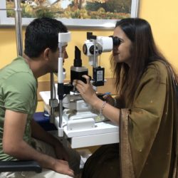

Comprehensive Eye Exam

A comprehensive eye exam does more than update your glasses prescription. It checks your eye pressure, optic nerve health, retina,…

Glaucoma: Risk Factors

All of us are at risk for glaucoma, which is why ophthalmologists recommend regular eye checks for everyone. In fact,…