Eye Pressure Test in Glaucoma is critical for diagnosing and monitoring the disease. The pressure inside the eye is an index of the pressure on the optic nerve. If this pressure rises, it can damage the optic nerve, and your vision. This disease is called glaucoma.
Eye pressure testing is one of the most common eye examinations. Many patients are told their pressure is “normal” or “a little high,” but the meaning of these numbers is often misunderstood. Moreover, it is important to understand eye pressure changes in order to understand glaucoma treatment.
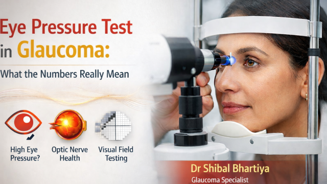
In glaucoma care, eye pressure is important — but it is not the whole story. Understanding how pressure relates to the optic nerve, visual field tests, and long-term risk is essential for protecting vision, explains Dr Bhartiya.
Dr Shibal Bhartiya is a fellowship-trained glaucoma specialist and Mayo Clinic Research Collaborator with over 25 years of experience. Her approach focuses on identifying risk before damage is irreversible, simplifying treatment decisions, and protecting vision long-term. Emphasis on early detection, risk assessment, and continuity of care. She is rated 5 stars across 1,500+ patient reviews on Google.
What Is Eye Pressure?
Eye pressure, also called intraocular pressure (IOP), refers to the pressure created by the fluid inside the eye.
The eye constantly produces and drains a clear fluid called aqueous humor. When this fluid does not drain properly, pressure inside the eye can increase.
Higher pressure can damage the optic nerve that carries visual information from the eye to the brain. This damage is the hallmark of glaucoma.
However, pressure alone does not define glaucoma.
Some patients develop optic nerve damage even with normal eye pressure, while others tolerate higher pressures without damage.
This is why glaucoma evaluation always goes beyond a single number.
What Is a Normal Eye Pressure?
For most people, eye pressure typically ranges between 10 and 21 mmHg.
But the concept of “normal” is more complicated than this range suggests.
Some patients develop normal-tension glaucoma, where optic nerve damage occurs even though eye pressure falls within the normal range. Others may have ocular hypertension, where pressure is elevated but the optic nerve remains healthy.
Because of this variation, glaucoma specialists interpret eye pressure in the context of the individual eye, not as an isolated number.
How Eye Pressure Is Measured
Measuring intraocular pressure accurately is not as simple as it sounds. The eye is not a rigid sphere, and every tonometry method makes assumptions about corneal thickness, curvature, and rigidity that can affect the reading. Understanding the main methods helps you interpret your own pressure numbers — and ask the right questions.
Goldmann Applanation Tonometry (GAT)
Goldmann applanation tonometry is the gold standard against which all other methods are measured. The doctor uses a small prism at the slit lamp to flatten a defined area of the cornea and calculates pressure from the force required to do so. It requires anaesthetic drops and yellow dye. It is accurate, reproducible, and used in virtually every glaucoma clinic worldwide.
Its limitation is that it assumes a standard corneal thickness of 520 microns. Corneas that are thinner than this will give falsely low readings; thicker corneas give falsely high ones. Corneal pachymetry — a quick test to measure corneal thickness — is therefore an important companion investigation in glaucoma assessment.
A Comparative evaluation of TonoPen AVIA, Goldmann applanation tonometry and non-contact tonometry by Dr Bhartiya and her colleagues at AIIMS, New Delhi, revealed that GAT remains the gold standard for measuring eye pressure.
Non-Contact Tonometry (Air Puff)
The air puff tonometer uses a brief pulse of air to indent the cornea and estimates pressure from the corneal response. No anaesthetic is needed and the instrument does not touch the eye. It is widely used for screening because it is fast, comfortable, and requires minimal training to operate.
It is less precise than Goldmann — readings can vary with blink reflex, corneal hydration, and patient anxiety — and is best used as a screening tool rather than for definitive pressure measurement in glaucoma patients already under treatment.
Perkins Applanation Tonometry
The Perkins tonometer uses the same applanation principle as Goldmann but is handheld. This makes it invaluable for measuring pressure in patients who cannot sit at a slit lamp — children under anaesthesia, patients in the operating theatre, or those who are bed-bound. Agreement between Perkins and Goldmann is good under standard conditions, though both are affected by altitude-related atmospheric pressure changes, as demonstrated in a study co-authored by Dr Bhartiya and colleagues across clinical centres in Mexico and the United States.
Dynamic Contour Tonometry (Pascal)
Dynamic contour tonometry (DCT) is a contact method that measures IOP by sensing the pressure within the eye directly, rather than by indenting or applanating the cornea. Because it is largely independent of corneal biomechanical properties, it is less affected by corneal thickness and curvature than Goldmann. It also provides an ocular pulse amplitude — a measure of the pressure fluctuation with each heartbeat — which carries prognostic value in glaucoma.
Two studies in which Dr Bhartiya was involved examined how atmospheric pressure changes at altitude affect DCT and Goldmann readings differently, with important implications for interpreting IOP measurements in patients living at or travelling to high altitudes.
TonoPen
The TonoPen is a portable, handheld electronic tonometer that applanates a very small area of the cornea. It is useful in scarred or irregular corneas where Goldmann readings are unreliable, and in theatre settings. Test-retest variability — how consistent the readings are on repeat measurement — was formally evaluated in a study by Dr Bhartiya and colleagues, confirming that while the device is clinically useful, a degree of measurement variability should be factored into clinical decisions.
Diaton (Transpalpebral Tonometry)
The Diaton measures IOP through the eyelid, without touching the cornea at all. This makes it attractive for patients with corneal disease or those who have had corneal surgery. A comparative evaluation by Dr Bhartiya’s group found that while Diaton readings correlated with Goldmann, the agreement was not close enough to use the two interchangeably in routine clinical management.
What Does This Mean for You as a Patient?
The method used to measure your pressure matters. If your glaucoma management is based on a borderline pressure reading from a screening air puff test, or if you have a thin cornea that has never been measured, the number your doctor is treating may not reflect your true intraocular pressure.
If you have any doubts about the accuracy of your pressure readings — particularly if you have an unusually thin or thick cornea, if readings have been inconsistent, or if you have had corneal surgery — it is worth asking your doctor which method was used and whether a second measurement with a different technique is warranted.
Why Eye Pressure Alone Cannot Diagnose Glaucoma
Glaucoma diagnosis requires looking at multiple pieces of information together.
A comprehensive glaucoma evaluation typically includes:
• optic nerve examination
• OCT scan of the optic nerve
• visual field testing
• corneal thickness measurement
• repeated pressure measurements over time
It is common for patients to be told their pressure is “normal,” only to later discover that subtle optic nerve damage was already present.
This is why glaucoma specialists focus on structure, function, and risk over time, rather than relying on pressure alone.
Understanding OCT and Visual Field Tests
Two tests are especially important in glaucoma care: OCT and Visual Fields
OCT (Optical Coherence Tomography) measures the thickness of the optic nerve fibers. It can detect structural damage even before vision changes occur.
Visual field testing measures how well the eye sees in different areas of the field of vision. It detects functional changes caused by optic nerve damage.
Often, structural changes on OCT appear years before noticeable symptoms. When both tests are interpreted together, they provide a clearer picture of glaucoma risk and progression.
Related Reading
Get an Online Glaucoma Consult
Why Do I Need a Visual Field Test?
Understanding Your OCT Report in Glaucoma
Visual Field and OCT: Structure & Function Correlation
Glaucoma Progression: What It Means and How to Slow It
Get a Glaucoma Second Opinion in Gurgaon
What Is Target Eye Pressure?
Once glaucoma is diagnosed, doctors often refer to a target eye pressure.
Target pressure is the level of eye pressure believed to be safe for that particular optic nerve.
However, target pressure is not a fixed number.
It is dynamic and individualized, based on several factors:
• stage of glaucoma
• age and life expectancy
• optic nerve vulnerability
• rate of progression
• response to treatment
For some patients, a modest reduction in pressure may be sufficient. Others may require a much lower pressure to protect the optic nerve.
You can read one of my research papers on this topic, indexed on Pubmed, on New perspectives on target intraocular pressure , which I co-authored with my colleagues from Sydney Eye Hospital, Sydney, Australia; The University of Sydney, Sydney, Australia; and Department of Clinical Neurosciences, University of Geneva, Switzerland.
Target Pressure and Quality of Life
An important part of glaucoma care is balancing disease control with quality of life.
Lowering eye pressure aggressively may sometimes require multiple medications, laser treatments, or surgery. While these treatments are effective, they can also affect daily comfort, cost, and convenience.
Modern glaucoma management therefore considers:
• stability of the optic nerve
• rate of disease progression
• treatment burden
• patient lifestyle and preferences
The goal is not simply to chase a number, but to maintain long-term visual stability while preserving quality of life.
Why Follow-Up Matters in Glaucoma
Glaucoma is a long-term condition, and eye pressure can change over time.
Regular follow-up allows doctors to monitor:
• pressure trends
• optic nerve changes
• visual field progression
• response to treatment
In many cases, glaucoma progression becomes clear only when tests are compared over time.
Consistent follow-up is therefore one of the most important parts of protecting vision.
When Should You See a Glaucoma Specialist?
A specialist evaluation or second opinion may be helpful if you:
• have elevated eye pressure
• have a family history of glaucoma
• were told your optic nerve looks suspicious
• have abnormal OCT or visual field results
• want a second opinion about glaucoma diagnosis or treatment
Early evaluation can help clarify risk and prevent long-term vision loss.
Frequently Asked Questions
What eye pressure is considered dangerous for glaucoma?
Eye pressure above 21 mmHg is traditionally considered higher than normal, but glaucoma can occur even when pressure is within the normal range. Some people develop normal-tension glaucoma, where optic nerve damage occurs despite normal pressure readings. Because of this, glaucoma specialists evaluate eye pressure together with optic nerve examination, OCT scans, and visual field tests rather than relying on a single number.
Can glaucoma occur if my eye pressure is normal?
Yes. This condition is called normal-tension glaucoma. In these cases, the optic nerve becomes damaged even though eye pressure appears normal. This is why glaucoma diagnosis depends on a combination of tests including optic nerve examination, OCT imaging, and visual field testing, not just pressure measurements.
How often should eye pressure be checked?
For healthy individuals without risk factors, eye pressure is usually checked during routine eye examinations. However, people with family history of glaucoma, suspicious optic nerves, elevated pressure, diabetes, or increasing age may require more detailed evaluation and regular follow-up. Patients diagnosed with glaucoma typically need periodic monitoring of eye pressure, OCT scans, and visual field tests to assess stability over time.
Why does my glaucoma doctor talk about target eye pressure?
Target eye pressure is the pressure level believed to be safe for a particular optic nerve. It represents the pressure at which further glaucoma damage is unlikely to occur. However, this target is not fixed. It may change over time depending on disease progression, response to treatment, and overall risk. The goal of treatment is to keep eye pressure around this safe range while maintaining comfort and quality of life.
Conclusion
Eye pressure is an important part of glaucoma care, but it is only one piece of a much larger puzzle.
Understanding how pressure relates to optic nerve health, visual field testing, and long-term risk allows glaucoma to be managed more thoughtfully and effectively.
The aim of modern glaucoma care is not simply to lower pressure, but to preserve vision over the long term while maintaining quality of life.
Read the research articles
This article was written by Dr Shibal Bhartiya, fellowship-trained glaucoma specialist and Mayo Clinic Research Collaborator, Clinical Director at Marengo Asia Hospitals, Gurugram, known for ethical, patient-centred glaucoma care and independent glaucoma second opinions. She is also the Program Director for Community Outreach & Wellness; and for the Marengo Asia International Institute of Neuro and Spine. This article was updated in April 2026.
She has published peer-reviewed research on glaucoma management, examining how treatment decisions should balance medical evidence, patient preferences, and long-term vision outcomes.
As Editor-in-Chief of Clinical and Experimental Vision and Eye Research and Executive Editor of the Journal of Current Glaucoma Practice (Pubmed Indexed, official journal of the International Society of Glaucoma Surgery), Dr Shibal Bhartiya brings editorial and research depth to every clinical decision. Her 200+ publications, including 90+ PubMed-indexed publications and 28 edited textbooks span glaucoma biology, surgical outcomes, health equity, and emerging diagnostics.
Access her work on Pubmed, Google Scholar, ResearchGate and ORCID.
Dr Shibal Bhartiya
Glaucoma • Second Opinion • Advanced Care
www.drshibalbhartiya.com
+91 88826 38735
1500+ Five Star Patient Reviews Google Business Profile
Upload your reports for a structured review.
If you are unable to come to Dr Bhartiya’s clinic: Read more about teleconsultation for glaucoma