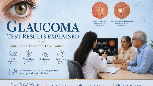
No single test confirms or rules out glaucoma. Doctors read your OCT, visual field and eye pressure together and track how they change over time.
Quick Interpretation Guide
Key rule: No single test confirms glaucoma; patterns + progression matter
OCT scan: Detects thinning of the optic nerve (early damage can appear here first)
Visual field test: Shows blind spots or peripheral vision loss (functional impact of disease)
Eye pressure (IOP): A risk factor, not a diagnosis, can be normal in glaucoma
Optic nerve exam: Assesses cupping and structural changes
If results are borderline or conflicting, progression over time, not a single test, determines diagnosis and treatment decisions. Dr Shibal Bhartiya, glaucoma specialist in Gurgaon, offers structured second opinions to interpret reports and guide treatment decisions.
Most patients reach my clinic holding something. A folder. A USB drive. Printouts from three different centres. They all ask the same thing. Is this serious? Do I need treatment?
That is the right question. The reports cannot answer it alone, because each one shows a single day. Glaucoma plays out over years.
Early glaucoma often causes no symptoms. Your vision can feel normal while the tests already show change. Small differences on a report matter more than how clearly you see.
Dr Shibal Bhartiya is a fellowship-trained glaucoma specialist and Mayo Clinic Research Collaborator with over 25 years of experience. Her approach focuses on identifying risk before damage is irreversible, simplifying treatment decisions, and protecting vision long-term. Emphasis on early detection, risk assessment, and continuity of care.
What Each Glaucoma Test Measures
Each test answers a different question. And the art of glaucoma practice is in interpreting them together, and not in isolation.
| Test | What it measures | What an abnormal result can mean | What can fool it |
|---|---|---|---|
| OCT scan | Thickness of the nerve fibre layer and optic nerve rim | Loss of nerve tissue, often before you notice any change | Myopia, tilted or large discs, poor signal, scan errors |
| Visual field (perimetry) | How well you see across your field, point by point | Blind spots or loss of side vision | Tiredness, learning effect, unreliable answers, cataract |
| Eye pressure (IOP) | Fluid pressure inside the eye | Higher risk of nerve damage | Corneal thickness, time of day, one-off readings |
| Optic nerve exam | Cup size, rim shape, notching | Visible structural damage | Naturally large cups, observer variation |
| Gonioscopy | The eye’s drainage angle | A narrow or closed angle, which changes the type of glaucoma | Needs clinical skill, so it never appears on a printout |
How to Read Your OCT Report
OCT scans the optic nerve and the nerve fibre layer of the retina. Your report shows thickness in microns, usually as an average and by sector.
Most reports colour each sector. Green means within normal limits. Yellow means borderline. Red means outside the normal range for that machine’s database.
A red sector is a flag. It is not a diagnosis.
Check the signal strength first. A poor scan can create false red areas. I also look at the ganglion cell layer, which helps in myopic eyes where the nerve fibre layer can mislead.
How to Read Your Visual Field Report
Check reliability first. Frequent fixation losses or false positive answers make a field hard to trust. A repeat test often gives a clearer answer.
Depending on the machine, your report may show three summary numbers:
- Mean deviation (MD) shows overall loss in decibels. A more negative number means more loss.
- Pattern standard deviation (PSD) shows how patchy the loss is.
- Visual Field Index (VFI) gives remaining function as a percentage.
The map matters more than the numbers. I look for loss that follows the nerve fibre pattern, such as an arc above or below the centre.
Then I check whether the map matches the OCT. Damage in the upper nerve should match loss in the lower field, and the reverse. Want the full walkthrough? Read what to expect and what the results mean.
How to Read Your Eye Pressure
Statistically normal eye pressure runs roughly 10 to 21 mmHg. Glaucoma can occur inside that range. Pressure can also sit above it without any nerve damage.
Several things shift the reading. Pressure changes through the day. A thick cornea can push the reading up, and a thin one can pull it down. One clinic reading captures none of that.
Pressure is a risk factor. It is also the one risk factor we can currently lower with treatment.
Why Change Over Time Matters More Than Any Number
One abnormal scan does not confirm glaucoma. One normal field does not rule it out.
A single report tells me where you are today. It cannot tell me where you are heading, or how fast.
Repeat tests also vary a little on their own. I look for change bigger than that normal noise. A reading that falls steadily across visits worries me more than a low reading that stays flat.
None of this works without a baseline. Many patients arrive with none. In that case, today’s tests become the baseline and we compare from there.
What Doctors Often Miss
- Machine mix-ups. Different OCT brands use different normal databases. Their numbers do not swap one for one.
- Scan quality. A red result on a poor scan can come from the scan, not the eye.
- Disagreement between tests. When structure and function disagree, that disagreement is itself a finding.
- Population differences. Many normal databases draw mostly on Western eyes. Studies suggest Asian eyes differ in disc size and nerve fibre thickness. A red flag may sit within normal variation for you. Read more on glaucoma in Indian eyes.
How I Review Your Reports
- Do the OCT and visual field agree? If not, which one reflects true disease?
- Is there a baseline, and what is the rate of change?
- Does the optic nerve I see match the measurements?
- What does your full risk profile show: age, family history, corneal thickness and systemic health?
- Do you need to act, wait, or watch?
What Actually Matters When Reading Glaucoma Test Results
Consistency across tests. Structure and function should agree. When they do not, that disagreement is itself a clinical finding.
Change over time. Progression, not an absolute number, is how glaucoma causes irreversible harm. A stable OCT at 80 microns is far less alarming than one that dropped from 100 to 80 over two years.
Correlation with clinical examination. Disc photographs, gonioscopy, pachymetry, and a detailed history all shape what the reports mean. Printouts do not replace an examination.
A baseline to compare against. Without a baseline reading, no one can determine whether your results are stable or worsening. Many patients have no baseline at all.
When to See a Doctor
See a Doctor Urgently
Go the same day for sudden eye pain with headache or nausea, halos around lights with a red eye, or sudden loss of vision. These can signal acute angle closure or another emergency. Do not wait for a routine slot.
See a Doctor
Book a visit, and consider a second opinion, if:
- Your OCT and visual field keep disagreeing on repeat testing.
- You were told surgery is needed but your vision feels unchanged.
- Your results differ across centres.
- You have no baseline and cannot tell if you are stable.
- You are a glaucoma suspect and nobody has set a follow-up plan.
What This Means for You
A single abnormal result is a reason to look closer, not a diagnosis.
Bring every report you have, as original files or printouts, plus your list of eye drops. Older tests matter most because they give me a baseline. See what to bring for your visit.
More tests rarely solve confusion. If your reports leave you unsure, you may need interpretation, not another scan. Upload your reports for a structured review, in person or through teleconsultation.
Book an Appointment:
📍 Gurgaon 📞 +91 88826 38735 | 🌐 Contact Us | Second Opinion Form for teleconsults
Frequently Asked Questions
Can I have glaucoma with normal eye pressure?
Yes. Normal tension glaucoma is well recognised and common in Asia. Many people with glaucoma have pressure in the statistically normal range. Pressure alone cannot rule it out.
What does a thin or red OCT result mean?
It means your nerve fibre layer measures below the database average at that point. It does not automatically mean glaucoma. Myopia, natural variation, past inflammation or a poor scan can cause the same result. I compare it with your field, your optic nerve and your history.
Can a normal visual field miss glaucoma?
Yes. Nerve damage often shows on OCT months or years before the field changes. A normal field means your function is intact today. Repeat testing shows whether that holds. See visual field testing in Gurgaon.
Why do my results differ between hospitals?
Machines differ in brand, software and normal database. Fields also vary with tiredness, technique and the learning effect. Some variation is normal. Comparing tests from the same machine type over time gives the most reliable picture.
How often should I repeat glaucoma tests?
It depends on your risk. With confirmed glaucoma, OCT and visual fields are often repeated every six to twelve months. Suspects usually need review once a year. Your specialist adjusts this if you progress.
Why do my results vary across different hospitals or centres?
OCT results vary across different machine brands, software versions, and normative databases. Visual field results vary with patient fatigue, technique, and learning effect. Variation across centres is common and does not always indicate a change in your condition. Comparing tests done on the same machine type, at the same centre, over time gives the most reliable information.
What is the difference between glaucoma and a glaucoma suspect?
A glaucoma suspect is someone who has one or more features that raise concern: elevated eye pressure, a suspicious optic nerve, a thin retinal nerve fibre layer, a family history, or an equivocal visual field, but who does not yet meet the criteria for a glaucoma diagnosis. Suspects require regular monitoring because some will convert to glaucoma over time and some will not. Distinguishing the two requires careful longitudinal review.
When should I seek a second opinion on my glaucoma reports?
Seek a second opinion if your OCT and visual field results disagree persistently, if you have been told surgery is needed but your vision seems unchanged, if your reports vary significantly across centres, or if you have no baseline and cannot determine whether your condition is stable. A second opinion from a fellowship-trained glaucoma specialist can clarify your diagnosis and give you confidence in your treatment plan.
Known for her structured approach to glaucoma risk assessment and progression analysis, Dr Shibal Bhartiya provides trusted second opinions in person and online.
This page is a part of the Glaucoma Hub. you may want to read about Glaucoma Progression, and Risk Stratification in Glaucoma. Other articles of interest could be Advanced Glaucoma Care in Gurgaon, What Good Glaucoma Care Actually Optimises For, What Happens If Glaucoma Is Left Untreated?, More Glaucoma Eye Drops is Not Better Glaucoma Care, 5 Mistakes Patients Make in Glaucoma Care and Do You Really Need Treatment for Glaucoma?
To know more about visual fields, read What to Expect and What the Results Mean, How To Do Visual Fields, Glaucoma Test Results Explained: OCT, Visual Fields and Eye Pressure, Why Do I Need a Visual Field.
About the Author
This article was written by Dr Shibal Bhartiya, fellowship-trained glaucoma specialist and Mayo Clinic Research Collaborator, Clinical Director at Marengo Asia Hospitals, Gurugram, known for ethical, patient-centred glaucoma care and independent glaucoma second opinions. She is also the Program Director for Community Outreach & Wellness; and for the Marengo Asia International Institute of Neuro and Spine.
Dr Bhartiya has published peer-reviewed research onglaucoma management, examining how treatment decisions should balance medical evidence, patient preferences, and long-term vision outcomes. She is also a committed patient advocate and mainstream medical columnist, translating complex ophthalmic evidence into actionable public health insights across leading national platforms including NDTV and OnlyMyHealth
As Editor-in-Chief of Clinical and Experimental Vision and Eye Research and Executive Editor of the Journal of Current Glaucoma Practice (Pubmed Indexed, official journal of the International Society of Glaucoma Surgery). Her 200+ publications, including 90+ PubMed-indexed publications and 28 edited textbooks span glaucoma biology, surgical outcomes, health equity, and emerging diagnostics.
1600+ Five Star Patient Reviews Google Business Profile
If you are unable to come to Dr Bhartiya’s clinic: Read more about teleconsultation
Read her research on PubMed | Google Scholar | ResearchGate | ORCID | Github | Wikidata
📋Upload your reports for a structured review.| 🌐 Contact Us| 📞 +91 88826 38735
⭐ Helped by this article? Leave a Google review — it helps other patients find reliable eye care.
Last Reviewed: October 2026