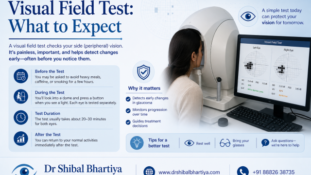
A visual field test maps the full extent of what you can see, including your peripheral vision, without moving your eyes. It is one of the most important tests in glaucoma care, and understanding what happens during and after it makes the experience significantly less stressful, says Dr Shibal Bhartiya.
Most patients find the test harder than they expected. That is normal, and it does not mean something is wrong.
Dr Shibal Bhartiya is a fellowship-trained glaucoma specialist and Mayo Clinic Research Collaborator with over 25 years of experience. Her approach focuses on identifying risk before damage is irreversible, simplifying treatment decisions, and protecting vision long-term. Emphasis on early detection, risk assessment, and continuity of care. She is rated 5 stars across 1,500+ patient reviews on Google.
Why the Visual Field Test Matters
Glaucoma damages vision from the periphery inward. By the time central vision is affected, significant and irreversible damage has already occurred. The visual field test, also called perimetry, detects this peripheral loss early, often before you are aware of any change yourself.
It is the closest thing glaucoma care has to a progress report. A single test gives a snapshot. A series of tests over time, your visual field history, tells your doctor whether your glaucoma is stable, slowly progressing, or changing faster than expected. This directly guides treatment decisions: whether your current pressure target is adequate, whether medication needs to be adjusted, and whether surgery needs to be considered.
No other test replaces it. OCT scans the structure of the optic nerve. Visual field testing measures function, what you actually see. Both are needed, and they do not always agree. When they diverge, understanding which is leading and which is lagging is a matter of specialist interpretation.
What Happens During the Test
You will sit in front of a large, white, bowl-shaped machine, most commonly a Humphrey Field Analyser, though other perimeters are used. One eye is tested at a time; the other is covered with a patch or occluder.
You will be asked to rest your chin on a chin rest, keep your head still, and focus on a small central fixation target, usually a yellow or white dot in the centre of the bowl. You must keep your gaze on this target throughout the test, even when lights appear in your peripheral vision.
A series of small lights of varying brightness will flash at different locations across your visual field. Your job is to press a handheld button each time you see a light. The machine presents lights at each location multiple times, at different intensities, to determine the threshold, the dimmest light you can reliably detect, at every point in your visual field.
The test takes approximately five to eight minutes per eye, though this varies by the programme used and the patient.
What Makes It Difficult — and Why That Is Normal
The visual field test is genuinely challenging. Several things catch patients off guard:
You will miss lights. This is expected and built into the algorithm. The test deliberately presents lights at intensities near your threshold, meaning you are not supposed to see all of them. Missing lights does not mean your vision is failing.
The lights are dimmest in the areas the machine is most uncertain about. If you have already been shown to see well in a given area, the machine will challenge that area less. Where it is uncertain, it will probe more, with dimmer stimuli that are harder to detect.
Fixation matters enormously. Looking away from the centre, even briefly, produces false results. If you lose fixation, pause, tell the technician, and resume. Most machines monitor fixation automatically and flag losses. A test with many fixation losses is unreliable and may need to be repeated.
Fatigue affects the second half of the test. Sustained attention is cognitively demanding. The second eye tested in a session is often performed slightly worse than the first, particularly in older patients. This is physiological, not pathological.
The first test is almost always the worst. Visual field testing has a steep learning curve. First-time patients almost universally perform below their true visual field capacity because the task is unfamiliar. By the second and third tests, performance stabilises. This is called the learning effect, and it is one of the reasons a single test result is rarely acted upon in isolation.
Types of Visual Field Tests
Standard automated perimetry (SAP) is the gold standard for glaucoma monitoring. The Humphrey Visual Field 24-2 tests 54 locations within the central 24 degrees of vision, the area most relevant for glaucoma detection and monitoring. The 30-2 extends this to 76 points and is used in specific situations.
Frequency doubling technology (FDT) perimetry uses a different stimulus, flickering gratings, to selectively test a subset of nerve fibres that are particularly vulnerable in early glaucoma. It is faster and easier to perform but provides less detailed information than SAP.
Goldmann perimetry is a manual test using a moving stimulus, performed by a trained technician. It is used in specific clinical situations including advanced glaucoma, neurological visual field defects, and functional visual loss assessment.
Supra-threshold screening tests whether you can see at a fixed brightness level above the expected threshold. It is faster than full threshold testing and used for screening rather than monitoring.
For glaucoma follow-up, standard automated perimetry, specifically the Humphrey 24-2, is the workhorse test.
How to Read Your Visual Field Printout
The Humphrey Field Analyser printout is dense, but the key elements are:
Greyscale map: The intuitive, visual representation of your field. Darker areas represent locations where sensitivity is reduced. This is the map most patients look at first, but it is the least statistically reliable representation.
Total Deviation plot: Compares your sensitivity at each point to age-matched normal values. Points shown in black are significantly below normal. This is more informative than the greyscale.
Pattern Deviation plot: Removes the effect of any generalised depression (such as from a cataract or media opacity) and shows localised defects. This is the most diagnostically useful plot for glaucoma because it isolates defects that are truly focal, the pattern of damage characteristic of glaucomatous nerve injury.
Mean Deviation (MD): A single number summarising the overall level of visual field sensitivity relative to normal. Normal is close to zero. Increasingly negative numbers (−3, −6, −10 and beyond) indicate progressively greater overall loss. Advanced glaucoma is typically defined as MD worse than −12 dB.
Pattern Standard Deviation (PSD): Measures the unevenness of loss across the field. A high PSD with a relatively normal MD suggests focal, localised damage, a pattern typical of early glaucoma. A low PSD with a very negative MD suggests uniform, generalised loss, seen in advanced glaucoma or in diffuse media opacities.
Visual Field Index (VFI): A percentage score 100% is normal, 0% is total loss. It is used to track progression over time and is less affected by cataract than MD.
Reliability indices: Fixation losses, false positives, and false negatives. High false positives (trigger-happy pressing) artificially inflate sensitivity. High false negatives (missing lights you previously detected) suggest fatigue or inattention. A test with poor reliability indices needs to be interpreted with caution, and sometimes repeated.
What Doctors Often Miss Telling You
- One abnormal result rarely changes management. Glaucoma decisions are based on a pattern across multiple tests, not a single printout. If your field looks worse than last time, your doctor will almost certainly want to confirm it before adjusting treatment. A single bad test is not a crisis.
- Cataract significantly affects visual field results. A dense cataract depresses overall sensitivity and can simulate or mask glaucomatous loss. If your visual field has deteriorated and you also have a developing cataract, your surgeon needs to know that both conditions exist, and your glaucoma specialist needs to be involved in the timing of cataract surgery.
- Your medications can affect the test. Certain sedating medications, including some antihistamines, antidepressants, and sleep aids, can blunt attention and worsen performance on perimetry. If you take any of these, try to time your test when their effect is minimal, or discuss this with your doctor.
- Miosis from glaucoma drops affects field testing. Some glaucoma eye drops, particularly older pilocarpine formulations, constrict the pupil significantly. A very small pupil reduces the light entering the eye and depresses field sensitivity. If you are on pilocarpine, your technician should be aware.
- The machine does not diagnose glaucoma, your doctor does. The printout flags statistical deviations from normal. It does not tell you whether those deviations represent glaucoma, another disease, or a test artefact. Interpretation requires clinical context.
When to Worry
The visual field test result itself is not an emergency. However, seek urgent review if:
- Your visual field shows a new, dense central scotoma, loss near the fixation point, that was not present before
- You notice a new area of missing vision in your daily life that has not been assessed
- Your MD has worsened by more than 3 dB compared to your previous test
- Your doctor uses the words “rapid progression” in relation to your field results
- You develop any sudden visual change between scheduled tests, do not wait for your next perimetry appointment
This page is a part of the Glaucoma Hub. you may want to read about Glaucoma Progression, and Risk Stratification in Glaucoma. Other articles of interest could be Advanced Glaucoma Care in Gurgaon, What Good Glaucoma Care Actually Optimises For, What Happens If Glaucoma Is Left Untreated?, More Glaucoma Eye Drops is Not Better Glaucoma Care, 5 Mistakes Patients Make in Glaucoma Care and Do You Really Need Treatment for Glaucoma?
Frequently Asked Questions
How long does a visual field test take?
Each eye takes approximately five to eight minutes with standard automated perimetry, depending on the programme used. Allow 20–30 minutes for the full appointment including setup, one rest break if needed, and discussion of results.
Does the visual field test hurt?
No. It is entirely non-invasive. Some patients find it tiring or mentally demanding, but there is no discomfort involved.
How often do I need a visual field test?
Most glaucoma patients need two visual field tests per year. Newly diagnosed patients, those with rapidly progressing disease, or those in whom treatment has recently changed may need more frequent testing. Stable, well-controlled patients may be tested once a year. Your doctor will advise the appropriate interval for your situation.
What if I perform badly on the test?
One poor test is not clinically meaningful in isolation. Tell your technician if you are struggling — a short break mid-test is allowed. Your doctor will look at your result in the context of your previous tests and your clinical picture. A single outlier result is generally not acted upon without confirmation.
Can I wear my glasses during the visual field test?
Yes, and you should. You will be given a trial lens to correct for your distance prescription and any reading addition appropriate for the test distance. Wear your glasses or contact lenses as you normally would; the technician will handle the necessary adjustments.
What is a visual field defect?
A visual field defect is an area of reduced or absent sensitivity in your visual field. In glaucoma, defects typically appear first in the mid-peripheral field, often as an arcuate scotoma (an arc-shaped area of loss) corresponding to the pattern of retinal nerve fibre bundles that glaucoma damages first.
Can the visual field improve?
In established glaucoma, visual field loss is generally permanent, the nerve fibres that have been destroyed do not regenerate. However, some apparent improvement can occur when a previous test was performed poorly, due to fatigue, learning effect, or inattention, and a subsequent test reflects true performance more accurately. This is one reason why a series of tests is more meaningful than any single result.
Speak to a Specialist
If your visual field results have confused or worried you, or if you have been told your field is changing and want a second opinion on what that means for your treatment, a specialist review of your full field history, alongside your OCT and clinical findings, is the right next step.
Book a consultation: +91 88826 38735 | www.drshibalbhartiya.com
Upload your visual field printouts and OCT reports through the website before your appointment — reviewing them in advance makes the consultation significantly more productive.
To know more about visual fields, read What to Expect and What the Results Mean, How To Do Visual Fields, Glaucoma Test Results Explained: OCT, Visual Fields and Eye Pressure, Why Do I Need a Visual Field.
About the Author
This article was written by Dr Shibal Bhartiya, fellowship-trained glaucoma specialist and Mayo Clinic Research Collaborator, Clinical Director at Marengo Asia Hospitals, Gurugram, known for ethical, patient-centred glaucoma care and independent glaucoma second opinions. She is also the Program Director for Community Outreach & Wellness; and for the Marengo Asia International Institute of Neuro and Spine.
She has published peer-reviewed research on glaucoma management, examining how treatment decisions should balance medical evidence, patient preferences, and long-term vision outcomes.
As Editor-in-Chief of Clinical and Experimental Vision and Eye Research and Executive Editor of the Journal of Current Glaucoma Practice (Pubmed Indexed, official journal of the International Society of Glaucoma Surgery), Dr Shibal Bhartiya brings editorial and research depth to every clinical decision. Her 200+ publications, including 90+ PubMed-indexed publications and 28 edited textbooks span glaucoma biology, surgical outcomes, health equity, and emerging diagnostics.
Access her work on Pubmed, Google Scholar, ResearchGate and ORCID.
Dr Shibal Bhartiya
Glaucoma • Second Opinion • Advanced Care
www.drshibalbhartiya.com
+91 88826 38735
1600+ Five Star Patient Reviews Google Business Profile
Upload your reports for a structured review.
If you are unable to come to Dr Bhartiya’s clinic: Read more about teleconsultation for glaucoma
PubMed Profile | Google Scholar | ResearchGate | ORCID
⭐ Helped by this article? Leave a Google review — it helps other patients find reliable eye care.
📋 Upload your reports for review before your appointment at www.drshibalbhartiya.com
📞 +91 88826 38735