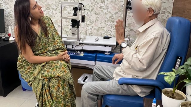
A visual field test checks your side (peripheral) vision and helps detect or monitor glaucoma and other optic nerve conditions. During the test, you look straight ahead and press a button whenever you notice lights appearing in different parts of your vision.
Automated static perimetry is the clinical gold standard for tracking glaucoma progression. Yet it is notoriously anxiety-inducing. High fixation losses and false positives corrupt diagnostic data when a patient is stressed. Active coaching before and during the test stabilises fixation, yields clean reproducible data, and transforms a feared exam into a collaborative clinical tool.
Please also read Visual Field Test (Perimetry) in Gurgaon.
Dr Shibal Bhartiya is a fellowship-trained glaucoma specialist and Mayo Clinic Research Collaborator with over 25 years of experience. Her approach focuses on identifying risk before damage is irreversible, simplifying treatment decisions, and protecting vision long-term. Emphasis on early detection, risk assessment, and continuity of care. She is rated 5 stars across 1,500+ patient reviews on Google.
How Patient-First Coaching Transforms Glaucoma Perimetry
Ask any glaucoma patient what part of their routine checkup they dread most. Nine out of ten will say the visual field test.
Sitting alone in a dark room, staring at a central yellow light, clicking a button for faint flashes you think you might be missing — it feels less like a diagnostic test and more like a high-stakes exam you are destined to fail.
A patient recently left a review that captured exactly why we approach this differently. They noted how other clinics seat you in the machine and tell you to press the clicker. No explanation. No preparation. Just anxiety and confusion. They described how, in our clinic, the entire experience was different. We walked them through what the visual field map actually shows. We explained the rhythm of the test before they started. They felt like a partner in their own care — not a passive subject.
You can read their experience here on Google.
When a patient understands that missing some flashes is a normal part of the machine’s threshold calculation, their heart rate drops. Their blinking stabilises. Their anxiety disappears.
That extra ten minutes of human coaching does not just produce a more comfortable patient. It produces pristine, accurate diagnostic data — the data we rely on to protect their optic nerve for decades.
What Actually Happens During a Visual Field Test
You sit with one eye covered and rest your chin on the machine. Your job is simple: keep looking at the central target and press the button whenever you notice a light anywhere in your side vision.
You are not expected to see every flash.
In fact, the machine deliberately presents lights that become increasingly faint to identify the threshold where vision transitions from “seen” to “not seen.” Missing some lights is not failure — it is how the test works.
Blink normally. Take short pauses if needed. If your attention drifts for a moment, do not panic and start clicking rapidly to catch up. The best visual field tests are usually not the fastest tests. They are the calmest.
The Most Common Mistake Patients Make
Patients often believe this is an intelligence test or a reaction-time test.
It is neither.
Trying too hard can sometimes reduce accuracy. Clicking every time you think a light might have appeared creates false positives. Chasing missed flashes leads to fatigue and fixation loss.
The goal is not perfection. The goal is honest responses.
Why One Visual Field Rarely Tells the Whole Story
A visual field is not interpreted in isolation.
Sleep, dry eye, anxiety, distraction, cataract, learning the machine, and even understanding instructions can influence a result.
That is why glaucoma decisions are usually made by combining visual fields with optic nerve examination, eye pressure, imaging, and change over time.
Protecting vision is rarely about one dramatic test result. It is about recognising patterns early and responding before change becomes irreversible.
This page is a part of the Glaucoma Hub. you may want to read about Glaucoma Progression, and Risk Stratification in Glaucoma. Other articles of interest could be Advanced Glaucoma Care in Gurgaon, What Good Glaucoma Care Actually Optimises For, What Happens If Glaucoma Is Left Untreated?, More Glaucoma Eye Drops is Not Better Glaucoma Care, 5 Mistakes Patients Make in Glaucoma Care and Do You Really Need Treatment for Glaucoma?
FAQs
How do I prepare for a visual field test?
No special preparation is usually needed. Wear your glasses if advised, stay relaxed, and try to rest your eyes before the test.
Is a visual field test painful?
No. A visual field test is non-contact, painless, and usually takes only a few minutes for each eye.
Why do visual field tests need to be repeated?
Visual field tests help monitor change over time. In glaucoma, repeated tests are often more useful than a single result because they help detect progression early.
Why is the visual field test for glaucoma so stressful?
The test is designed to find the absolute limit of your peripheral vision. It presents flashes that are intentionally very faint, so feeling like you are missing lights or guessing is completely normal. This design triggers anxiety when the process is not explained beforehand. Preparation changes the entire experience.
How does anxiety affect the accuracy of a glaucoma perimetry test?
High anxiety leads to irregular blinking, rapid head movements, and false-positive clicking. These introduce significant noise into the results. An ophthalmologist cannot reliably distinguish true disease progression from a stressful test day. A coached, relaxed patient produces far more clinically reliable data.
What if I think I did badly on my visual field test?
Many patients feel they performed poorly, especially during early tests. A difficult test does not automatically mean glaucoma has worsened. Ophthalmologists interpret reliability measures, compare previous results, and look for repeatable patterns over time.
Am I Doing My Visual Field Test Wrong?
Most patients worry they are doing badly because they miss flashes or feel uncertain during the test. That feeling is normal. Visual field testing is designed to find the edge of what you can see, so missing lights is expected and does not mean you have failed.
Why Do I Keep Missing Lights on My Glaucoma Test?
The machine deliberately shows lights that become fainter and fainter to calculate your visual threshold. Missing some lights helps the test work properly. Trying to click for every possible flash often makes results less reliable than staying relaxed and responding naturally.
Read about the full spectrum of glaucoma diagnosis and treatment. To know more about visual fields, read What to Expect and What the Results Mean, How To Do Visual Fields, Glaucoma Test Results Explained: OCT, Visual Fields and Eye Pressure, Why Do I Need a Visual Field.
About the Author
This article was written by Dr Shibal Bhartiya, fellowship-trained glaucoma specialist and Mayo Clinic Research Collaborator, Clinical Director at Marengo Asia Hospitals, Gurugram, known for ethical, patient-centred glaucoma care and independent glaucoma second opinions. She is also the Program Director for Community Outreach & Wellness; and for the Marengo Asia International Institute of Neuro and Spine.
She has published peer-reviewed research on glaucoma management, examining how treatment decisions should balance medical evidence, patient preferences, and long-term vision outcomes.
As Editor-in-Chief of Clinical and Experimental Vision and Eye Research and Executive Editor of the Journal of Current Glaucoma Practice (Pubmed Indexed, official journal of the International Society of Glaucoma Surgery), Dr Shibal Bhartiya brings editorial and research depth to every clinical decision. Her 200+ publications, including 90+ PubMed-indexed publications and 28 edited textbooks span glaucoma biology, surgical outcomes, health equity, and emerging diagnostics.
1500+ Five Star Patient Reviews Google Business Profile
If you are unable to come to Dr Bhartiya’s clinic: Read more about teleconsultation
Read her research on PubMed | Google Scholar | ResearchGate | ORCID
Upload your reports for a structured review.| www.drshibalbhartiya.com | +91 88826 38735
Leave a review on Google