Gonioscopy in Gurgaon: Why This Test Explains Your Glaucoma Risk

Gonioscopy is the clinical gold standard that directly examines the eye’s drainage angle to determine whether your glaucoma risk stems from an open, narrow, or physically blocked fluid pathway. Performed under dimmed lighting with dynamic indentation, it detects occludable angles and permanent scarring that standard slit-lamp exams miss—identifying whether you need preventive laser iridotomy or medical pressure control before irreversible nerve damage occurs. Dr Shibal Bhartiya performs gonioscopy in Gurgaon with the room lights dimmed rather than under bright slit-lamp illumination. This is because bright light constricts the pupil and can make a narrow angle falsely appear open.

Gonioscopy is an eye test that uses a special mirrored lens placed on the eye’s surface to directly view the drainage angle. The drainage angle is the structure that determines whether glaucoma is open-angle or angle-closure. That decision determines whether your treatment is drops, laser, or urgent intervention. Which is why it is extremely important.

If you’ve been told you need a gonioscopy in Gurgaon, it’s worth knowing that not all gonioscopy is done the same way, and the difference affects whether a narrow or closing angle gets caught at all.

Dr Shibal Bhartiya is a fellowship-trained glaucoma specialist and Mayo Clinic Research Collaborator with over 25 years of experience. Her approach focuses on identifying risk before damage is irreversible, simplifying treatment decisions, and protecting vision long-term. Emphasis on early detection, risk assessment, and continuity of care. She is rated 5 stars across 1,600+ patient reviews on Google. Her PubMed-indexed research on angle-closure diagnosis speaks to her extensive work and experience in gonioscopy.


Does Gonioscopy Hurt? What to Expect During the 5-Minute Test

No, gonioscopy is not painful, but you may feel a little pressure on the eye. Your eye is numbed with anaesthetic drops, so the test itself doesn’t hurt. You may feel mild pressure but not pain. A small amount of gel is placed on a contact lens. The lens is then gently rested on the surface of your eye. You’ll be asked to look in a few different directions while I view your drainage angle through the microscope. The whole test takes about five minutes. Your vision may stay slightly blurry for 15–20 minutes afterward as the gel clears.

What Your Findings Mean: Open Angle vs. Occludable Angle vs. Angle Closure

  • Open angle: Your drainage channel is unobstructed. If you have glaucoma, it’s open-angle glaucoma — typically managed with drops, laser, or surgery aimed at lowering pressure over time.
  • Narrow or occludable angle: Your drainage channel is at risk of closing, even if it hasn’t yet. This changes the conversation from “treat glaucoma” to “prevent an acute attack” — often with a same-visit or near-term laser iridotomy.
  • Closed angle: The drainage channel is blocked. This is urgent. Untreated angle closure can cause a sudden, painful pressure spike and permanent vision loss within hours to days.
  • Other findings — pigment deposits, scarring (synechiae), or abnormal blood vessels. These can point to pigment dispersion syndrome, past trauma, or diabetic eye changes. Each redirects your treatment plan differently.

In short: Gonioscopy doesn’t just confirm glaucoma. It decides which kind you have, and that decision determines whether your treatment is drops, laser, or urgent intervention.


Why is Gonioscopy Important?

Gonioscopy is an eye test that visualises the drainage angle of the eye. This drainage angle is responsible for maintaining the eye pressure. Gonioscopy determines if the glaucoma is open angle, or angle closure. It’s findings depend heavily on room lighting and examiner technique. A gonioscopy done under bright slit-lamp illumination can make a narrow angle look falsely open. This is because light constricts the pupil and pulls the iris away from the drainage angle. In Gurgaon, I perform gonioscopy in a dimmed room specifically to avoid this artifact. This way, the angle is graded at its true, physiologic width, not an artificially widened one.


Why Doing Gonioscopy in the Dark Matters

This is one of the most under-discussed variables in glaucoma diagnosis.

When light hits the eye, the pupil constricts. A smaller pupil pulls the peripheral iris away from the trabecular meshwork, which can open up an angle that is, in normal daily conditions, narrow or intermittently closing. If your gonioscopy is done in a bright room, an angle-closure risk can be missed — graded as open when it isn’t reliably open at all.

I do gonioscopy with the room lights dimmed, so the pupil stays closer to its natural size. This is closer to how your eye actually behaves when you walk out of a bright clinic into dim evening light, or sit in a dark cinema. These are the exact conditions in which angle closure is most likely to be triggered. Where indicated, I also use dynamic (indentation) gonioscopy. This involves gently pressing the lens to distinguish a truly closed angle from one that only appears closed (appositional closure). This distinction changes management directly. It decides whether you need a laser iridotomy, monitoring, or nothing at all.


Why Experience in This Specific Test Matters

Gonioscopy is a skill-dependent test — the same eye can be graded differently by two examiners. It takes practiced technique to hold the lens steady, avoid corneal compression that distorts the view, and read subtle angle structures consistently.

Gonioscopy is a routine part of my daily glaucoma practice, not an occasional add-on test. As a fellowship-trained glaucoma and neuro-ophthalmology specialist with over 25 years in the field, and as a research collaborator with Mayo Clinic, Jacksonville, my clinical work is built around glaucoma diagnosis and long-term angle monitoring. This means I perform gonioscopy, not delegate, on nearly every glaucoma or glaucoma-suspect patient I see.


Gonioscopy vs. Anterior Segment OCT (AS-OCT)

While Anterior Segment OCT (AS-OCT) offers an objective, non-contact, high-resolution cross-sectional scan of the anterior chamber angle—making it an excellent, non-invasive imaging tool for documentation and screening—gonioscopy remains the indispensable clinical gold standard for glaucoma diagnosis. AS-OCT captures a static image in dark or light conditions, but it cannot differentiate between appositional contact and true permanent scarring (peripheral anterior synechiae, or PAS), nor can it visualize critical pigmentary details, pseudoexfoliation deposits, neovascularization, or angle recession.

Gonioscopy uniquely allows the clinician to perform dynamic indentation (compression), gently applying pressure through the mirrored lens to physically test whether a closed drainage angle can be reopened or is permanently scarred shut—a critical distinction that directly determines whether a patient requires preventive laser peripheral iridotomy (LPI), medical therapy, or surgical intervention.


Dr Shibal Bhartiya’s Published Research on Angle Closure

My angle-closure glaucoma work has been published in peer-reviewed journals and is indexed on PubMed, including:

You can view these and my other publications on PubMed and Google Scholar.


Frequently Asked Questions

Does room lighting really change my gonioscopy result?

Yes. Bright light constricts the pupil and can pull the iris away from the drainage angle, making a narrow angle look artificially open. Gonioscopy done in dim lighting reflects your angle’s true, physiologic width.

What is dynamic (indentation) gonioscopy, and do I need it?

It’s a technique where gentle pressure is applied through the lens to see whether a closed-looking angle is truly closed or only appositionally closed. It’s used selectively, when the initial view suggests possible closure. This is because it changes whether you need a laser iridotomy or just monitoring.

Is gonioscopy available in Gurgaon?

Yes. I perform gonioscopy routinely in Gurgaon as part of every comprehensive glaucoma evaluation. I specifically do it in a dark room, and do the dynamic indentation where indicated.

How is this different from gonioscopy done elsewhere?

The test itself is standardised, but technique varies. Room lighting, indentation technique, and examiner experience all affect the reading. These are the specific variables I control for in every gonioscopy I perform.

How long will my vision stay blurry after a gonioscopy, and can I drive myself home?

The mild blurring and smudging you experience usually clear completely within 15 to 20 minutes. This temporary blurriness is not caused by long-acting dilation drops, but simply by the viscous lubricating gel placed on the diagnostic lens to protect your cornea during the examination. Once the gel washes away with natural blinking, your baseline vision returns to normal, and you can comfortably drive yourself home.

Can a gonioscopy test actually prevent an acute glaucoma attack?

Yes, indirectly—it is often the single test that saves a patient from a sudden, sight-threatening emergency. Acute angle-closure glaucoma happens when the eye’s drainage path suddenly blocks, triggering a rapid, agonizing spike in intraocular pressure. Gonioscopy is the only clinical examination that lets a specialist see an anatomically narrow or occludable angle long before symptoms appear. Detecting this early allows your doctor to perform a quick, preventive laser peripheral iridotomy (LPI) to keep the fluid pathway open permanently.


Book a Gonioscopy or Glaucoma Evaluation in Gurgaon

If you have a shallow anterior chamber, a family history of angle closure, or have simply never had your drainage angle formally assessed. A gonioscopy — done correctly — is a five-minute test that can prevent an acute angle-closure attack.

If you have been diagnosed with glaucoma, but have never had a gonioscopy, Request a Glaucoma Second Opinion.

Book an Appointment At my clinic in Gurgaon→

📍 Gurgaon 📞 +91 88826 38735 | 🌐 Contact Us | Second Opinion Form for teleconsults

Please also read about Comprehensive Eye Exam.


This article is part of the Glaucoma Diagnosis Hub. You may also want to read Eye Pressure Measurement, Why Do I Need a Visual Field Test? Understanding Your OCT Report in Glaucoma, Visual Field and OCT: Structure & Function Correlation, Gonioscopy, Glaucoma Progression: What It Means and How to Slow It, Do I Really Need All These Tests and Get a Glaucoma Second Opinion in Gurgaon


About the Author

This article was written by Dr Shibal Bhartiya, fellowship-trained glaucoma specialist and Mayo Clinic Research Collaborator, Clinical Director at Marengo Asia Hospitals, Gurugram, known for ethical, patient-centred glaucoma care and independent glaucoma second opinions. She is also the Program Director for Community Outreach & Wellness; and for the Marengo Asia International Institute of Neuro and Spine.

Dr Bhartiya has published peer-reviewed research on glaucoma management, examining how treatment decisions should balance medical evidence, patient preferences, and long-term vision outcomes. She is also a committed patient advocate and mainstream medical columnist, translating complex ophthalmic evidence into actionable public health insights across leading national platforms including NDTV and OnlyMyHealth

As Editor-in-Chief of Clinical and Experimental Vision and Eye Research and Executive Editor of the Journal of Current Glaucoma Practice (Pubmed Indexed, official journal of the International Society of Glaucoma Surgery). Her 200+ publications, including 90+ PubMed-indexed publications and 28 edited textbooks span glaucoma biology, surgical outcomes, health equity, and emerging diagnostics.

1670+ Five Star Patient Reviews Google Business Profile

If you are unable to come to Dr Bhartiya’s clinic: Read more about teleconsultation

Read her research on PubMed | Google Scholar | ResearchGate | ORCID | Github | Wikidata

📋Upload your reports for a structured review.| 🌐 Contact Us| 📞 +91 88826 38735

⭐ Helped by this article? Leave a Google review — it helps other patients find reliable eye care.

Last Reviewed: October 2026
What Happens During a Glaucoma Consultation?

A glaucoma consultation in my clinic follows a structured five-step process. Detailed history and vision assessment, comprehensive eye examination, glaucoma-specific testing (including corneal thickness, eye pressure, gonioscopy, OCT, and visual fields when needed), pupil dilation if required, and a personalized discussion of findings.

Every consultation ends with practical education on how to use eye drops correctly and simple strategies to improve treatment adherence. Successful glaucoma care depends on both accurate diagnosis and consistent treatment.

Dr Shibal Bhartiya is a fellowship-trained glaucoma specialist and Mayo Clinic Research Collaborator with over 25 years of experience. Her approach focuses on identifying risk before damage is irreversible, simplifying treatment decisions, and protecting vision long-term. Emphasis on early detection, risk assessment, and continuity of care. She is rated 5 stars across 1,500+ patient reviews on Google.

What Happens During a Glaucoma Consultation? A Doctor’s Walkthrough

Most patients walk into a glaucoma consult expecting a quick pressure check and a prescription. What they get instead, in my clinic, is a sequence. History, vision, anterior segment, a deliberate order of imaging and gonioscopy, baseline pressure testing across more than one visit. The final ten minutes that I consider non negotiable, teaching you how to actually use your drops.

I have refined this sequence over years of glaucoma practice because the disease itself demands it. You cannot feel your eye pressure changing. You cannot feel your nerve fibre layer thinning. The only way to catch glaucoma early and keep it from progressing is a structured, repeatable, slightly unglamorous process. Repeated the same way every single time.

This page walks through that process exactly as it happens in my clinic. So that you know what to expect. It also helps you plan your day better.

Quick Answer: A glaucoma consultation in my clinic moves through five stages. First, the optometrist takes a detailed history and checks vision. This includes uncorrected vision, best corrected vision, and vision with your current glasses. Second, I review that history myself and examine the front of your eye. Third, I run structural and functional tests in a specific order. Corneal thickness, then pressure measurement, then gonioscopy, with OCT and visual field testing done before gonioscopy when they are needed. Fourth, if your pupils need to be dilated, you wait about forty five minutes. Fifth, no consult ends without me personally teaching you how to instil your eye drops correctly and how to remember whether you have taken them.

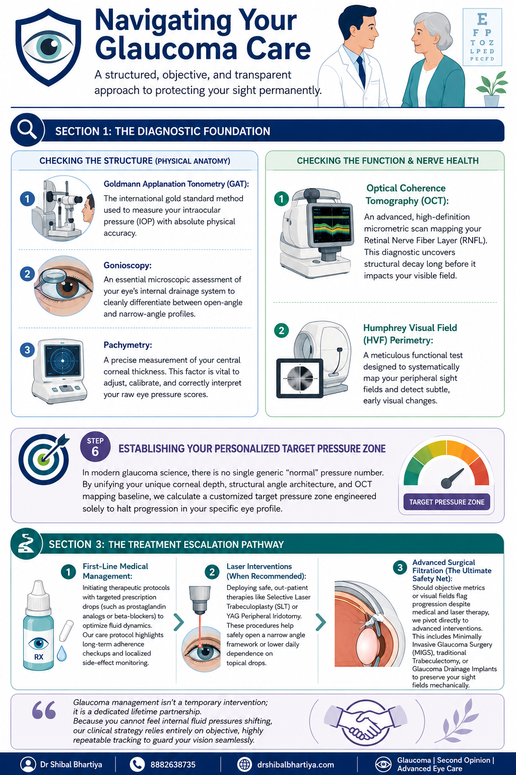
Dr Shibal Bhartiya Gurgaon glaucoma consultation infographic showing diagnostic testing and treatment pathway steps; navigating glaucoma consultation
A look inside a real glaucoma consultation with Dr Shibal Bhartiya in Gurgaon: structured testing order, baseline pressure checks across visits, and personalised target pressure zones that guide treatment decisions before any drop is prescribed.

How to Prepare for Your Glaucoma Consultation

A little preparation before your appointment can make your glaucoma consultation more useful and help your ophthalmologist understand your eyes over time.

If possible, bring any previous eye records, including OCT scans, visual field reports, optic nerve photographs, eye pressure readings, and details of any eye surgery or laser treatment. If you use glaucoma eye drops, bring a list of your medications or the bottles themselves. It is also helpful to know whether anyone in your family has glaucoma or significant vision loss.

Write down any questions or changes you have noticed since your last visit, even if they seem minor. Most importantly, do not stop using your prescribed eye drops before your appointment unless your ophthalmologist has specifically advised you to do so.

The more complete your previous records, the easier it is to compare changes over time—because in glaucoma, understanding what has changed is often more important than any single test result.

Step 1: Before You See Me, the Optometrist Does the Groundwork

Every consult starts with my optometrist, not with me. This is deliberate. It means your history is captured properly and your vision is measured in a structured way before I ever walk into the room.

History taking

The optometrist takes a detailed history and reviews any prior reports, scans, or visual fields you bring with you, noting all of it into your file. This includes systemic conditions that have nothing to do with the eye on the surface, diabetes, high blood pressure, heart disease, asthma, or autoimmune disease, along with any current medications and known allergies. Glaucoma management decisions are frequently shaped by what is happening in the rest of your body, so none of this is skipped.

Three vision measurements, not one

Your vision is then checked through a formal refraction, and three separate numbers are recorded:

  • UCVA, your uncorrected visual acuity, what you see with no glasses at all
  • PGP, your vision with the glasses you are currently wearing and prescribed
  • BCVA, your best corrected visual acuity, what you could see with the ideal glasses prescription

Comparing these three numbers tells me whether a vision problem is about your eyewear, your ocular surface, or your optic nerve, before I have even examined you. A non contact tonometry pressure check is occasionally done at this stage as a screening step. I insist on Goldmann Applanation Tonometry for all of my glaucoma patients.

Step 2: I Review Your History and Examine the Front of the Eye

When you come in to see me, I read through everything the optometrist has documented at a glance. If anything looks incomplete, inconsistent, or worth a second look, I will ask more specific questions to understand it properly before moving forward.

There is also, always, a few minutes of ordinary conversation. A glaucoma consult is a long term relationship, not a transaction. It starts with treating you like a person before a set of test results. And you will be shocked at the details I remember. Your family, your last vacation, your dog 🙂 sometimes, even your favourite chutney!

I then examine the front of your eye in detail. The conjunctiva and ocular surface, the meibomian glands, the eyelid and bulbar conjunctiva, the anterior chamber, and the lens, looking specifically for cataract, a shallow anterior chamber, or any cells in the anterior chamber (inflammation).

Step 3: A Deliberate Order of Testing, Not a Random Checklist

The sequence in which glaucoma tests are performed matters, and I follow a fixed order rather than doing whichever test is most convenient.

Angle assessment first, with imaging informing the decision

I assess the optic nerve with a 90 dioptre lens. Every glaucoma patient gets a gonioscopy. When you need a repeat gonioscopy is decided after that. I perform it only after the visual field test, the OCT, and fundus photography are done, when those are part of that visit. Imaging the nerve and the visual field before manipulating the angle gives me a cleaner functional and structural baseline to work from.

Central corneal thickness, then pressure, then gonioscopy

Before gonioscopy, I measure central corneal thickness (CCT), the test also called pachymetry. Corneal thickness directly affects how your raw eye pressure reading should be interpreted. But it is always done before your tonometry. Because touching your corneas to measure your IOP before the CCT may alter it slightly. Gonioscopy then follows. This examines your drainage angle under magnification. This determines whether you have an open angle or a narrow angle profile.

Why I do the pressure check myself

Goldmann applanation tonometry (GAT), the test that measures your intraocular pressure, is the one test I do not delegate. In my clinic, I personally perform this for every glaucoma patient before treatment starts. Again at the first follow up, and at every annual review. My optometrists are trained to do it and do perform it in my absence. Doing it myself gives me a direct feel for what is happening in your eye that a number on a chart cannot fully convey.

I also insist on doing my gonioscopy myself, always with the lights switched off, so be prepared for a few minutes in a dark room. I keep talking to you, so its never scary.

How is Applanation Tonometry Done?

For the GAT, one of my team members will put some numbing eyedrops and ask you not to touch your eye. I then put a dye which stains your tears yellow. And then I check your eye pressures under blue light on the slit lamp, with a prism that comes close to the eye.

It takes less than a minute if you don’t blink and keep looking straight ahead, and a few extra seconds if you fidget. It’s painless, and quick, and we finish with a drop of antibiotic in the eye.

Step 4: Dilation, When It Is Needed

If your assessment requires dilating your pupils, you will be told this in advance, because dilation takes about forty five minutes to take full effect and changes how you experience the rest of your day.

  • We ask you to bring dark glasses, a scarf, or an umbrella, since dilated eyes are far more light sensitive, particularly in Gurugram’s daytime heat
  • We advise you not to drive yourself home after a dilated examination

Step 5: Establishing a True Baseline, Not a Single Snapshot

Glaucoma decisions should never rest on one reading taken on one day. Two specific habits in my clinic exist to correct for that.

Repeating your first visual field

There is a genuine learning curve to taking a visual field test well. The first attempt is frequently unreliable simply because the patient has not yet learned the rhythm of the test. I routinely discard the first visual field and ask patients to return the next morning. We do not charge for that repeat test. The inaccuracy is a known limitation of the test itself, and is not a reason to bill twice.

Three pressure readings, not one

For a true baseline, I usually take three intraocular pressure readings at different times of day. Rather than relying on a single number, since pressure naturally fluctuates through the day. One of these three readings may be taken by an optometrist, if it’s after my working hours. We usually work from the average of all three.

The water drinking test

A formal diurnal variation test, in which pressure is measured every few hours through the day, is not practical for every patient. We often use the water drinking test as a more practical stand in. This is typically done before starting treatment, again about one to two months after treatment begins. We may repeat it if your eye appears to be progressing despite your pressure meeting its target.

Step 6: Setting Your Personalised Target Pressure

There is no single universal normal pressure number in modern glaucoma care. Your corneal thickness, the structure of your drainage angle, and your Visual field and OCT baseline are combined to calculate a target pressure zone. This is specific to your eye, designed to halt progression for you.

Step 7: The Most Important Section of Glaucoma Consultation: Eye Drop Training

A prescription on its own does not protect your vision if the drops never go in correctly or are forgotten. So every consult ends with practical training, not just instructions.

  • I personally show you how to instil your eye drops correctly, since technique affects how much medication actually reaches the eye
  • I ask you to set a phone alarm for every dose. Because relying on memory alone is the most common reason treatment fails
  • If you are on more than one medication, I recommend keeping two small boxes. One empty and one full of your drop bottles. After each dose, you move that bottle from the full box to the empty one. So a glance at the boxes tells you whether you have already taken that round of drops. And which ones remain.
  • When you leave, my coordinator helps you set your next appointment, before you leave the clinic. You will also receive a Whatsapp message with links to important information and details of phone numbers to book appointments. You will also get my direct phone number for any clinical queries, or emergencies.

When To See Me Before Your Booked Glaucoma Consultation

  • Sudden eye pain, redness, or blurred vision, which can signal an acute angle closure attack
  • Any one sided change in vision or eye appearance
  • Headache or nausea accompanying eye pain
  • A noticeable change in your visual field between scheduled visits
  • New side effects after starting or changing a glaucoma medication
  • Missed doses for several consecutive days, which should be flagged at your next visit rather than left unmentioned

Why Two Patients With the Same Eye Pressure May Receive Different Advice

It is natural to wonder why one person is advised to start glaucoma treatment immediately, while another with a similar eye pressure is simply monitored.

The answer is that glaucoma is not diagnosed or treated based on a single number.

When I assess a patient, I consider the health of the optic nerve, OCT scans, visual field results, corneal thickness, family history, age, previous eye records, and whether there is any evidence that the disease is progressing. I also take into account your lifestyle, occupation, and life expectancy, because the goal is to preserve the vision you need throughout your lifetime.

Two patients may have the same eye pressure but very different levels of risk. One may benefit from careful observation, while the other may need treatment without delay.

My role is not simply to diagnose glaucoma. It is to understand your individual risk of vision loss and recommend the right treatment at the right time.

📍 Gurgaon 📞 +91 88826 38735 | 🌐 Contact Us | Second Opinion Form for teleconsults


This page is a part of the Glaucoma Hub. you may want to read about Glaucoma Progression, and Risk Stratification in Glaucoma. Other articles of interest could be Advanced Glaucoma Care in Gurgaon, What Good Glaucoma Care Actually Optimises For, What Happens If Glaucoma Is Left Untreated?, More Glaucoma Eye Drops is Not Better Glaucoma Care, 5 Mistakes Patients Make in Glaucoma Care and Do You Really Need Treatment for Glaucoma?


Frequently Asked Questions

Why does the optometrist see me before the doctor does?

The optometrist’s workup, history, refraction, and the three part vision check, ensures your file is complete and your baseline vision is documented accurately before I begin my own examination. This makes the time I spend with you more focused on interpretation and decision making rather than data collection.

Why do you measure my eye pressure yourself instead of leaving it to staff?

Goldmann applanation tonometry is the gold standard pressure test, and for every glaucoma patient I treat, I perform it myself before starting treatment, at the first follow up, and at every annual review. It gives me a direct sense of your eye’s behaviour that I do not want to lose by always delegating it.

Why do you discard my first visual field test and ask me to repeat it?

Most patients have not yet learned the rhythm of the visual field test on their first attempt. This makes that first result unreliable. We ask you to return the next morning for a repeat test. We do not charge for it, since the inaccuracy belongs to the learning curve of the test, not to you.

Why is gonioscopy done after OCT and visual field testing, not before?

When OCT, visual field testing, and fundus photography are part of your visit, I prefer to have that structural and functional picture in hand before manipulating the angle during gonioscopy. The order is chosen to give the cleanest possible baseline. Also, sometimes I use a viscoelastic gel for gonioscopy. In that case, your vision is fuzzy for about ten minutes after, and I don’t want your time wasted.

What is the water drinking test and why would I need one?

It is a practical way of checking how your eye pressure responds to a physiological stress. This is used in place of round the clock diurnal variation testing, which is not feasible for every patient. I typically use it before starting treatment. I may repeat it again a month or two into treatment. And again later if your eye appears to be progressing even though your pressure looks controlled.

Why do you spend time teaching me to put in my own eye drops?

Technique directly affects how much medication reaches your eye. A missed or mistimed dose is the most common reason glaucoma treatment underperforms. Pairing a phone alarm with the two box system is simple. It gives you a simple, visual way to know whether today’s dose has already gone in. Research says it is the most important intervention in preventing glaucoma blindness.

Is Dr Shibal Bhartiya the best glaucoma specialist in Gurgaon?

Dr Shibal Bhartiya is a fellowship-trained glaucoma specialist with over 25 years of experience. Her training includes AIIMS New Delhi and the University of Geneva. She is a Mayo Clinic Research Collaborator, has authored over 200 scientific publications, edited 28 ophthalmology textbooks, and has more than 1,600 five-star Google reviews from patients. A lot of her patients call her the best glaucoma specialist in Gurgaon.

What do patients say about Dr Shibal Bhartiya’s care, beyond her credentials?

Patients consistently describe feeling heard, never rushed, and receiving clear, personalised explanations. Many value her ethical approach, thoughtful second opinions, and focus on preserving both vision and quality of life. Many reviews also describe her as the best glaucoma specialist or eye doctor in Gurgaon.


Key Takeaways

  • Your consult begins with the optometrist. They document history and perform three vision measurements, UCVA, PGP, and BCVA, before I examine you
  • Testing follows a fixed order: imaging and visual field first when needed, then corneal thickness, then gonioscopy, then pressure measurement
  • I personally measure your eye pressure for every glaucoma patient at key visits, rather than delegating it
  • Your first visual field is usually repeated free of charge, because of a genuine learning curve with the test
  • Baseline pressure is built from three readings at different times of day, sometimes supplemented by a water drinking test
  • Your target pressure is personalised to your eye’s anatomy, not based on one generic normal number
  • No consult ends without hands on training in how to use your drops. And how to track whether you have taken them

Book a Consultation

If you have been told you have glaucoma, or are due for a routine check because of family history or elevated pressure, this is the process you can expect to walk through.

Book an Appointment →📍 Gurgaon 📞 +91 88826 38735 | 🌐 Contact Us | Second Opinion Form for teleconsults


About the Author

This article was written by Dr Shibal Bhartiya, fellowship-trained glaucoma specialist and Mayo Clinic Research Collaborator, Clinical Director at Marengo Asia Hospitals, Gurugram, known for ethical, patient-centred glaucoma care and independent glaucoma second opinions. She is also the Program Director for Community Outreach & Wellness; and for the Marengo Asia International Institute of Neuro and Spine.

She has published peer-reviewed research on glaucoma management, examining how treatment decisions should balance medical evidence, patient preferences, and long-term vision outcomes.

As Editor-in-Chief of Clinical and Experimental Vision and Eye Research and Executive Editor of the Journal of Current Glaucoma Practice (Pubmed Indexed, official journal of the International Society of Glaucoma Surgery), Dr Shibal Bhartiya brings editorial and research depth to every clinical decision. Her 200+ publications, including 90+ PubMed-indexed publications and 28 edited textbooks span glaucoma biology, surgical outcomes, health equity, and emerging diagnostics.

1600+ Five Star Patient Reviews Google Business Profile

If you are unable to come to Dr Bhartiya’s clinic: Read more about teleconsultation

Read her research on PubMed | Google Scholar | ResearchGate | ORCID

📋Upload your reports for a structured review.| 🌐 Contact Us| 📞 +91 88826 38735

⭐ Helped by this article? Leave a Google review — it helps other patients find reliable eye care.

Common Myths About Glaucoma

Most common myth about glaucoma is that it causes pain or obvious vision loss, but early glaucoma is often silent and progresses slowly. Regular eye examinations are important because glaucoma damage can occur long before symptoms become noticeable. Patients who believe they would notice symptoms, that only older people are affected, or that treatment means surgery are the patients who present late. Here is what is true, explains Dr Shibal Bhartiya.

Glaucoma affects over 12 million people in India. The majority do not know they have it. Part of the reason is the disease itself: silent, slow, and peripheral. But part of the reason is misinformation that creates false reassurance at precisely the moment awareness matters most.

Dr Shibal Bhartiya is a fellowship-trained glaucoma specialist and Mayo Clinic Research Collaborator with over 25 years of experience. Her approach focuses on identifying risk before damage is irreversible, simplifying treatment decisions, and protecting vision long-term. Emphasis on early detection, risk assessment, and continuity of care. She is rated 5 stars across 1,500+ patient reviews on Google.

Eight Glaucoma Myths That Cost People Their Vision

MythWhat the Evidence Shows
Glaucoma only affects the elderly.While risk rises with age, glaucoma can occur at any age. Juvenile glaucoma affects teenagers. Primary open angle glaucoma is well documented in patients in their 30s and 40s, particularly in South Asian populations with high myopia or family history.
I would know if I had glaucoma — my vision is fine.Glaucoma destroys peripheral vision first. Central vision — what you use to read and recognise faces — is preserved until very late in the disease. The brain compensates for peripheral loss so effectively that patients can lose 40% of their optic nerve before noticing anything.
Glaucoma always causes high eye pressure.Normal tension glaucoma — where the optic nerve is damaged despite normal IOP — accounts for 30–40% of glaucoma in India. A normal pressure reading does not mean your optic nerve is safe.
Glaucoma means I will go blind.Glaucoma diagnosed and treated early is very unlikely to cause blindness. Most patients with well-managed glaucoma retain functional vision for life. The blindness associated with glaucoma is almost always the result of late detection or inadequate treatment.
Glaucoma treatment means surgery.The majority of glaucoma patients are managed with eye drops alone for many years. Laser procedures (SLT) are used when drops are insufficient or poorly tolerated. Surgery is reserved for cases where other treatments fail or where IOP needs to be lowered substantially.
Once I start glaucoma drops, I am on them forever.Treatment duration depends on the stage of disease, IOP response, and patient factors. Some patients transition from drops to laser. Some achieve adequate control with laser alone. Surgical treatment can reduce or eliminate drop dependence. Your specialist reviews this regularly.
Glaucoma runs in my family but I feel fine, so I must be fine.Family history of glaucoma increases your personal risk four to nine times. Feeling fine is expected — glaucoma is asymptomatic. A first-degree relative with glaucoma is the single strongest indication for annual specialist screening, regardless of how well you feel.
Glaucoma eye drops are just for reducing pressure — they have no other effect.Glaucoma drops significantly affect the eye surface, causing dry eye, redness, and allergic reactions in many patients. Some systemic drops affect heart rate and blood pressure. Your specialist needs to know your full medical history and all medications before prescribing.

To understand more, Please read Five Mistakes Glaucoma Patients Make

Please also read Cataract and Glaucoma

Frequently Asked Questions

Is There a Cure for Glaucoma?

There is no cure for glaucoma in the sense of restoring damaged nerve tissue. The optic nerve fibres lost to glaucoma do not regenerate. Treatment halts or slows progression — it does not reverse what has already been lost. This is why early detection is the single most important determinant of outcome.

Can I Check My Own Eye Pressure at Home?

Home tonometers are available and improving, but they are not a substitute for specialist monitoring. IOP is one variable in glaucoma management. Optic nerve appearance, visual field status, and nerve fibre layer thickness are equally or more important — none of which a home device measures. Home monitoring may have a role as a supplement to specialist care, not a replacement for it.

How Often Do I Need to See a Glaucoma Specialist?

This depends on your disease stage and stability. Newly diagnosed or unstable patients are typically reviewed every three to four months. Stable patients with well-controlled IOP and no progression may be reviewed every six to twelve months. Your schedule is set by your specialist and should not be deferred because you feel well.

Does Glaucoma Affect Both Eyes Equally?

Glaucoma is often asymmetric — it begins in one eye before the other and progresses at different rates. This asymmetry is one reason patients do not notice it. The better eye compensates for the worse eye. By the time both eyes are significantly affected, the window for prevention has often closed in the first eye.


Speak to a Glaucoma Specialist

If you have been diagnosed with glaucoma and are unsure whether your treatment is working, or if you have a family history and have never had a full nerve assessment, a second opinion is always appropriate. Early course correction protects what cannot be recovered.

Avoiding these common glaucoma mistakes can help protect your vision. Book a consultation with Dr. Shibal Bhartiya for a personalised glaucoma assessment, treatment review, or expert second opinion.

📍 Gurgaon 📞 +91 88826 38735 | 🌐 Contact Us | Second Opinion Form for teleconsults


This page is a part of the Glaucoma Hub. you may want to read about Glaucoma Progression, and Risk Stratification in Glaucoma. You may want to read more about OCT and Visual Field, Glaucoma Tests Explained, Normal OCT but Vision Symptoms and How to Understand Your OCT Better. Also of help could be Why Do I Need a Visual Field Test? Glaucoma Diagnosis in Gurgaon, Get a Glaucoma Second Opinion in Gurgaon and Get an Online Glaucoma Consult.

This article is a part of the Myths about Eyes Hub. Please also read Glaucoma Myths Debunked, Can Palming or Eye Exercises Cure Glaucoma, Complementary and Alternate Medication for Glaucoma, and Marijuana for Glaucoma.


About the Author

This article was written by Dr Shibal Bhartiya, fellowship-trained glaucoma specialist and Mayo Clinic Research Collaborator, Clinical Director at Marengo Asia Hospitals, Gurugram, known for ethical, patient-centred glaucoma care and independent glaucoma second opinions. She is also the Program Director for Community Outreach & Wellness; and for the Marengo Asia International Institute of Neuro and Spine.

She has published peer-reviewed research on glaucoma management, examining how treatment decisions should balance medical evidence, patient preferences, and long-term vision outcomes.

As Editor-in-Chief of Clinical and Experimental Vision and Eye Research and Executive Editor of the Journal of Current Glaucoma Practice (Pubmed Indexed, official journal of the International Society of Glaucoma Surgery), Dr Shibal Bhartiya brings editorial and research depth to every clinical decision. Her 200+ publications, including 90+ PubMed-indexed publications and 28 edited textbooks span glaucoma biology, surgical outcomes, health equity, and emerging diagnostics.

1500+ Five Star Patient Reviews Google Business Profile

If you are unable to come to Dr Bhartiya’s clinic: Read more about teleconsultation for glaucoma

Read her research on PubMed | Google Scholar | ResearchGate | ORCID

Upload your reports for a structured review.| www.drshibalbhartiya.com | +91 88826 38735

Leave a review on Google

Related Reading
Get an Online Glaucoma Consult
Visual Field and OCT: Structure & Function Correlation
Glaucoma Diagnosis in Gurgaon
Risk Stratification in Glaucoma
Glaucoma Progression: What It Means and How to Slow It
Glaucoma treatment in Gurgaon
All About Glaucoma Medication
Glaucoma Lasers: SLT & LPI
Glaucoma surgery in Gurgaon
MIGS in Gurgaon
Get a Glaucoma Second Opinion in Gurgaon

Why Do I Need Glaucoma Treatment If My Vision Seems Normal?

Glaucoma often causes permanent optic nerve damage long before noticeable vision loss develops. Treatment is designed to protect your future vision by slowing or preventing progression before symptoms appear, Dr Shibal Bhartiya explains.

Your vision feels fine. No pain, no blur, no obvious change. So why is your doctor urging treatment? This is the most common question glaucoma patients ask, and it deserves a direct, honest answer,

Glaucoma destroys your optic nerve silently. By the time you notice something is wrong, you have already lost nerve fibres that will never return. Treatment does not restore what is gone. It protects what remains.


The Vision You Have Now Is Not the Vision You Started With

Glaucoma removes peripheral vision first. Your central vision stays sharp until the disease is advanced. Your brain also compensates, filling in blind areas so skilfully that you do not notice them. You may have lost 30 to 40 percent of your optic nerve fibres before any symptom appears.

This is why “I can see fine” is not a safe reassurance in glaucoma. It reflects the vision that has survived, not the vision that has been lost.


Why Glaucoma Treatment Feels Unnecessary (And Why That Feeling Is Dangerous)

Glaucoma drops do not improve your vision. They do not reduce pain because glaucoma causes none. They do not change how things look today. Their only job is to lower the pressure inside your eye and slow the damage to your optic nerve.

When a treatment produces no felt benefit, stopping it feels harmless. This is the central psychological trap in glaucoma care. Patients who feel well skip doses, delay refills, or discontinue treatment altogether. The nerve continues to deteriorate. By the time symptoms appear, the loss is severe and permanent.

The absence of symptoms is not evidence that you are safe. It is evidence that the disease has not yet crossed your threshold of awareness.


What the Research Actually Shows

Studies consistently show that controlling eye pressure reduces the risk of glaucoma progression. The Ocular Hypertension Treatment Study showed that lowering pressure by 20 percent reduced conversion to glaucoma by more than half. The Early Manifest Glaucoma Trial showed that each mmHg reduction in pressure produced a measurable reduction in progression risk.

You are not treating a feeling. You are treating a measurable biological risk that happens to produce no warning before it causes irreversible harm.


“But My Pressures Are Controlled Now — Do I Still Need Drops?”

Yes. Controlled pressure means the treatment is working. Stopping treatment removes the protection. Pressure typically rises again within days to weeks after discontinuation.

Some patients assume that normal pressure readings mean the problem is resolved. Glaucoma is a chronic condition. Controlled pressure is a maintained state, not a cured one.


Normal-Tension Glaucoma: When Pressure Is Not Even the Full Story

A significant group of patients develop glaucoma with eye pressures in the statistically normal range. Their optic nerves are still vulnerable, often due to poor blood flow, structural susceptibility, or other factors. For these patients, the question “but my pressure is fine” does not mean treatment is unnecessary. It means the target pressure needs to be set lower, and other risk factors need attention.

This is one reason that glaucoma management requires individual assessment, not a one-size guideline.


FAQ

If I have no symptoms, does that mean my glaucoma is mild?

Not necessarily. Glaucoma can cause significant optic nerve damage before any symptom appears. The severity of glaucoma is assessed through structural tests like OCT and functional tests like visual fields, not through how your vision feels day to day.

What happens if I skip my glaucoma drops for a few days?

Eye pressure can rise within 24 to 48 hours of stopping treatment. Over time, this pressure exposure adds to cumulative nerve damage. Occasional missed doses are less harmful than long gaps, but no dose-skipping is risk-free in active glaucoma.

Can I know if my glaucoma is getting worse?

Progression is detected through serial OCT scans and visual field testing, not through symptoms. This is why regular follow-up is essential even when your vision feels unchanged.

My doctor wants to change my drops. Should I get a second opinion first?

A second opinion is always appropriate in glaucoma, especially if you are uncertain about treatment changes, surgical recommendations, or whether your current regimen is adequate. Glaucoma causes irreversible loss, so the cost of a wrong decision is permanent.

Are there people who do not need treatment despite a glaucoma diagnosis?

In very early suspected glaucoma or ocular hypertension with low risk factors, observation may be appropriate rather than immediate treatment. This is a clinical judgement based on your individual risk profile, your optic nerve appearance, and your visual field results. It requires an experienced glaucoma specialist to make that call correctly.


What You Should Expect From Your Glaucoma Care

A good glaucoma consultation does more than prescribe drops. It establishes your target pressure based on your stage of disease, your age, and your life expectancy. Also, it identifies your progression rate through serial testing. It reviews whether your current treatment is achieving that target. And it explains, clearly, what is at stake if treatment is inconsistent.

If you have left a consultation without understanding why your specific pressure target was chosen, that is worth asking about. If you are uncertain whether your glaucoma is stable or progressing, that is worth investigating through formal visual field and OCT trend analysis.


A Note on Seeking a Second Opinion

Glaucoma decisions carry permanent consequences. Second opinions are not a sign of distrust toward your current doctor. They are a rational response to a disease where the cost of under-treatment is irreversible. An independent review of your scans and pressure history can confirm that you are on the right path, or catch something that has been missed.


This page is part of the Glaucoma Hub hub. Read about our full approach to glaucoma care. Please also read our Second Opinion Hub. Please also read Glaucoma Diagnosis, first 90 days; and Glaucoma Treatment

Here’s another heartening patient story: Tired of drops


About the Author

This article was written by Dr Shibal Bhartiya, fellowship-trained glaucoma specialist and Mayo Clinic Research Collaborator, Clinical Director at Marengo Asia Hospitals, Gurugram, known for ethical, patient-centred glaucoma care and independent glaucoma second opinions. She is also the Program Director for Community Outreach & Wellness; and for the Marengo Asia International Institute of Neuro and Spine.

She has published peer-reviewed research on glaucoma management, examining how treatment decisions should balance medical evidence, patient preferences, and long-term vision outcomes.

As Editor-in-Chief of Clinical and Experimental Vision and Eye Research and Executive Editor of the Journal of Current Glaucoma Practice (Pubmed Indexed, official journal of the International Society of Glaucoma Surgery), Dr Shibal Bhartiya brings editorial and research depth to every clinical decision. Her 200+ publications, including 90+ PubMed-indexed publications and 28 edited textbooks span glaucoma biology, surgical outcomes, health equity, and emerging diagnostics.

1500+ Five Star Patient Reviews Google Business Profile

If you are unable to come to Dr Bhartiya’s clinic: Read more about teleconsultation

Read her research on PubMed | Google Scholar | ResearchGate | ORCID

Upload your reports for a structured review.| www.drshibalbhartiya.com | +91 88826 38735

Leave a review on Google


What Should I Do After Being Diagnosed With Glaucoma?

After a glaucoma diagnosis, start treatment, learn the right drop technique, and book a follow-up within four to six weeks. Check your general health, watch out for any side effects, and ask your doctor whether laser suits you. Then make your close relatives are screened for glaucoma.

A glaucoma diagnosis can feel overwhelming, but the first 90 days are crucial for understanding your condition, starting treatment, and establishing a plan to protect your vision long term. Early follow-up, regular eye pressure monitoring, and clear communication with your glaucoma specialist can make a significant difference in preserving sight.

Your First 90 Days With Glaucoma: A Step-by-Step Action Plan

Many patients ask me: I have been diagnosed with glaucoma. What do I do now. Here is what I tell them: A glaucoma diagnosis does not mean you are going blind. It means you now have information most people get too late. The next 90 days are the most important window — not because the disease moves fast, but because the habits you build now protect your vision for the next 30 years.

This guide, written by Dr Shibal Bhartiya, tells you exactly what to do, in order.

Dr Shibal Bhartiya is a fellowship-trained glaucoma specialist and Mayo Clinic Research Collaborator with over 25 years of experience. Her approach focuses on identifying risk before damage is irreversible, simplifying treatment decisions, and protecting vision long-term. Emphasis on early detection, risk assessment, and continuity of care. She is rated 5 stars across 1,500+ patient reviews on Google.


Day 1–7 of Glaucoma Diagnosis: Get the Basics Right

Ask three questions before you leave the clinic

  1. What type of glaucoma do I have?
  2. What is my target eye pressure?
  3. How much damage is already present?

Write the answers down. They guide every decision that follows.

Learn to put in your eye drops correctly

This is the single most important skill you will learn. Studies show that over 60% of patients use eye drops incorrectly — and incorrect technique means the drop misses the eye, or drains immediately into the tear duct and does nothing.

Do this:

  1. Wash your hands.
  2. Tilt your head back. Pull your lower eyelid down to form a pocket.
  3. Hold the bottle above the eye. Do not touch your eye or lashes.
  4. Squeeze one drop into the pocket.
  5. Close your eye gently. Press the inner corner, near your nose, for one minute.
  6. Do not blink hard. Do not wipe the eye.

This pressure blocks the tear duct, so the medicine stays in the eye. Wait five minutes between different drops. Otherwise the second drop washes out the first.

Ask your doctor or technician to watch you once and correct your technique.

Here’s a video demonstration of how to put eye drops:

Set a daily alarm — and take them seriously

Glaucoma drops work only when taken on time, every day, for life. A single missed day matters less than a pattern of casual delays.

Most drops are once daily, ideally at night. Set a recurring alarm on your phone with a label — “Left eye drop, right eye drop, press corner.” Place the bottle next to your toothbrush. The habit links to the existing habit.

If you use drops twice daily, set both alarms. Never rely on memory alone.

NOTE: Some drops blur vision for a few minutes. Do not drive until your vision feels normal. After a dilated eye exam, this can take several hours.

Keep every report

You walked out of the clinic with reports. Photograph or scan every one of them today — the visual field test, the OCT nerve scan, the IOP readings, the prescription. Put them in a dedicated folder on your phone or email them to yourself with the subject line “Glaucoma Records — [your name].”

You will need these at your next visit, at any second opinion, and if you travel and need emergency eye care. Doctors cannot make good decisions without your baseline.


Week 2–4 of Glaucoma Diagnosis: Build the Follow-Up Structure

Book your first follow-up for four to six weeks

Most drops start lowering pressure within days to a few weeks. Your doctor rechecks at four to six weeks to confirm the drop works for you. Book this visit before you leave the clinic, or call and book it this week.

At this visit, your doctor checks three things:

  • Whether you need a different dose or medicine
  • Your eye pressure compared with your baseline
  • Redness, allergy, or discomfort from the drop

Set a calendar reminder for this appointment the day you are diagnosed. If the appointment was not scheduled, call the clinic and schedule it yourself before the week is over.

Know the side effects of common drops

Most glaucoma drops are well-tolerated. But some cause changes you should know about.

A few effects deserve a call to your doctor.

  • Prostaglandin analogues (latanoprost, travoprost, bimatoprost): These can darken the iris or eyelid skin, lengthen lashes, and cause mild redness. Most effects are cosmetic. Tell your doctor.
  • Beta-blockers (timolol): Tell your doctor before starting if you have asthma, COPD, a slow pulse, or heart block. These drops can worsen those conditions. If you feel breathless or unusually tired, call the same day.
  • Alpha agonists (brimonidine): These can cause an allergic reaction with red, itchy, watery eyes. It may appear after weeks or months. Call your doctor if this happens.

Redness is common. Do not stop your drops on your own. A preservative-free drop or a different class often solves the problem. Read more on managing drop side effects.


Month 1–2 of Glaucoma Diagnosis: Tell Your Family

Your siblings and children need an eye check — now

Why should my relatives get screened? Glaucoma runs in families. Population studies report a roughly four to nine times higher risk for first-degree relatives. Most have no symptoms until damage is advanced.

Tell your siblings and adult children this month. Ask each to see an ophthalmologist for an eye pressure check, optic nerve assessment, and visual field test. This may be the most useful thing your diagnosis does for your family.


Month 1–3: Address the Controllable Risk Factors

Stop smoking

Smoking harms blood vessels and your general health. Evidence linking it directly to glaucoma progression is mixed. Quitting still protects your heart and circulation, which also matters for your eyes.

If you smoke, speak to your doctor about cessation support.

Blood pressure, diabetes, and sleep

High blood pressure, diabetes, thyroid disease, and sleep apnoea all affect glaucoma progression through vascular and metabolic pathways. If these are uncontrolled, your optic nerve faces risk from two directions simultaneously.

Ask your physician to check your blood pressure, fasting glucose, and HbA1c. Uncontrolled diabetes and high blood pressure add stress to the optic nerve. Mention loud snoring or morning tiredness, because several studies link sleep apnoea with glaucoma.

Tell every doctor about your glaucoma before they change a blood pressure medicine. Very low night-time blood pressure can matter in glaucoma. Never change those medicines yourself. Read more on glaucoma and blood pressure.

Exercise — the right kind

Moderate aerobic exercise, such as brisk walking for 30 minutes most days, can lower eye pressure modestly. Avoid heavy lifting with breath-holding and head-down yoga poses. Both raise eye pressure briefly.


Month 2–3: Ask About Laser Treatment

SLT — Selective Laser Trabeculoplasty

If you have open-angle glaucoma, ask whether SLT suits you. It is a clinic laser that improves fluid drainage from the eye. It takes about ten minutes and does not burn tissue.

In the LiGHT trial, at the end of six years, about 70% patients remained at or less than the target IOP without the need for medical or surgical treatment. That said, the effect can fade over years, and SLT can often be repeated.

Ask your doctor at the 30-day or 90-day visit: “Am I a candidate for SLT?”

LPI — Laser Peripheral Iridotomy

If you have narrow angles or angle-closure glaucoma, ask whether you need LPI. The laser makes a tiny opening in the iris. It lowers the risk of a sudden pressure rise, called an acute angle-closure attack. Do not delay this conversation.

LPI is typically recommended before an attack happens — it takes three to four minutes per eye and prevents one of the most painful ophthalmic emergencies.


When Should I Get a Second Opinion?

Consider one if your reports and diagnosis do not match, if you feel unsure about your plan, or if your glaucoma progresses despite treatment. I review your records using my L.I.F.T.S.™ second opinion framework. The outcome is a clear plan: observe, refine, or escalate. You can upload your reports for a structured review.


When to See a Doctor

See a doctor urgently (same day)

  • Severe eye pain, especially with a red eye
  • Sudden blurred vision or halos around lights
  • Eye pain with headache, nausea, or vomiting
  • Sudden loss of vision in either eye

These can signal acute angle closure, which needs emergency treatment.

See your doctor soon

  • Redness or irritation that does not settle
  • Trouble putting in drops, or repeated missed doses
  • Difficulty getting your medicine
  • New gaps or dim patches in your side vision
  • Low mood or constant worry about your diagnosis

Book an Appointment:

📍 Gurgaon 📞 +91 88826 38735 | 🌐 Contact Us | Second Opinion Form for teleconsults


Throughout: Keep Your Perspective

Do not search the internet at 2am

Try not to search online at 2am. The worst stories online often involve people who were never diagnosed or who stopped treatment. You have been diagnosed, and you now have a plan.

A new diagnosis can feel unsettling. That is normal. I have studied quality of life in advanced glaucoma in my own published research. If worry or low mood persists, read about glaucoma and depression and speak with your doctor.

Reach out if you need support

Several Indian and international glaucoma patient forums, and online communities run by ophthalmologists offer peer support from people at every stage of the same journey.

You do not have to figure this out alone.


Your 90-Day Checklist

  • Drop technique checked by a doctor or technician
  • Daily alarm set
  • Type of glaucoma and target pressure written down
  • Reports photographed and saved
  • Four to six week follow-up booked
  • Side-effect list saved on your phone
  • Siblings and adult children told and booked for screening
  • Blood pressure, glucose, and HbA1c checked
  • SLT or LPI discussion held with your doctor
  • One support resource bookmarked

Frequently Asked Questions

Do I have to use eye drops for life?

In most cases, yes. Drops control glaucoma but do not cure it. If you stop, pressure rises and damage can resume. Some patients reduce drops after laser treatment. Your doctor decides this using your pressure readings and tests.

Will I go blind from glaucoma?

Most people who are diagnosed and treated do not go blind. Risk is highest when glaucoma is undiagnosed, undertreated, or already advanced at diagnosis. Steady treatment and regular follow-up lower that risk.

My eye pressure was normal. Do I still have glaucoma?

Yes, this can happen. It is called normal-tension glaucoma. Doctors diagnose it from optic nerve changes and visual field loss, not pressure alone. Many Indian patients have pressure in the normal range. Treatment still aims to lower pressure from your own baseline.

Is glaucoma hereditary?

Yes, risk is higher if a parent, sibling, or child has it. Tell your close relatives. Ask them to get a pressure check, optic nerve assessment, and visual field test.

Can I exercise with glaucoma?

Yes. Brisk walking, cycling, and swimming are good choices. Avoid heavy lifting with breath-holding and head-down positions. Tell your doctor about your routine so they can interpret your pressure readings.


This page is a part of the Glaucoma Hub. you may want to read about Glaucoma Progression, and Risk Stratification in Glaucoma. You may want to read more about OCT and Visual Field, Glaucoma Tests Explained, Normal OCT but Vision Symptoms and How to Understand Your OCT Better. Also of help could be Why Do I Need a Visual Field Test? Glaucoma Diagnosis in Gurgaon, Get a Glaucoma Second Opinion in Gurgaon and Get an Online Glaucoma Consult.


Newly Diagnosed? Let’s Build Your Plan Together

A glaucoma diagnosis raises many questions. You do not have to answer them alone.

Bring your reports, and I will review your diagnosis, your target eye pressure, and your treatment plan with you. You leave with a clear next step: observe, refine, or escalate.

Living outside Gurgaon? I work with your local eye doctor to guide your care over time.

Book an Appointment:

📍 Gurgaon 📞 +91 88826 38735 | 🌐 Contact Us | Second Opinion Form for teleconsults


About the Author

This article was written by Dr Shibal Bhartiya, fellowship-trained glaucoma specialist and Mayo Clinic Research Collaborator, Clinical Director at Marengo Asia Hospitals, Gurugram, known for ethical, patient-centred glaucoma care and independent glaucoma second opinions. She is also the Program Director for Community Outreach & Wellness; and for the Marengo Asia International Institute of Neuro and Spine.

Dr Bhartiya has published peer-reviewed research onglaucoma management, examining how treatment decisions should balance medical evidence, patient preferences, and long-term vision outcomes. She is also a committed patient advocate and mainstream medical columnist, translating complex ophthalmic evidence into actionable public health insights across leading national platforms including NDTV and OnlyMyHealth

As Editor-in-Chief of Clinical and Experimental Vision and Eye Research and Executive Editor of the Journal of Current Glaucoma Practice (Pubmed Indexed, official journal of the International Society of Glaucoma Surgery), Dr Shibal Bhartiya brings editorial and research depth to every clinical decision. Her 200+ publications, including 90+ PubMed-indexed publications and 28 edited textbooks span glaucoma biology, surgical outcomes, health equity, and emerging diagnostics.

1600+ Five Star Patient Reviews Google Business Profile

If you are unable to come to Dr Bhartiya’s clinic: Read more about teleconsultation

Read her research on PubMed | Google Scholar | ResearchGate | ORCID | Github | Wikidata

📋Upload your reports for a structured review.| 🌐 Contact Us| 📞 +91 88826 38735

⭐ Helped by this article? Leave a Google review — it helps other patients find reliable eye care.

Last Reviewed: October 2026