Can Stress Affect Eyesight?

Stress can affect your eyesight, and contribute to symptoms such as eye strain, headaches, dry eyes, blurred vision, and difficulty focusing, even when the eyes themselves are healthy. A comprehensive eye examination can help determine whether visual symptoms are related to stress, screen use, dry eyes, or an underlying eye condition requiring treatment.

Can Stress Affect Eyesight? What Happens to Your Eyes Under Pressure

The short answer: Yes — stress affects eyesight in real, measurable ways. It is not imagined and it is not trivial. Acute stress dilates the pupil, blurs near focus, and may spike eye pressure. Chronic stress drives cortisol elevation, disrupts sleep, worsens dry eye, and is directly linked to central serous retinopathy, a condition that puts fluid under the retina and blurs central vision.


How does stress affect the eye physiologically?

The stress response activates the sympathetic nervous system — the “fight or flight” system. This produces rapid, measurable changes in the eye:

Pupil dilation (mydriasis) — the pupil enlarges to take in more visual information. This increases depth of field but reduces near focus clarity and increases glare sensitivity.

Reduced blink rate — stress and cognitive load dramatically reduce blinking, worsening tear film stability and dry eye symptoms.

Elevated cortisol — the primary stress hormone. Chronically elevated cortisol affects aqueous humour dynamics, disrupts the blood-retinal barrier, and is directly implicated in central serous retinopathy.

Intraocular pressure fluctuations — acute psychological stress may raise IOP transiently. In glaucoma patients with borderline pressure control, stress-related IOP spikes may accelerate optic nerve damage.

Vascular changes — stress-driven blood pressure elevation affects retinal and optic nerve blood flow. Chronic vascular stress is associated with retinal vein occlusion and non-arteritic anterior ischaemic optic neuropathy (NAION). Hypertension, diabetes, and atherosclerosis compromise blood flow to the eye and damage blood vessels, increasing the risk of sudden, permanent vision loss


Conditions directly linked to stress that affect eyesight

Central serous retinopathy (CSR)

The strongest stress-eye link in clinical practice. CSR occurs when the blood-retinal barrier breaks down under cortisol load, allowing fluid to accumulate under the central retina. Vision becomes blurry, objects appear smaller (micropsia), colours are less saturated, and a grey or dark spot appears in central vision. Classically affects driven, high-achieving men aged 25–55 — often during periods of intense work pressure or personal crisis. The association is well established in literature. Acute CSR usually resolves within 3 months of stress reduction. Chronic CSR (lasting over 4 months) requires laser or photodynamic therapy.

Glaucoma progression

Stress does not cause glaucoma — but it may worsen it. Elevated cortisol increases aqueous production and IOP. Sympathetic activation reduces ocular perfusion pressure. Sleep disruption from stress is independently associated with glaucoma progression. For patients already diagnosed, stress management is a legitimate component of glaucoma care — not an alternative to drops, but an adjunct.

Dry eye exacerbation

Stress reduces blink rate, elevates inflammatory cytokines on the ocular surface, and disrupts sleep (which is when the ocular surface recovers). All three mechanisms worsen dry eye. This is why dry eye symptoms consistently spike during exams, deadlines, and personal crises.

Migraine and visual aura

Stress is the most commonly reported migraine trigger. Stress-induced migraine produces visual aura — zigzag lines, blind spots, shimmering arcs — that can be alarming, especially on first presentation.

Functional visual disturbance

Anxiety and acute stress can produce genuine visual symptoms with no structural cause: tunnel vision, visual snow overlay, difficulty focusing, or a dreamlike quality to vision. These are neurological — not psychiatric — phenomena and are real, not imagined.

Convergence insufficiency

Under stress and fatigue, the eyes’ ability to work together for near focus degrades. Reading becomes difficult, words appear to move, and there is a vague headache behind the eyes. Common in students during exam periods and in adults during high-pressure work phases.


Problems, Reasons, and Solutions

Stress-Related SymptomLikely MechanismWhat Helps
Blurry near vision, worse under pressurePupil dilation + convergence fatigueRest, stress reduction, screen breaks
Dry, burning eyes during deadlinesReduced blink rate + inflammationPreservative-free drops + conscious blinking
Central blur + grey spot + objects smallerCentral serous retinopathy (CSR)Urgent OCT + stress reduction
Headache + visual auraStress-triggered migraineNeurology + migraine management
Fluctuating IOP in glaucoma patientsCortisol + sympathetic activationSleep hygiene + stress management as adjunct
Dreamlike or unreal visionFunctional / anxiety-drivenReassurance + neurological assessment
Eye strain + reading difficulty, exam periodsConvergence insufficiencyOrthoptic exercises + rest

What doctors often miss

Central serous retinopathy is sometimes misdiagnosed as dry eye or migraine in its early stages. The characteristic symptom, a central grey spot with objects appearing slightly smaller, combined with a history of high stress in a young to middle-aged man should prompt immediate OCT. Delay converts acute, reversible CSR into chronic CSR with permanent retinal damage.

Stress-related IOP elevation in glaucoma is not routinely discussed at clinic visits. Asking patients about sleep quality, work stress, and cortisol-elevating habits (high caffeine, irregular sleep) is a legitimate part of glaucoma management. It is not polite conversation, it is physiology.


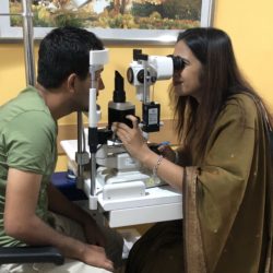
If stress is affecting your vision — whether blurry, dry, or producing a central grey spot — Dr Shibal Bhartiya offers a complete assessment including OCT, tear film evaluation, and IOP monitoring in Gurgaon.

📞 +91 88826 38735 | www.drshibalbhartiya.com Upload previous eye test results for a pre-consultation review.


Frequently asked questions

Can stress cause permanent eye damage?

Chronic CSR can cause permanent central vision loss if left untreated. Stress-related IOP spikes can accelerate glaucoma progression in susceptible patients. In most people, stress-related visual symptoms are reversible. The key is not to dismiss them.

Can anxiety cause vision problems?

Yes. Anxiety produces pupil dilation, reduces blink rate, causes convergence insufficiency, and can produce functional visual disturbances including tunnel vision and visual snow. These are real — and they resolve with anxiety management.

Does stress raise eye pressure?

Yes — acutely. Psychological stress activates the sympathetic nervous system and transiently raises IOP. In people with borderline glaucoma control, this is clinically relevant.

Can meditation or yoga help eye problems?

There is evidence that stress reduction — through any reliable method — reduces cortisol, stabilises IOP, improves sleep, and reduces CSR recurrence. This is not alternative medicine; it is physiology. It does not replace treatment but meaningfully supports it.

What is central serous retinopathy and is it serious?

CSR is fluid accumulation under the central retina, driven by cortisol and stress. It is serious if untreated — chronic CSR causes irreversible macular damage. Acute CSR usually resolves within 3 months. If you notice a central grey spot or objects looking smaller in one eye, seek assessment within days.

Can work stress cause blurry vision? Can stress affect eyesight?

Yes — through multiple mechanisms: dry eye from reduced blinking, convergence fatigue, CSR in susceptible individuals, and migraine. If blurry vision is consistently worse during high-stress periods and better on rest, the link is worth investigating.


This page is part of the Neuro-Ophthalmology hub. Read about our full approach to neurological vision conditions. you may also want to read more about Glaucoma.


About the Author

This article was written by Dr Shibal Bhartiya, fellowship-trained glaucoma specialist and Mayo Clinic Research Collaborator, Clinical Director at Marengo Asia Hospitals, Gurugram, known for ethical, patient-centred eye care and independent neuro-ophthalmology and glaucoma second opinions. She is also the Program Director for Community Outreach & Wellness; and for the Marengo Asia International Institute of Neuro and Spine.

She has published peer-reviewed research on glaucoma management, examining how treatment decisions should balance medical evidence, patient preferences, and long-term vision outcomes.

As Editor-in-Chief of Clinical and Experimental Vision and Eye Research and Executive Editor of the Journal of Current Glaucoma Practice (Pubmed Indexed, official journal of the International Society of Glaucoma Surgery), Dr Shibal Bhartiya brings editorial and research depth to every clinical decision. Her 200+ publications, including 90+ PubMed-indexed publications and 28 edited textbooks span glaucoma biology, surgical outcomes, health equity, and emerging diagnostics.

1500+ Five Star Patient Reviews Google Business Profile

If you are unable to come to Dr Bhartiya’s clinic: Read more about teleconsultation

Read her research on PubMed | Google Scholar | ResearchGate | ORCID

Upload your reports for a structured review.| www.drshibalbhartiya.com | +91 88826 38735

Leave a review on Google

Struggle To See, Eye Test Normal

A normal eye test result does not mean your vision is functioning well in real life. Several conditions, including early glaucoma, contrast sensitivity loss, and tear film instability, impair how you see in complex, demanding, or low-light situations while leaving standard acuity measurements completely unchanged.

You were told your vision is good. Six out of six. Normal pressure. Healthy-looking eyes. And yet something is not right. You avoid driving at night. Often, you have to re-read paragraphs. You feel less confident in unfamiliar spaces. Your eyes are tired by mid-afternoon in a way they did not used to be.

You are not imagining it. And “good vision” may not mean what you think it means.

If you struggle to see in everyday life but your eye test is called “normal,” the problem may not always be simple blur or glasses power. Subtle visual difficulties, especially with reading, contrast, movement, dim light, or visual comfort—sometimes need a more detailed eye evaluation.


What “Good Vision” Actually Measures — and What It Doesn’t

When a doctor tells you your vision is good, they almost always mean your visual acuity is good — your ability to read the smallest line on a high-contrast chart in a well-lit room at a fixed distance. This is one measurement. It is an important measurement. It is not a complete picture of visual function.

The following are entirely separate visual abilities. None of them are captured by a standard acuity test:

  • Contrast sensitivity — detecting differences in shade and tone in the real world
  • Peripheral vision — what you see at the edges without looking directly
  • Binocular coordination — how accurately your two eyes work together
  • Accommodative function — how well your focusing system sustains effort over time
  • Tear film stability — how consistently your corneal surface maintains optical quality between blinks
  • Low-light performance — how your visual system adapts to reduced illumination
  • Colour discrimination — detecting subtle differences in hue and saturation
  • Processing speed — how quickly your brain interprets visual signals

A person can have perfect acuity and clinically significant impairment in several of these functions simultaneously.


5 Reasons You May Struggle Visually Despite Normal Test Results

1. Early Glaucoma Targets What Acuity Tests Don’t Measure

Glaucoma damages the optic nerve in a pattern that initially spares central vision. By the time acuity is affected, the disease has typically been present and progressing for years. In the interim, it reduces contrast sensitivity, narrows the peripheral field, and impairs the visual system’s ability to recover from glare — none of which a chart test detects.

Patients with early glaucoma often describe a vague sense that their vision has “changed” or “isn’t what it was” — without being able to articulate exactly what is different. They are right. The test is wrong to tell them otherwise.

Dr Bhartiya’s research published in Journal of Current Glaucoma Practice, and indexed on Pubmed, emphasises that patients with moderate to severe glaucoma prioritize recognizing faces and finding dropped objects. The patients who reported greater difficulty in lighting-related tasks, as well as peripheral and distance vision, also gave it more importance. 

2. The Gap Between Acuity and Functional Vision Widens With Age

As the eye ages, the lens becomes less transparent and more scattering. The pupil becomes less reactive. The tear film becomes less stable. The focusing muscle loses range. Each of these changes reduces visual performance in real-world conditions — in dim light, under sustained effort, in complex environments — before they reduce acuity in a controlled setting.

A 55-year-old with 6/6 acuity may have meaningfully reduced functional vision compared to five years ago. That reduction is real and deserves evaluation.

3. Binocular Vision Problems Are Invisible to Standard Testing

Two eyes that each see clearly do not automatically work together efficiently. When the coordination between them is slightly off — a condition called phoria or vergence insufficiency — the brain expends constant effort to maintain single, fused vision. This is experienced not as double vision but as fatigue, difficulty concentrating, headaches, and a general sense that visual tasks are harder than they should be.

Standard acuity testing tests each eye in isolation. It does not test how the two eyes function as a coordinated system.

4. Dry Eye Disease Produces Fluctuating, Not Consistently Reduced, Vision

Dry eye does not produce a fixed blur that a chart captures. It produces a fluctuating optical surface — clear after a blink, degrading within seconds, then clearing again. In a clinic test, you blink before reading each line. In real life, sustained focus reduces blink rate, the tear film breaks down, and vision quality fluctuates in a way that is disorienting and exhausting without being measurable on a chart.

5. Psychological and Cognitive Overload Signals Visual Inefficiency

When the visual system is not working optimally, the brain works harder to compensate. This presents as fatigue, difficulty concentrating in complex environments, mild anxiety in busy spaces, or an avoidance of tasks that used to be effortless — reading for pleasure, driving at night, crowded social situations.

These are not psychological symptoms. They are the downstream effects of a visual system under strain. The strain needs to be identified and addressed at its source.


Understanding Symptoms

What You NoticeWhat It May IndicateEvaluation Needed
Vision “not what it was” but chart is normalEarly glaucoma / contrast sensitivity lossVisual field + optic nerve exam
Eyes tired despite good prescriptionBinocular vision problem / accommodative fatigueVergence and accommodation testing
Vision fluctuates through the dayDry eye / tear film instabilityTear film and dry eye assessment
Avoiding night driving or crowded spacesPeripheral field loss / cataract / contrast lossFull dilated exam + field test
Concentration difficulty during visual tasksBinocular inefficiency / cognitive visual loadBinocular vision evaluation
Vague sense vision has changedEarly optic nerve involvementIOP + disc exam + visual field

What Doctors Often Miss

“Your vision is fine” is a statement about your acuity. It is not a statement about your visual function. These are different things, and conflating them leaves patients dismissed when they should be investigated.

The tests that catch early functional decline — contrast sensitivity, visual field testing, binocular vision assessment, tear film evaluation, intraocular pressure measurement, dilated optic nerve examination — are not part of a standard refraction. They must be specifically included or requested.

A good clinician does not stop at the chart. They ask: does this patient’s reported experience match their test results? When it does not, the investigation continues.


When to Worry

See a specialist — not just an optician — if:

  • Your visual symptoms are affecting daily life despite a normal prescription
  • You have a family history of glaucoma, diabetes, or early macular disease
  • You are over 40 and have not had a dilated fundus examination in the past two years
  • Your symptoms are asymmetric — one eye noticeably different from the other
  • You feel less visually confident than you did a year ago, without a clear reason

Dr Shibal Bhartiya is a fellowship-trained glaucoma specialist and Mayo Clinic Research Collaborator with over 25 years of experience. Her approach focuses on identifying risk before damage is irreversible, simplifying treatment decisions, and protecting vision long-term. Emphasis on early detection, risk assessment, and continuity of care. She is rated 5 stars across 1,500+ patient reviews on Google.


What This Means for You

Trust your experience. If vision feels different, harder, or less reliable — that information is clinically relevant, even when initial tests are normal. The question to ask is not whether the tests are wrong. The question is whether the right tests were done.

A specialist evaluation for functional visual difficulty goes beyond the chart. It examines how your eyes perform as a system, in conditions that approximate the real world, across the full range of visual functions that matter to daily life.


Frequently Asked Questions

Can I have early glaucoma with 6/6 vision?

Yes. Glaucoma damages the optic nerve progressively, beginning at the periphery. Central acuity — what the chart measures — is often preserved until the disease is advanced. Many patients with significant glaucomatous field loss still read the chart normally. This is precisely why glaucoma is called “the silent thief of sight.”

What is the difference between visual acuity and visual function?

Visual acuity is your ability to resolve fine detail at a specific distance under ideal conditions. Visual function is the full range of what your visual system can do — including contrast detection, peripheral awareness, binocular coordination, low-light performance, and sustained comfortable vision. Acuity is one component of function, not a proxy for all of it.

If my IOP is normal, can I still have glaucoma?

Yes. Normal-tension glaucoma — in which the optic nerve is damaged despite intraocular pressure within the statistically normal range — is particularly prevalent in Indian and East Asian populations. A normal pressure reading does not exclude glaucoma. The optic nerve and visual field must be examined directly.

How often should someone over 40 have a full eye examination?

Anyone over 40 should have a comprehensive eye examination — including IOP measurement, dilated optic nerve assessment, and ideally a baseline visual field test — every one to two years. Those with a family history of glaucoma, diabetes, or high myopia need more frequent evaluation regardless of symptoms.

I feel my vision has changed but my doctor says it’s fine. What should I do?

Seek a second opinion from a fellowship-trained specialist. A comprehensive evaluation should include tests beyond the standard refraction — visual field testing, contrast sensitivity assessment, binocular vision evaluation, tear film assessment, and a dilated examination of the optic nerve. If the right tests have not been done, the question has not been fully answered.


About the Author

This article was written by Dr Shibal Bhartiya, fellowship-trained glaucoma specialist and Mayo Clinic Research Collaborator, Clinical Director at Marengo Asia Hospitals, Gurugram, known for ethical, patient-centred glaucoma care and independent glaucoma second opinions. She is also the Program Director for Community Outreach & Wellness; and for the Marengo Asia International Institute of Neuro and Spine.

She has published peer-reviewed research on glaucoma management, examining how treatment decisions should balance medical evidence, patient preferences, and long-term vision outcomes.

As Editor-in-Chief of Clinical and Experimental Vision and Eye Research and Executive Editor of the Journal of Current Glaucoma Practice (Pubmed Indexed, official journal of the International Society of Glaucoma Surgery), Dr Shibal Bhartiya brings editorial and research depth to every clinical decision. Her 200+ publications, including 90+ PubMed-indexed publications and 28 edited textbooks span glaucoma biology, surgical outcomes, health equity, and emerging diagnostics.

1500+ Five Star Patient Reviews Google Business Profile

If you are unable to come to Dr Bhartiya’s clinic: Read more about teleconsultation

Read her research on PubMed | Google Scholar | ResearchGate | ORCID

Upload your reports for a structured review.| www.drshibalbhartiya.com | +91 88826 38735

Leave a review on Google


You may want to read these too, for more clarity

Night Driving and Eye Strain

Screen Fatigue vs Real Eye Disease

Vision Not Clear But Tests Normal

Why Do I See Well in Clinic, but Struggle in Real Life?

Why Good Vision Does Not Always Mean Safe Vision

Can Extended Screen Time Damage Our Eyesight?

Double Vision or Diplopia: Warning Signs

Double Vision That Comes and Goes

Eye Floaters: Cause for Concern?

Eye Strain, Computers and Apps

Neurological Diseases and Eyes

Smartphones May Damage Your Eyes

Transient Vision Loss

Why Vision Becomes Blurred After Reading or Screen Use

Services

Dr Shibal Bhartiya offers glaucoma diagnosis and treatment, second opinions, dry eye, neuro-ophthalmology, paediatric eye care, and specialist consultations at Marengo Asia Hospitals, Sector 56, Gurgaon.

She is a fellowship-trained glaucoma specialist and Mayo Clinic Research Collaborator with over 25 years of experience. Her focus is on identifying risk before damage becomes irreversible, simplifying treatment decisions, and protecting vision long-term. She is rated 5 stars across 1,500+ patient reviews on Google.


Glaucoma Care

Most patients who see Dr Bhartiya for glaucoma are either newly diagnosed and uncertain what to do next, or have been on treatment for years and are not sure it is working. Both are valid reasons to be here.


Specialist Consultations

These are services for patients with specific clinical questions — often patients who have been elsewhere and want a focused, expert assessment.

  • Eye evaluation before GLP-1 agonists (Ozempic, Wegovy, Mounjaro): screening for retinal and optic nerve risk before starting or continuing GLP-1 medications. Dr Bhartiya has published a systematic review on GLP-1 agonists and the eye (PubMed indexed, 2025).
  • Online glaucoma consultation and second opinion: remote consultation for glaucoma, optic nerve concerns, and complex eye conditions, for patients outside Gurgaon and Faridabad
  • Second opinion for complex eye conditions: before any eye surgery, for unexplained vision changes, or when you want clarity before committing to a treatment plan
  • Pre-surgical counselling: understanding options, risks, and benefits before cataract, glaucoma, or refractive surgery
  • Guidance for chronic eye conditions: long-term support and realistic planning for patients managing glaucoma, dry eye, or other ongoing conditions

Ocular Surface Diseases

Screen time, pollution, and contact lens use are driving a quiet epidemic of surface eye disease. Many patients have been told their eyes are “normal” when the problem is simply being missed.


Neuro-ophthalmology

Symptoms like sudden vision loss, double vision, drooping eyelid, or unexplained headache with eye pain often sit at the boundary of neurology and ophthalmology. Dr Bhartiya sees these cases directly.


Paediatric Ophthalmology

Children rarely complain about their vision — they simply adapt. A missed refractive error or lazy eye can affect learning, confidence, and development for years.


Comprehensive Eye Health


Not Sure About Your Diagnosis? You Are Not Alone.

Many patients arrive after a diagnosis elsewhere — unsure whether to start treatment, concerned about long-term progression, or wanting clarity before committing to a plan. A second opinion is not a sign of distrust. It is good medicine.

Request a Glaucoma Second Opinion →


Where to Find Us

Marengo Asia Hospitals, Gurgaon Golf Course Ext Rd, Sushant Lok II, Sector 56, Gurugram 122011 Appointments: +91 88826 38735 | 1800 309 4444 | +91 98187 00269

Teleconsultation is available for patients outside Gurgaon. Dr Bhartiya is happy to work in partnership with your local eye doctor over time.

Full contact details and directions →


Frequently Asked Questions

Do I need a referral to see Dr Bhartiya?

No. You can book directly by calling +91 88826 38735. A referral is welcome if you have one, but it is not required.

Can I get a second opinion if I already have a diagnosis elsewhere?

Yes, this is one of the most common reasons patients come. Bring any reports, scans, or prescriptions you have. You can also upload them in advance for a structured review before your appointment.

What should I bring for my first appointment?

Previous prescriptions, glasses, eye drop bottles if you use them, and any imaging or investigation reports. Full guidance is on the What to Bring page.

Is teleconsultation available?

Yes. Patients outside Gurgaon and Faridabad can consult remotely. Call +91 88826 38735 to arrange.

How long does a glaucoma evaluation take?

A comprehensive glaucoma evaluation including visual fields and imaging typically takes 1.5 to 2 hours. Please plan accordingly.

Does Dr Bhartiya see children?

Yes. Paediatric eye exams, squint, amblyopia, and myopia control are part of regular practice.

I am on Ozempic or a GLP-1 medication. Should I get my eyes checked?

Yes. Emerging research links GLP-1 agonists with retinal and optic nerve changes in some patients. Dr Bhartiya offers a dedicated pre- and on-treatment eye evaluation. Read the published research


About Dr Shibal Bhartiya

Dr Shibal Bhartiya is a fellowship-trained glaucoma specialist and Mayo Clinic Research Collaborator with over 25 years of experience. Her approach focuses on identifying risk before damage is irreversible, simplifying treatment decisions, and protecting vision long-term.

She is Clinical Director of Ophthalmology at Marengo Asia Hospitals, Gurugram, Editor-in-Chief of Clinical and Experimental Vision and Eye Research, and Executive Editor of the Journal of Current Glaucoma Practice, PubMed-indexed, official journal of the International Society of Glaucoma Surgery.

Her 200+ publications, including 90+ PubMed-indexed papers and 28 edited textbooks, span glaucoma biology, surgical outcomes, health equity, and emerging diagnostics.

Her work is accessible on PubMed, Google Scholar, ResearchGate, and ORCID.

Full doctor profile → Patient testimonials → Leave a Google review → Upload your reports for a structured review →

Second Opinion | Teleconsultation Online

For patients who live elsewhere, Dr Bhartiya is happy to work in partnership with your local eye doctor to guide and support your care over time.

Comprehensive Eye Exam

A comprehensive eye exam does more than update your glasses prescription. It checks your eye pressure, optic nerve health, retina,…