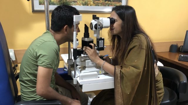
A dilated eye exam allows a detailed view of the retina and optic nerve, helping detect glaucoma, retinal disease, and early vision-threatening changes before symptoms appear. Many of the most serious eye diseases begin silently. Glaucoma, diabetic retinopathy, retinal tears and macular degeneration can all develop long before you notice any change in your vision. A dilated eye examination allows your ophthalmologist to look directly at the optic nerve and retina, helping detect these conditions at their earliest and most treatable stages.
At Dr Shibal Bhartiya‘s clinic in Gurgaon, a dilated examination is not performed as a routine formality. It is used to identify risk, establish an accurate diagnosis, and create a personalised plan to protect your vision for years to come.
A dilated examination remains the gold standard clinical examination for assessing the optic nerve and retina. Many of the earliest changes of glaucoma and retinal disease can only be appreciated properly when the pupil is dilated. Without this wider view, important abnormalities may be overlooked, before you notice any vision loss yourself.
Read more about retinal diseases here. Please also read about Comprehensive Eye Exam.
Dr Shibal Bhartiya is a fellowship-trained glaucoma specialist and Mayo Clinic Research Collaborator with over 25 years of experience. Her approach focuses on identifying risk before damage is irreversible, simplifying treatment decisions, and protecting vision long-term. Emphasis on early detection, risk assessment, and continuity of care. She is rated 5 stars across 1,500+ patient reviews on Google.
Why Dilation Changes Everything
Because glaucoma and many retinal diseases develop silently, this wider view often allows disease to be detected years before it affects vision. The pupil is the eye’s doorway. In its natural state, it is small, typically 3 to 4 millimetres in bright light. Eye drops used for dilation widen the pupil to 6 to 8 millimetres, opening a clear, unobstructed window to the posterior segment of the eye.
This wider view allows a specialist to assess:
- The optic nerve head: its shape, colour, cup-to-disc ratio, and rim tissue
- The retinal nerve fibre layer, where early glaucoma damage first appears
- The macula, which governs central and detailed vision
- The peripheral retina, where tears, holes, and detachments begin
- Retinal blood vessels, which reflect systemic conditions including diabetes, hypertension, and cardiovascular disease
Without dilation, many of these structures either cannot be seen at all or can only be partially evaluated.
Who Needs a Dilated Eye Exam
While everyone benefits from periodic dilated examinations, some people are at significantly higher risk of developing sight-threatening disease. A dilated examination is recommended for every adult over 40 years of age. The need is more urgent if you have any of the following:
- A family history of glaucoma (first-degree relatives increase your risk three to nine times)
- Diabetes or pre-diabetes
- High blood pressure or cardiovascular disease
- High myopia (short-sightedness greater than -3.00 dioptres)
- Previous eye trauma or surgery
- Long-term use of steroids: inhaled, topical, or oral
- Symptoms such as loss of peripheral vision, halos around lights, or unexplained headaches
If you are in any of these groups and have not had a dilated examination in the last year, the risk of undetected disease is real.
What Happens During the Exam
Your examination begins with eye drops that gently enlarge the pupil. These usually take around 20–30 minutes to work. During this time, your vision, eye pressure and the front part of your eyes are also assessed. The drops used include tropicamide, cyclopentolate, homatropine, phenylephrine or a combination.
Once the pupils are fully dilated, the specialist examines the fundus using a slit lamp with a high-powered lens, a binocular indirect ophthalmoscope, or both. The optic nerve is examined stereoscopically, meaning in three dimensions, which allows subtle changes in nerve fibre layer or cup architecture to be detected that a photograph alone cannot capture.
The entire examination, including dilation time, takes approximately 45 to 60 minutes.
After the Exam: What to Expect
Dilation causes temporary blurring of near vision and increased light sensitivity. These effects typically resolve within 4 to 6 hours, though in some individuals with lightly pigmented irides it may last slightly longer.
You will be able to see distances reasonably well but reading and close work will be difficult for several hours. Driving is not recommended immediately after dilation. Bring a pair of sunglasses for comfort in bright light. If possible, arrange for someone to accompany you or plan your schedule to allow recovery time before driving.
The blurring is temporary and does not damage the eye. Most people are able to return to their usual routine later the same day.
What the Exam Can and Cannot Tell You
A single dilated examination provides a cross-sectional view: a snapshot of your eye health at that moment. It cannot by itself determine whether glaucoma is progressing. For that, serial examinations comparing optic nerve appearance over time are required, alongside perimetry (visual field testing) and imaging such as OCT (optical coherence tomography).
This is why regularity matters as much as the examination itself. A dilated exam done once and never repeated offers far less protection than annual examinations that allow a trained specialist to identify subtle change over time.
Please also read Are My Eye Floaters and Flashing Lights a Cause for Concern?
The Difference a Specialist Makes
Although the examination itself is straightforward, accurately interpreting what is seen requires experience. Early glaucoma, subtle optic nerve changes and peripheral retinal abnormalities can be extremely difficult to recognise, particularly before symptoms develop.
A fellowship-trained glaucoma specialist examining your optic nerve is not performing the same examination as a routine screening. The questions being asked are different: Is this cup configuration within normal variation, or does it carry risk? Is there notching in the inferior or superior rim? Is the pattern of change consistent with early glaucomatous damage?
Distinguishing normal anatomical variation from the earliest signs of disease often depends on experience gained through years of subspecialty practice, research and longitudinal follow-up of patients.
Known for her structured approach to glaucoma risk assessment and progression analysis, Dr Shibal Bhartiya provides trusted second opinions for patients seeking clarity before major treatment decisions. She also provides comprehensive second opinions, both in person and online, for patients seeking clarity before making important treatment decisions.
📍 Gurgaon 📞 +91 88826 38735 | 🌐 Contact Us | Second Opinion Form for teleconsults
Read Dr Bhartiya’s article on NDTV Can Your Eyes Predict A Heart Attack? What Your Retinal Scan Actually Reveals
Frequently Asked Questions
Is a dilated eye exam painful?
No. Most people experience only a brief stinging sensation when the drops are placed in the eye. This lasts only a few seconds. During the examination you may notice bright lights, but the procedure itself is painless and does not damage the eye.
How often should I have a dilated eye exam?
Once a year if you are over 40, have a family history of glaucoma, diabetes, or high myopia. Every two years may be appropriate for younger adults with no risk factors, though your specialist will guide frequency based on your individual profile.
Can I have a dilated eye exam even if I wear contact lenses?
Yes, but remove your contact lenses before the examination and bring your glasses. Contacts should not be reinserted for at least four to six hours after dilation.
Will I need a dilated exam if I have already had an OCT scan?
OCT provides structural information and is highly valuable, but it does not replace clinical examination. A dilated fundus examination by a specialist adds stereoscopic assessment of the optic nerve and macula; and the ability to detect lesions that imaging alone may miss. However, the final decision, in this case, rests with your doctor.
I have no symptoms. Do I still need a dilated exam?
Yes. This is perhaps the most important point in eye care: several diseases causes no pain and no visible symptoms until significant damage. By the time a patient notices something is wrong, the damage is often advanced and permanent. The entire purpose of a dilated examination is to find disease before symptoms begin. This is especially true for the following diseases:
- Glaucoma
- Retinal diseases like diabetic retinopathy, hypertensive retinopathy, age related macular degeneration
- Cataract
- Refractive errors, especially in children
Can a dilated exam be done during pregnancy?
If you are pregnant or breastfeeding, tell your ophthalmologist before the examination. In many situations, a dilated examination can still be performed safely when clinically necessary, but the decision should always be individualised after discussing the benefits and any potential considerations with both your ophthalmologist and obstetrician.
Why Early Detection Matters
Most people associate eye examinations with checking glasses. In reality, one of the greatest benefits of a comprehensive dilated examination is detecting disease before vision is affected.
By the time glaucoma causes noticeable symptoms, nearly 40–50% of the optic nerve may already be damaged. Similarly, diabetic retinopathy, retinal tears and age-related macular degeneration often begin without pain or obvious warning signs.
Detecting these conditions early gives patients the best opportunity to preserve vision, reduce the need for complex treatment, and maintain independence throughout life.
Book a Comprehensive Eye Examination
Many sight-threatening eye diseases can be detected years before symptoms appear. A comprehensive dilated eye examination provides the opportunity to identify problems early, monitor changes over time, and begin treatment when it can make the greatest difference.
Whether you need a routine eye health assessment, have risk factors for glaucoma or retinal disease, or would simply like the reassurance of a specialist evaluation, Dr Shibal Bhartiya provides evidence-based, personalised care focused on protecting your vision for the long term.
Book an Appointment:
📍 Gurgaon 📞 +91 88826 38735 | 🌐 Contact Us | Second Opinion Form for teleconsults
This article is part of the Retina Hub. For a deeper look at any of these conditions, see our dedicated pages: Dilated Eye Exam, AMD, Diabetic Retinopathy, CSR, Are My Eye Floaters and Flashing Lights a Cause for Concern? and Myopia. Also read Vision Not Clear After Cataract Surgery, Low Vision, Can Eye Injections Save Vision? and Amsler Grid.. If you are managing childhood myopia and want to understand the long-term retinal risk it carries into adulthood, our Children’s Eye Care Hub covers myopia control separately.
About the Author
This article was written by Dr Shibal Bhartiya, fellowship-trained glaucoma specialist and Mayo Clinic Research Collaborator, Clinical Director at Marengo Asia Hospitals, Gurugram, known for ethical, patient-centred glaucoma care and independent glaucoma second opinions. She is also the Program Director for Community Outreach & Wellness; and for the Marengo Asia International Institute of Neuro and Spine.
She has published peer-reviewed research on glaucoma management, examining how treatment decisions should balance medical evidence, patient preferences, and long-term vision outcomes.
As Editor-in-Chief of Clinical and Experimental Vision and Eye Research and Executive Editor of the Journal of Current Glaucoma Practice (Pubmed Indexed, official journal of the International Society of Glaucoma Surgery), Dr Shibal Bhartiya brings editorial and research depth to every clinical decision. Her 200+ publications, including 90+ PubMed-indexed publications and 28 edited textbooks span glaucoma biology, surgical outcomes, health equity, and emerging diagnostics.
1600+ Five Star Patient Reviews Google Business Profile
If you are unable to come to Dr Bhartiya’s clinic: Read more about teleconsultation
Read her research on PubMed | Google Scholar | ResearchGate | ORCID
📋Upload your reports for a structured review.| 🌐 Contact Us| 📞 +91 88826 38735
⭐ Helped by this article? Leave a Google review — it helps other patients find reliable eye care