A comprehensive eye exam does more than update your glasses prescription. It checks your eye pressure, optic nerve health, retina, and cornea, catching silent conditions like glaucoma before they cause permanent damage. Most exams take 30 to 60 minutes. If your pupils are dilated, allow up to 90 minutes.
For patients with a family history of glaucoma, raised eye pressure, or unexplained vision changes, a comprehensive eye exam is the first and most critical step toward protecting your sight. Dr Shibal Bhartiya explains more.
Dr Shibal Bhartiya is a fellowship-trained glaucoma specialist and Mayo Clinic Research Collaborator with over 25 years of experience. Her approach focuses on identifying risk before damage is irreversible, simplifying treatment decisions, and protecting vision long-term.
What Does a Comprehensive Eye Exam Include?
Vision Tests
Visual Acuity
This test measures how sharply you see at distance and near. You will read letters from an eye chart projected on a screen (distance) and from a small hand-held card (near). The result tells your doctor whether you have a refractive error and whether you need glasses or a change in prescription.
Colour Vision
Your doctor uses Ishihara charts — a small booklet of coloured dot patterns — to check your colour vision and screen for colour blindness.
Eye Alignment Tests
Cover and Alternate Cover Test
Your doctor asks you to focus on a target while covering one eye, then the other, and then alternating. This test detects squint (strabismus) and lazy eye (amblyopia).
Ocular Motility
With your head still, you follow a moving light or pen in different directions. Your doctor checks for double vision (diplopia) and weakness in the muscles that control eye movement.
Refraction
You rest your chin in an autorefractometer, a machine that estimates your glasses power automatically. Your doctor may follow this with a retinoscopy (a light-based lens check in a dim room) and then a phoropter or trial frame to fine-tune your prescription. Contact lens wearers may need additional measurements.
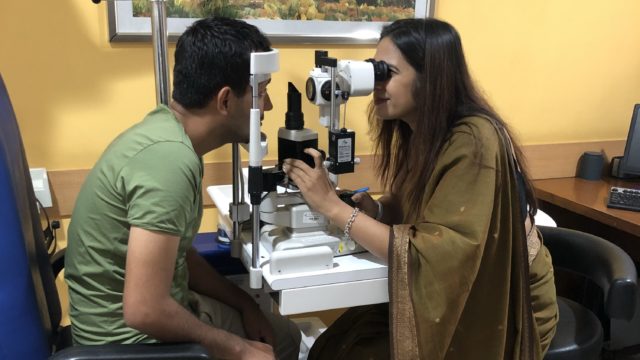
Slit Lamp Examination
The slit lamp is a binocular microscope. You rest your chin and forehead against the frame while your doctor examines the front structures of the eye — eyelids, cornea, conjunctiva, iris, and lens. A hand-held lens then allows a view of the vitreous, retina, and optic nerve. The slit lamp detects cataracts, corneal ulcers, macular degeneration, and diabetic retinopathy, among other conditions.
Eye Pressure Test (Glaucoma Screening)
Raised intraocular pressure is the main risk factor for glaucoma. Two methods are used:
Non-Contact Tonometry (Air Puff Test)
You look at a light inside the machine. A short puff of air measures the pressure without touching your eye. It is painless, though the sound may startle you.
Applanation Tonometry
Anaesthetic drops numb the eye. Yellow dye is added to your tear film. At the slit lamp, your doctor uses a blue light and a small prism to measure pressure precisely. The prism gently touches the eye surface. You will not feel it.
For a detailed explanation of what eye pressure means and how it relates to glaucoma risk, visit the glaucoma diagnosis page.
Pupil Dilation
Your doctor may instil dilating drops to widen your pupils and get a clearer view of the structures at the back of the eye. The drops are given two or three times and take about 30 minutes to work. Dilation causes light sensitivity and blurs near vision for two to three hours. Do not drive after a dilated exam. Bring sunglasses.
Dilated Retinal Evaluation
After dilation, your doctor examines the retina and optic nerve in detail using indirect ophthalmoscopy — a head-mounted light. You will be asked to look at your outstretched thumb in different positions. The light is bright but the test is brief.
What Additional Tests Might Be Ordered?
If your doctor needs more information, she may request:
Retinal photography, gonioscopy, corneal pachymetry, corneal topography, macular or optic nerve OCT, visual field testing, ultrasound, or biometry.
These tests rule out or confirm specific conditions. If you have received a glaucoma diagnosis elsewhere and want an independent review of your results, you can request a second opinion here.
How Long Does an Eye Exam Take?
A routine exam without dilation takes 15 to 20 minutes. With dilation, allow 60 to 90 minutes in total.
Questions to Ask Before You Leave
Is my vision fully corrected with glasses or contact lenses?
Ask your doctor to confirm that your prescription gives you the best possible vision.
Is my eye pressure within the normal range?
Normal pressure is roughly 10 to 21 mmHg, but target pressure varies by individual. Ask what your numbers mean for you.
Are my retina, cornea, and optic nerve healthy?
Get a clear yes or no — and ask what to watch for between visits.
Do I need any further tests?
If tests are recommended, ask what each one checks and when to schedule it.
When do I need to come back?
Frequency depends on your age, risk factors, and whether any conditions were found. Ask specifically.
How do I reach you in an emergency?
Confirm the after-hours contact before you leave.
Do you need a comprehensive eye examination?
If your last eye test only checked your glasses number, it may have missed early disease.
If you’d like a more detailed assessment, you can book a consultation or request a structured second opinion.
What we look for in a comprehensive eye examination, which can be missed in a general test:
- Early optic nerve changes
- Silent glaucoma risk
- Structure-function mismatch
- Long-term risk, not just current vision
Read the research articles
This article was written by Dr Shibal Bhartiya, fellowship-trained glaucoma specialist and Mayo Clinic Research Collaborator, Clinical Director at Marengo Asia Hospitals, Gurugram, known for ethical, patient-centred glaucoma care and independent glaucoma second opinions. This article was edited in April 2026.
She has published peer-reviewed research on glaucoma management, examining how treatment decisions should balance medical evidence, patient preferences, and long-term vision outcomes.
As Editor-in-Chief of Clinical and Experimental Vision and Eye Research and Executive Editor of the Journal of Current Glaucoma Practice (Pubmed Indexed, official journal of the International Society of Glaucoma Surgery), Dr Shibal Bhartiya brings editorial and research depth to every clinical decision. Her 200+ publications, including 90+ PubMed-indexed publications and 28 edited textbooks span glaucoma biology, surgical outcomes, health equity, and emerging diagnostics.
Her work can be accessed on Pubmed, Google Scholar, ResearchGate and ORCID.
Dr Shibal Bhartiya
Glaucoma • Second Opinion • Advanced Care
www.drshibalbhartiya.com
+91 88826 38735
1500+ Five Star Patient Reviews Google Business Profile