Sleep apnoea can worsen glaucoma by reducing oxygen supply and causing night-time blood flow instability to the optic nerve. If glaucoma progresses despite controlled eye pressure, underlying obstructive sleep apnea should be evaluated as part of long-term risk management, says Dr Shibal Bhartiya.
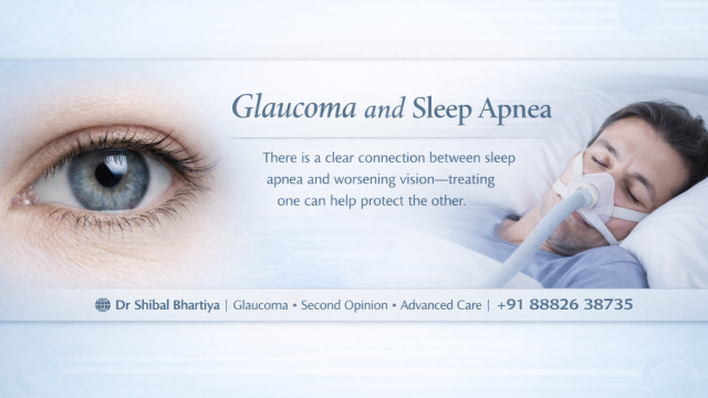
Most people with obstructive sleep apnoea know about the cardiovascular risks. They know about daytime sleepiness, blood pressure, and the heart. Very few are told that sleep apnoea is an independent risk factor for glaucoma, and that it may be driving optic nerve damage in people whose eye pressure appears to be perfectly controlled.
This is not a fringe association. The evidence linking obstructive sleep apnoea to glaucoma, particularly normal tension glaucoma, is substantial. It is consistent across multiple studies, and mechanistically well understood. It is also almost entirely absent from the conversation between patients and their eye specialists.
That gap matters. Because a patient with glaucoma progressing despite controlled eye pressure, who has never been asked about snoring, morning headaches, or unrefreshing sleep, may be missing the most significant modifiable risk factor in their management. Dr Shibal Bhartiya explains more.
Dr Shibal Bhartiya is a fellowship-trained glaucoma specialist and Mayo Clinic Research Collaborator with over 25 years of experience. Her approach focuses on identifying risk before damage is irreversible, simplifying treatment decisions, and protecting vision long-term. Emphasis on early detection, risk assessment, and continuity of care. She is rated 5 stars across 1,500+ patient reviews on Google.
Known for her structured approach to glaucoma risk assessment and progression analysis, Dr Shibal Bhartiya provides trusted second opinions for patients seeking clarity before major treatment decisions.
What Obstructive Sleep Apnoea Does to the Body at Night
Obstructive sleep apnoea (OSA) is characterised by repeated episodes of partial or complete upper airway obstruction during sleep. Each episode causes oxygen saturation to fall. Carbon dioxide rises. The autonomic nervous system triggers a response that wakes you up, and restores breathing. This cycle: obstruction, desaturation, arousal, recovery, may repeat dozens or hundreds of times per night.
The consequences are not limited to sleep disruption. Each desaturation episode causes intermittent hypoxia, a transient drop in oxygen delivery to every tissue in the body. Each arousal causes a spike in blood pressure. Over months and years, this intermittent hypoxia and autonomic dysregulation causes systemic damage: to the cardiovascular system, the cerebrovascular system, the kidneys- and the optic nerve.
The Optic Nerve at Night: Where the Risk Concentrates
The optic nerve is exquisitely sensitive to fluctuations in oxygen delivery and blood flow. It is supplied by the posterior ciliary arteries. Their autoregulation is impaired by chronic hypoxia, elevated carbon dioxide, and autonomic dysregulation. All three are hallmarks of untreated sleep apnoea.
During apnoeic episodes, oxygen saturation can fall to 80 percent or below in severe cases. At the same time, carbon dioxide rises. The perfusion pressure in the optic nerve circulation fluctuates. The repeated pattern of hypoxia and perfusion again, generates oxidative stress and neuroinflammation in the retinal ganglion cells and their axons.
This is the same biological mechanism that drives neurodegeneration in other hypoxic conditions. In the optic nerve, it translates to retinal ganglion cell loss. This loss defines glaucoma.
Sleep Apnoea and Normal Tension Glaucoma
The association between sleep apnoea and glaucoma is strongest for normal tension glaucoma. In NTG, optic nerve damage occurs without elevated intraocular pressure.
This makes biological sense. Normal tension glaucoma has long been understood as a vascular disease as much as a pressure disease. The optic nerve in NTG patients is more susceptible to reduced blood flow. Sleep apnoea, which causes exactly this pattern of nocturnal vascular insult to the optic nerve, is a compelling mechanism for NTG development and progression.
For a patient with normal tension glaucoma whose pressure is well controlled but whose visual field continues to worsen, undiagnosed sleep apnoea should be one of the first things investigated.
Intraocular Pressure During Sleep: The Nocturnal Spike
There is a second mechanism. Intraocular pressure is not static through the night. It follows a circadian rhythm, typically peaking in the early morning hours, between 2am and 6am, when patients are supine and when aqueous humour production is at its highest relative to outflow.
In patients with sleep apnoea, this nocturnal IOP peak is amplified. Studies using continuous IOP monitoring devices have documented significantly higher nocturnal IOP peaks in OSA patients compared to controls. These peaks occur entirely outside the window of the clinic visit where pressure is measured.
This is clinically important for glaucoma patients whose office IOP appears well controlled. If the damage-causing IOP peaks are happening at 3am during apnoeic episodes, clinic measurements at 10am will never capture them. The patient appears stable. The optic nerve is not.
The Floppy Eyelid Syndrome Connection
There is a third, more direct mechanical pathway. Floppy eyelid syndrome, a condition characterised by lax, easily everted upper eyelids, is strongly associated with obstructive sleep apnoea. In floppy eyelid syndrome, the upper lid everts spontaneously during sleep, exposing the palpebral conjunctiva to the pillow surface and causing chronic mechanical irritation, papillary conjunctivitis, and ocular surface disease.
Floppy eyelid syndrome is present in a significant proportion of OSA patients and may be the ocular finding that first suggests the diagnosis. An eye specialist who identifies floppy eyelid syndrome should routinely enquire about sleep symptoms and consider referral for a sleep study.
The relevance to glaucoma is that OSA patients with floppy eyelid syndrome have ocular surface inflammation that may compound the pressure and vascular risks already present.
What This Means in the Clinic
For glaucoma patients, the implications are practical and immediate.
If you have glaucoma and your disease is progressing despite controlled pressure, sleep apnoea should be part of the clinical conversation. A sleep study, polysomnography or a validated home-based sleep study, is the appropriate investigation. It is non-invasive, widely available, and may identify a treatable cause of progression that no change in eye drops will address.
If you snore, wake unrefreshed, have morning headaches, or have been told you stop breathing during sleep, your eye specialist should know. These are not incidental details. They are clinically relevant to both your glaucoma risk and your glaucoma management.
If you have been diagnosed with sleep apnoea and are not currently under glaucoma surveillance, a baseline optic nerve assessment is appropriate. The risk elevation is significant enough to warrant proactive monitoring.
CPAP therapy, the primary treatment for moderate to severe OSA, improves nocturnal oxygen saturation, and stabilises nocturnal IOP fluctuation. Several studies have documented slower glaucoma progression in OSA patients who are compliant with CPAP compared to those who are not. Treating the sleep apnoea is part of treating the glaucoma.
The Integrated Pathway at Marengo Asia Hospitals
Sleep medicine, neurology, and ophthalmology converge in the assessment of a glaucoma patient with suspected sleep apnoea. At Marengo Asia Hospitals, Gurugram, this pathway is available under one roof.
Dr Shibal Bhartiya, as Clinical Director of Ophthalmology and Program Director of the Marengo Asia International Institute of Neuro and Spine, works in direct collaboration with neurologists and sleep medicine specialists. The diagnostic infrastructure includes video EEG with 24-hour brain monitoring, and access to full polysomnographic and respiratory sleep assessment. For a glaucoma patient with progressive field loss despite controlled pressure, this means the vascular, neurological, and sleep-related contributors to progression can all be investigated in one coordinated pathway, without the referral delays and fragmented reporting that characterise the standard model.
Clinical Reality (What’s not always obvious)
- obstructive sleep apnea is linked with glaucoma, but the relationship is complex and often under-recognised.
- Night-time oxygen dips and blood flow fluctuations can stress the optic nerve—even when daytime eye pressure seems controlled.
- Some patients continue to progress despite “normal” pressures—this is where systemic factors like sleep apnoea matter.
- Treating sleep apnoea (e.g., CPAP) may help stabilise risk—but does not replace glaucoma treatment.
- Poor sleep quality, fatigue, and vascular health all influence long-term disease behaviour—this is not just an eye problem.
What You Must Know
| Aspect | What It Means for You |
|---|---|
| Key link | Sleep apnoea can increase risk and progression of glaucoma |
| Why it matters | Reduced oxygen + blood flow instability affects optic nerve health |
| Who should be evaluated | Glaucoma patients with snoring, daytime sleepiness, obesity, resistant progression |
| Warning signs | Loud snoring, pauses in breathing, morning headaches, fatigue |
| Effect on glaucoma | Progression despite controlled eye pressure |
| Diagnosis | Sleep study (polysomnography) |
| Treatment | CPAP therapy, weight management, sleep optimisation |
| Does treatment cure glaucoma? | No—supports stability but does not replace eye treatment |
| What your doctor monitors | Optic nerve, OCT, visual fields, progression patterns |
| Big picture | Glaucoma care sometimes requires looking beyond the eye to protect vision long-term |
Frequently Asked Questions
Does sleep apnoea cause glaucoma?
Sleep apnoea is an independent risk factor for glaucoma, particularly normal tension glaucoma. It does not cause glaucoma in the way a blocked drainage angle does, but through repeated nocturnal hypoxia, nocturnal IOP spikes, and impaired optic nerve blood flow, it creates the conditions in which glaucomatous damage occurs and progresses.
I have glaucoma and my eye pressure is controlled, but my field is still getting worse. Could sleep apnoea be involved?
Yes, and this is one of the most important clinical scenarios in which sleep apnoea should be investigated. Progression despite controlled IOP suggests a vascular or neurological contributor. Sleep apnoea is the most common treatable vascular risk factor in this context. A sleep study is a reasonable next investigation.
What are the symptoms of sleep apnoea I should watch for?
Loud snoring, witnessed apnoeic episodes (stopping breathing during sleep, noted by a partner), waking unrefreshed despite adequate sleep hours, morning headaches, excessive daytime sleepiness, poor concentration, and nocturia. Not all patients with OSA have all symptoms. Some have none beyond the physiological consequences.
Does treating sleep apnoea help glaucoma?
Studies suggest CPAP therapy, the primary treatment for OSA, slows glaucoma progression in patients with both conditions. It stabilises nocturnal oxygen levels, reduces IOP fluctuation, and improves optic nerve perfusion. Treating OSA does not replace glaucoma treatment but is an important adjunct in patients with both conditions.
Should I have a sleep study if I have glaucoma?
If you have glaucoma, particularly normal tension glaucoma, and any symptoms of sleep apnoea, a sleep study may be appropriate. If your glaucoma is progressing despite well-controlled pressure, a sleep study should be actively considered regardless of symptom burden. The test is non-invasive and the finding, if positive, is directly actionable.
Can CPAP use affect intraocular pressure?
The effect of CPAP on IOP is primarily beneficial, by reducing the nocturnal peaks caused by apnoeic episodes. Some studies have noted a modest direct IOP-lowering effect of CPAP treatment. There is no evidence of clinically significant IOP elevation from CPAP use in the majority of patients.
Contact Lens Monitor for Continuous IOP Monitoring
Triggerfish® contact lens sensor is a specialised diagnostic contact lens used in glaucoma care to monitor intraocular pressure (IOP)–related changes over 24 hours. Unlike routine pressure measurements taken during clinic hours, the Triggerfish lens (Sensimed Triggerfish) helps detect pressure fluctuations that may occur at night or outside OPD visits, which can sometimes explain progression despite apparently controlled readings. It does not measure pressure directly in mmHg but records circumferential corneal changes related to IOP patterns, helping glaucoma specialists better understand individual risk profiles and treatment needs in selected patients.
Dr Shibal Bhartiya was the first doctor in India to use the Triggerfish® contact lens sensor for Continuous IOP Monitoring in clinical practice. Her initial experiences on Intraocular pressure (IOP) related pattern in patients with primary angle closure (PAC) and primary angle closure glaucoma (PACG) before and after laser peripheral iridotomy (LPI) was presented at ARVO, in Orlando Florida in 2014.
Her seminal works 24-hour Intraocular pressure monitoring: the way ahead, The Need to maintain Intraocular Pressure over 24 Hours, Diurnal Intraocular Pressure Fluctuation in Eyes with Angle-closure, and Diurnal Variation of IOP in Angle Closure Disease: Are We Doing Enough?
About the Author
This article was written by Dr Shibal Bhartiya, fellowship-trained glaucoma specialist and Mayo Clinic Research Collaborator, Clinical Director at Marengo Asia Hospitals, Gurugram, known for ethical, patient-centred glaucoma care and independent glaucoma second opinions. She is also the Program Director for Community Outreach & Wellness; and for the Marengo Asia International Institute of Neuro and Spine. This article was updated in April 2026.
She has published peer-reviewed research on glaucoma management, examining how treatment decisions should balance medical evidence, patient preferences, and long-term vision outcomes.
As Editor-in-Chief of Clinical and Experimental Vision and Eye Research and Executive Editor of the Journal of Current Glaucoma Practice (Pubmed Indexed, official journal of the International Society of Glaucoma Surgery), Dr Shibal Bhartiya brings editorial and research depth to every clinical decision. Her 200+ publications, including 90+ PubMed-indexed publications and 28 edited textbooks span glaucoma biology, surgical outcomes, health equity, and emerging diagnostics.
Access her work on Pubmed, Google Scholar, ResearchGate and ORCID.
Dr Shibal Bhartiya
Glaucoma • Second Opinion • Advanced Care
www.drshibalbhartiya.com
+91 88826 38735
1500+ Five Star Patient Reviews Google Business Profile
Upload your reports for a structured review.
If you are unable to come to Dr Bhartiya’s clinic: Read more about teleconsultation for glaucoma