Yes. Prostaglandin analogue eye drops, the most commonly prescribed glaucoma drops, can darken the skin around the eye, deepen the eyelid fold, lengthen lashes, and in some patients darken the iris. These changes are real, recognised, and should have been explained before your prescription was written. They are not dangerous. They do not mean you should stop the drop.
Prostaglandin analogues are the first-line treatment for most patients with glaucoma. They work by improving drainage of fluid from the eye, lowering pressure, and protecting the optic nerve. They are effective, well-tolerated, and taken once daily, usually at night.
Common examples include latanoprost, bimatoprost, travoprost, and tafluprost. If your glaucoma drop ends in -prost, it is almost certainly in this class.
What Changes Around the Eye and Why
Prostaglandin analogues stimulate melanin-producing cells around the eye. Over months of use, this produces several visible changes.
The skin of the eyelids and the area around the eye socket darkens gradually. This is called periorbital pigmentation. It happens slowly enough that many patients do not notice it themselves until someone else points it out, or until they see a photograph.
The eyelashes grow longer, thicker, and darker. Some patients welcome this. Others find it uneven or unexpected.
In some patients, particularly those with lighter or mixed-colour irises, the iris itself can darken permanently. In brown-eyed patients, this change is usually not visible.
There is also a structural change called prostaglandin-associated periorbitopathy. The fatty tissue around the eye socket thins. The upper eyelid develops a deeper fold. The eye may appear more sunken. This is subtle in most patients but noticeable in some, particularly after years of use.
Why Were You Not Told
Glaucoma consultations carry a great deal of information. Pressure numbers. Nerve scans. Field tests. Drop instructions. Cosmetic side effects that carry no medical risk sometimes do not make it into the conversation.
Patients who notice an unexpected change in their appearance and do not know why it is happening sometimes stop their drops. Stopping glaucoma drops without guidance can cause serious, irreversible vision loss.
You are entitled to know everything a treatment may do. Not just the part that fixes the problem. So please discuss with your doctor, and she will talk to you about managing the side effects.
Should You Stop the Drop
No. Not without speaking to your glaucoma specialist first.
Uncontrolled glaucoma causes permanent optic nerve damage. These cosmetic changes do not. The risk comparison is not close.
What you should do is tell your doctor. There are other classes of glaucoma drops that do not carry this effect. Beta blockers, alpha agonists, and carbonic anhydrase inhibitors work differently and do not cause periorbital pigmentation. In some patients, switching is appropriate. In others, the prostaglandin is the best option for controlling pressure and the cosmetic effect is manageable.
That conversation should happen with your specialist, with your actual pressure readings and nerve scans on the table. Not as a self-managed decision.
What to Do If You Have Already Noticed This
Bring it up at your next appointment. If it is significantly affecting you, request an earlier one. Ask specifically whether an alternative drop or laser would control your pressure equally well.
If you are newly diagnosed and being started on a prostaglandin drop, ask your doctor to walk you through what changes to expect and over what timeframe. This is not an unreasonable question. It is your face, and your treatment.
FAQs:
Will the Skin Darkening Go Away If I Stop the Drop?
Periorbital skin pigmentation partially reverses after stopping the drop, but this takes months and may not fully resolve. The structural changes of periorbitopathy are slower to reverse. Iris darkening, if it has occurred, is permanent. This is another reason why the conversation about side effects should happen before starting treatment, not after changes have already appeared.
Does This Happen to Everyone on Prostaglandin Drops?
No. The degree of change varies considerably between patients. Some patients use prostaglandin drops for years with minimal visible effect. Others notice changes within a few months. Patients with lighter skin tones may notice periorbital pigmentation more readily. Those with mixed-colour irises are more likely to develop iris darkening. Your specialist can discuss your individual risk based on your eye colour, skin tone, and the specific drop prescribed.
Can I Use the Drop in Only One Eye to Avoid Asymmetric Changes?
If your glaucoma affects only one eye, your doctor may prescribe the drop for that eye only. In this case, asymmetric cosmetic changes are possible — the treated eye may develop darker periorbital skin or longer lashes than the untreated side. This is worth discussing with your specialist before starting, so you can make an informed decision about treatment options.
Are There Glaucoma Drops That Do Not Cause This?
Yes. Beta blockers such as timolol, alpha agonists such as brimonidine, and carbonic anhydrase inhibitors such as dorzolamide do not cause periorbital pigmentation or periorbitopathy. Combination drops that include these classes are also available. Whether they are suitable for you depends on your pressure control needs, your general health, and other medications you may be taking. Your glaucoma specialist can advise.
Is This a Sign That the Drop Is Harming My Eye?
No. These are surface and structural changes around the eye, not damage to the optic nerve or retina. The drop is doing its job inside the eye. The cosmetic effects are a separate matter. Stopping the drop because of skin darkening, without medical guidance, risks the thing that actually matters: your vision.
She has published peer-reviewed research on glaucoma management, examining how treatment decisions should balance medical evidence, patient preferences, and long-term vision outcomes.
Coffee raises intraocular pressure temporarily in some people, but for most glaucoma patients, moderate coffee consumption does not appear to meaningfully worsen the disease. The relationship is more nuanced than a simple yes or no, and the answer depends on how much you drink, your genetic profile, and the type of glaucoma you have, explains Dr Shibal Bhartiya.
Here is what the research actually shows, and what to do with it.
Intraocular pressure (IOP) is the primary modifiable risk factor in glaucoma. Anything that raises IOP, even temporarily, is a reasonable concern for someone whose optic nerve is already under threat. Coffee contains caffeine, a known vasoactive compound, and patients frequently ask whether their morning cup is quietly making things worse.
It is a fair question. And it deserves a proper answer rather than a blanket reassurance or an unnecessarily alarming restriction.
What Caffeine Does to Eye Pressure
Caffeine causes a modest, transient rise in intraocular pressure in most people. Studies measuring IOP before and after caffeine consumption consistently show a rise of approximately 1–3 mmHg, peaking around 60–90 minutes after consumption and returning to baseline within two to three hours.
For context: normal IOP ranges from 10 to 21 mmHg. A rise of 1–3 mmHg in someone whose pressure is already well controlled, say, at 14 mmHg on treatment, takes them to 15–17 mmHg. This is unlikely to be clinically significant.
Where it becomes more relevant is in someone whose pressure is already at the upper end of their target range, or in someone who drinks four to five cups per day and maintains a sustained caffeine effect through most of their waking hours. In these cases, the cumulative IOP burden deserves consideration.
What Large Studies Tell Us
The relationship between coffee and glaucoma has been studied in large population cohorts, and the findings do not support a simple causal link between moderate coffee consumption and glaucoma development or progression.
The Nurses’ Health Study and Caffeine consumption and the risk of primary open-angle glaucoma study, two of the largest and longest-running cohort studies in medicine, found no significant association between caffeinated coffee consumption and the risk of primary open-angle glaucoma overall. Some analyses found a modest increased risk with very high consumption (five or more cups per day), but this was not consistent across subgroups.
A more specific and interesting finding has emerged around exfoliation glaucoma, a type more common in certain populations, where some studies have suggested that high coffee intake may be associated with increased risk. The mechanism is not yet fully understood, and the association requires further investigation before firm clinical recommendations can be made.
The Genetics of Caffeine and IOP
One of the more clinically useful recent findings comes from genome-wide association studies examining how caffeine affects IOP differently across individuals. Variants in genes involved in caffeine metabolism, particularly CYP1A2, which governs how quickly the liver clears caffeine, appear to influence how much IOP rises in response to coffee.
Fast metabolisers clear caffeine quickly and show less sustained IOP effect. Slow metabolisers maintain higher caffeine blood levels for longer and may show a more pronounced and prolonged IOP response.
This genetic variability explains why two patients can drink the same amount of coffee and have very different IOP responses, and why blanket advice to all glaucoma patients is an oversimplification.
Decaffeinated Coffee: A Useful Comparison
Studies comparing caffeinated and decaffeinated coffee in glaucoma patients provide a useful natural experiment. Decaffeinated coffee does not produce the same IOP rise as caffeinated coffee, supporting caffeine, rather than coffee itself, as the active agent. However, decaffeinated coffee is not entirely without vasoactive compounds, and some studies have found small IOP changes with decaf as well, though consistently smaller than with regular coffee.
If you are concerned about coffee and your pressure, switching to decaf is a reasonable, low-risk step that preserves the ritual without the caffeine load.
What About Other Caffeinated Drinks?
The same logic applies to tea, energy drinks, and cola. Caffeine from any source will produce a similar transient IOP effect. Green tea is a partial exception, it contains caffeine but also catechins with antioxidant properties that some research suggests may be beneficial for the optic nerve, though this evidence is preliminary and not a basis for clinical recommendation.
Energy drinks, which combine high caffeine with other stimulants, are a different category. There is no good reason for a glaucoma patient to consume energy drinks, and several reasons to avoid them.
What Doctors Often Miss Telling You
The timing of your coffee relative to your IOP measurement matters. If you routinely have two cups of coffee before your clinic appointment and your IOP is measured 90 minutes later, your reading may be artificially elevated. If you are concerned about this, note the time of your last cup before clinic visits.
Drinking coffee quickly raises IOP more than drinking it slowly. Rapid consumption of large volumes of any fluid, not just coffee, can transiently raise IOP through a volume effect. Sipping rather than gulping is a simple habit change with a real, if modest, physiological rationale.
The IOP effect of coffee is additive with other lifestyle factors. Elevated stress, poor sleep, and caffeine together represent a cumulative IOP burden on the optic nerve that is greater than any one factor alone. Coffee in isolation is rarely the decisive variable.
Exfoliation glaucoma patients may warrant extra caution. If you have been told your glaucoma is the exfoliation type, characterised by flaky white material in the eye, the coffee-IOP relationship in your population is less reassuring than in primary open-angle glaucoma. Discuss specific limits with your specialist.
Lowering coffee intake does not replace your medication. Some patients, after reading about coffee and IOP, experiment with cutting caffeine and monitoring their pressure at home, occasionally using this as a reason to reduce drops. This is not safe. The IOP reduction from cutting coffee is modest and inconsistent. Your medication is not optional.
When to Worry
Coffee consumption itself is not an emergency. But if you notice any of the following, seek assessment regardless of how much coffee you drink:
Headache or eye pain within one to two hours of drinking coffee
Blurred vision or coloured haloes after caffeinated drinks
A consistent pattern of worse vision in the mornings — when IOP tends to peak naturally — that you have not discussed with your specialist
Any new visual symptom that is not explained by your current follow-up plan
The Practical Bottom Line
One to two cups of coffee per day is unlikely to be clinically significant for most glaucoma patients with well-controlled disease. Three or more cups per day, particularly if consumed rapidly and in the morning before an IOP peak, represents a more meaningful cumulative effect worth discussing. Five or more cups per day, especially in patients with exfoliation glaucoma, poorly controlled pressure, or a family history of severe glaucomatous vision loss, warrants a direct conversation with your specialist.
You do not need to give up coffee. You need to understand your specific risk profile and make an informed decision, ideally with your glaucoma doctor.
Frequently Asked Questions
Does coffee cause glaucoma?
Large population studies have not established coffee as a direct cause of primary open-angle glaucoma in moderate consumers. Very high intake — five or more cups per day — has shown a modest association in some studies, particularly for exfoliation glaucoma. For most people, moderate coffee consumption is not a significant glaucoma risk factor.
How much does coffee raise eye pressure?
Caffeine raises intraocular pressure by approximately 1–3 mmHg in most people, peaking around 60–90 minutes after consumption and returning to baseline within two to three hours. The magnitude varies based on genetics, habitual caffeine intake, and baseline pressure.
Should I give up coffee if I have glaucoma?
Not necessarily. One to two cups per day is unlikely to be harmful for most patients with well-controlled glaucoma. If your pressures are at the upper end of your target range, if you have exfoliation glaucoma, or if you drink more than three cups per day, discuss your intake with your specialist. Switching to decaffeinated coffee is a reasonable middle option.
Is decaf coffee safe for glaucoma patients?
Decaffeinated coffee produces a smaller and less consistent IOP rise than caffeinated coffee and is a reasonable alternative for patients who want to reduce their caffeine load without giving up coffee entirely.
Does tea affect glaucoma the same way as coffee?
Tea contains caffeine and will produce a similar transient IOP effect, though typically at lower doses than a standard cup of coffee. Green tea contains additional antioxidant compounds (catechins) with preliminary evidence of benefit for the optic nerve, though this is not yet a basis for clinical recommendation.
Can I drink coffee before my IOP check?
Ideally, try to maintain your usual routine before clinic visits so that your IOP measurement reflects your typical pattern. If you are concerned about the effect of caffeine on your reading, note the time and amount of coffee consumed and mention it to your doctor. For accurate baseline readings, your specialist may sometimes ask you to hold caffeine before a specific pressure measurement.
What type of glaucoma is most affected by coffee?
The available evidence suggests exfoliation glaucoma may be more sensitive to the effects of high coffee intake than primary open-angle glaucoma. If you have been diagnosed with exfoliation syndrome or exfoliative glaucoma, this is worth a specific discussion with your glaucoma specialist.
Speak to a Specialist
Lifestyle questions in glaucoma, coffee, sleep, exercise, supplements, deserve the same careful, evidence-based answer as medication questions. If you are managing glaucoma and want to understand how your daily habits interact with your treatment, that conversation is a legitimate and important part of your care.
She has published peer-reviewed research on glaucoma management, examining how treatment decisions should balance medical evidence, patient preferences, and long-term vision outcomes.
Fellowship-trained glaucoma specialist. 3 years dedicated clinical training in glaucoma and cornea, plus concurrent role as Senior Research Associate, AIIMS, New Delhi
Structured fellowship, Senior Clinical Research Fellow, Department of Clinical Neurosciences, University of Geneva. Special focus: 24 hour IOP monitoring, Glaucoma lasers, SLT, MIGS )
One of few glaucoma specialists also specifically trained in optic neuropathies
Mayo Clinic Research Collaborator (current)
Executive Editor, Journal of Current Glaucoma Practice
1,580+ five-star patient reviews on Google, Gurgaon practice
Quick Answer: Dr Shibal Bhartiya is a fellowship-trained glaucoma specialist in Gurgaon with over 25 years of experience in ophthalmology. Her training included three years of dedicated glaucoma and cornea clinical training, alongside a concurrent role as Senior Research Associate, at AIIMS New Delhi. This was followed by a structured fellowship in glaucoma in the Department of Clinical Neurosciences at the University of Geneva, with a focus on MIGS. This combination makes her one of a small number of glaucoma specialists also specifically trained in optic neuropathies. Dr Bhartiya is also uniquely positioned to manage glaucoma-related ocular surface disease and dry eye, given her parallel cornea training.
Dr Shibal Bhartiya is a current Mayo Clinic Research Collaborator. She has published more than 200 peer-reviewed papers, and has edited over 28 ophthalmology textbooks. She serves as Executive Editor of the Journal of Current Glaucoma Practice. Across her Gurgaon practice’s 1,580+ verified five-star reviews, patients consistently describe feeling heard and treated as individuals, not just diagnoses, with tests, treatment plans, and disease explained in plain language.
Best Glaucoma Specialist in Gurgaon: What Sets Fellowship-Trained Glaucoma Care Apart
Searching for “best glaucoma specialist Gurgaon” usually surfaces a mix of general ophthalmologists, multi-specialty hospital listings, and very few fellowship-trained subspecialists. The difference matters more in glaucoma than almost any other eye condition. It is a disease that is silent until vision is already lost. The treatment decisions made early determine how much sight is preserved over a lifetime. Also, glaucoma rarely exists in isolation. Glaucoma patients frequently develop dry eye from long-term drop us. Some may present with overlapping optic nerve conditions that a purely glaucoma-trained eye can miss. This page sets out, plainly, what fellowship-level glaucoma training, cross-disciplinary training, and active research involvement actually mean for a patient sitting in the chair.
Dr Shibal Bhartiya is a fellowship-trained glaucoma specialist and Mayo Clinic Research Collaborator with over 25 years of experience. Her approach focuses on identifying risk before damage is irreversible, simplifying treatment decisions, and protecting vision long-term. Emphasis on early detection, risk assessment, and continuity of care.
Important: Glaucoma has no symptoms until significant, irreversible vision loss has already occurred in most cases. The qualifications and ongoing research engagement of the specialist you choose is important. It directly affects how early disease is caught and how treatment is sequenced.
What Fellowship-Level Glaucoma Training Looks Like
Training Component
What It Means
Why It Matters for Patients
Glaucoma + cornea clinical training, plus Senior Research Associate, AIIMS New Delhi
3 years of concurrent clinical and research immersion across glaucoma and cornea, alongside high volume, complex glaucoma case exposure; ongoing research collaborations with Prof Tanuj Dada, Head of the Glaucoma Unit at AIIMS
Uniquely positioned to manage glaucoma-related ocular surface disease and dry eye, which most glaucoma-only specialists are not specifically trained to treat
Structured fellowship, Senior Clinical Research Fellow, Dept of Clinical Neurosciences, University of Geneva
Fellowship under Prof Tarek Shaarawy, a leading international authority on MIGS, within a neurosciences department rather than a standard ophthalmology unit
One of a small number of glaucoma specialists also specifically trained to manage other optic neuropathies, not just glaucoma in isolation
Mayo Clinic Research Collaborator (current)
Active, ongoing collaboration with one of the world’s leading glaucoma research groups
Treatment recommendations are informed by current international research, not outdated protocols
Executive Editor, Journal of Current Glaucoma Practice
Reviews and shapes published glaucoma research globally
First-hand, early access to emerging evidence and treatment shifts
Sustained contribution to the field’s evidence base, not just clinical practice
Indicates depth of subject mastery beyond routine patient care
1,585+ five-star Google reviews, Gurgaon practice
Sustained, high-volume patient satisfaction across years of practice, not a handful of recent reviews
Real-world evidence that credentials translate into consistent, trusted patient experience — patients consistently describe feeling heard, having unhurried conversations, and treatment explained in terms of quality of life, not just disease numbers
When To See a Glaucoma Specialist (Not a General Ophthalmologist)
A family history of glaucoma, especially in a parent or sibling
High eye pressure found on a routine check, even without symptoms
Diabetes, high myopia, or long-term steroid use
Already diagnosed with glaucoma and considering a second opinion before surgery
Vision loss that your current doctor has not been able to fully explain
Persistent dryness, burning, or irritation alongside long-term glaucoma drop use
Considering newer surgical options like MIGS before agreeing to traditional surgery
Age over 40 with no eye pressure check in the last two years
What 1,580+ Five-Star Reviews Reflect
Patient tip: A high review volume matters less than what reviews consistently describe. Look for patterns, not just star counts. Most patients call her the best eye doctor, or the best glaucoma specialist in Gurgaon! Most also appreciate how friendly she, and how she especially takes care of children. Several patients mention how much her second opinions, as well as teleconsultations, helped them.
Patients consistently talk of her clear, unhurried explanations. They describe two being told clearly what stage their glaucoma is at. They also say that treatment decisions were explained rather than simply prescribed. In a condition where lifelong monitoring and trust matter as much as any single procedure, that consistency across nearly 1,600 reviews is itself a clinical signal.
Book a Consultation
If you’ve been told you have glaucoma, are at risk, or want a second opinion before surgery. Getting a clear, evidence- based, research-informed assessment early protects vision that cannot be regained later. Book an Appointment → contact us | +91 8882638735
Frequently Asked Questions
What makes a glaucoma specialist different from a general eye doctor?
A glaucoma specialist has completed dedicated subspecialty training beyond a general ophthalmology residency. This typically including a fellowship focused entirely on glaucoma diagnosis, surgical management, and long-term monitoring. This means deeper experience with complex cases, newer surgical techniques like MIGS, and treatment decisions. In Dr Bhartiya’s case, her clinical work grounded in evidence based medicine rather than general practice.
Is Dr Shibal Bhartiya the best glaucoma specialist in Gurgaon?
Dr Shibal Bhartiya is a fellowship-trained glaucoma specialist with over 25 years of experience, including dedicated glaucoma and cornea training at AIIMS New Delhi and a structured fellowship at the University of Geneva. She is a Mayo Clinic Research Collaborator, has authored over 200 publications. She has over 1,585 five-star patient reviews on google, most of which call her the best glaucoma specialist or best eye doctor!
What do patients say about Dr Shibal Bhartiya’s care, beyond her credentials?
Across more than 1,580 five-star reviews on google, patients consistently mention the human experience of care. They always mention being heard without feeling rushed, having questions answered in plain language. Patients appreciate that treatment is explained in terms of daily quality of life rather than just numbers and scans. They also appreciate her ethical, personalised care, with no unnecessary tests or surgeries.
Most patients also call her the best glaucoma specialist, or the best eye doctor in Gurgaon. Most also appreciate how friendly she, and how she especially takes care of children. Several patients mention how much her second opinions, as well as teleconsultations, helped them.
Why does training in cornea matter for a glaucoma specialist?
Long-term glaucoma management almost always involves years of preservative-containing eye drops. This commonly cause ocular surface disease and dry eye over time. A glaucoma specialist with concurrent cornea training is able to recognise and manage this overlap directly. Which means Dr Bhartiya does not have to refer patients elsewhere for a problem that the glaucoma treatment itself helped cause.
What does training in optic neuropathies add to glaucoma care?
Glaucoma is itself a type of optic neuropathy. Some patients have overlapping or atypical optic nerve conditions that can be mistaken for glaucoma or missed alongside it. Specialists trained specifically in optic neuropathies, in addition to glaucoma, are better equipped to catch these atypical presentations early and avoid misdiagnosis.
What is MIGS and why does fellowship training in it matter?
MIGS (Minimally Invasive Glaucoma Surgery) is a newer category of glaucoma surgery. This is less invasive than traditional procedures, with faster recovery and fewer complications for suitable candidates. Specialists trained directly with international MIGS researchers are better positioned to judge which patients are good candidates and to perform it safely.
Should I get a second opinion before glaucoma surgery?
Yes, particularly for procedures that are not reversible. Glaucoma surgery decisions benefit from input by a specialist with broad surgical and research experience. The right choice depends on disease stage, risk stratification and patient preference. Dr Shibal Bhartiya explains options, eye anatomy, and how the disease is likely to progress over decades. Her second opinion is not based just current eye pressure.
Key Takeaways
Fellowship-level glaucoma training (AIIMS, University of Geneva) means deeper exposure to complex cases and advanced techniques like MIGS
Concurrent cornea training at AIIMS uniquely positions her to manage glaucoma-related dry eye and ocular surface disease
Geneva fellowship was within a Department of Clinical Neurosciences, adding specific training in optic neuropathies beyond glaucoma alone
Active research collaboration (Mayo Clinic) keeps treatment decisions current with global evidence
1,580+ five-star reviews show consistency in patient trust and communication over time
Second opinions matter most before irreversible surgical decisions
Dr Bhartiya has published peer-reviewed research on glaucoma management, examining how treatment decisions should balance medical evidence, patient preferences, and long-term vision outcomes.
Flying with glaucoma is safe for the vast majority of patients, but there are specific situations where air travel carries real risk, and your eye doctor needs to know about your travel plans before you board, explains Dr Shibal Bhartiya.
Can You Fly With Glaucoma? What Does Every Glaucoma Patient Needs to Know Before Travelling? The key is knowing which category you fall into.
Why Glaucoma Patients Ask About Flying
The question comes up in almost every glaucoma clinic. Patients worry that cabin pressure will affect their eye pressure, that altitude will worsen their condition, or that long-haul flights will somehow accelerate damage. Most of these fears are unfounded, but not all of them.
The truth is layered. For most people with well-controlled glaucoma, flying presents no meaningful risk. For a small but important subset, those who have had recent eye surgery, those with advanced disease and very high pressures, or those with certain angle configurations; the picture is more complex and deserves a specific conversation with your specialist before travel.
What Happens to Eye Pressure at Altitude
Commercial aircraft cabins are pressurised, but not to sea-level pressure. The standard cabin pressure is equivalent to an altitude of approximately 6,000 to 8,000 feet. This mild reduction in ambient pressure has been studied in glaucoma patients, and the findings are reassuring for most.
Research has not shown a consistent or clinically significant rise in intraocular pressure (IOP) in glaucoma patients during commercial air travel when their disease is well controlled and they are on treatment. The pressure changes that occur in a pressurised cabin are modest, and the eye largely compensates.
Where altitude does matter is in unpressurised aircraft: small planes, high-altitude trekking, or glider flights, where ambient pressure drops more substantially. At true high altitude (above 3,500–4,000 metres), some studies suggest IOP can rise, though the evidence is not uniform.
When Flying With Glaucoma Needs Extra Care
After eye surgery — this is the most important restriction
If you have had glaucoma surgery: trabeculectomy, tube shunt implantation, or any procedure involving a gas bubble in the eye such as retinal surgery, flying is potentially dangerous until your surgeon clears you.
Gas bubbles used in certain retinal procedures expand at altitude. Even the modest pressure change in a commercial cabin can cause a gas bubble to expand significantly, raising intraocular pressure to dangerous levels and risking vision loss. Your surgeon will give you a specific no-fly period, typically few weeks, and this instruction must be followed precisely.
Carry a card or letter from your surgeon if you are within the post-operative window, even if you believe you are cleared, in case of questions at the airport.
After LASIK or corneal procedures
The evidence on flying after LASIK is generally permissive after the first 24 hours, but corneal healing and dry eye are relevant. Cabin air is very dry, humidity in aircraft cabins can fall to 10–20 percent, far below the 40–60 percent that eyes are comfortable in. For glaucoma patients who also have dry eye, a common combination, partly due to long-term use of preserved eye drops, this can cause significant discomfort and blur during flight.
Narrow-angle glaucoma or untreated angle closure
If you have been told you have a narrow drainage angle, or if you have a history of angle-closure episodes that have not been treated with a laser iridotomy, discuss flying with your specialist before travel. Changes in lighting conditions, particularly the dimmer cabin environment during night flights, cause the pupil to dilate, which can precipitate angle closure in susceptible eyes.
A prophylactic laser iridotomy, where appropriate, resolves this risk entirely. If you have already had bilateral iridotomies, this concern no longer applies.
Very advanced glaucoma with poorly controlled pressure
For patients with severe optic nerve damage and pressures that remain elevated despite maximum treatment, any additional stress on the optic nerve deserves consideration. This is not a blanket contraindication to flying, but it is a reason to have a specific conversation with your glaucoma specialist before a long journey, and to ensure your pressure is as well controlled as possible before departure.
Practical Advice for Flying With Glaucoma
Do not skip your eye drops. This is the most important practical point. Crossing time zones disrupts medication schedules. Plan your drop timing before you travel — work out when to take each drop relative to both departure and destination time zones, and set reminders. Missing doses, particularly on long-haul flights, can cause IOP to rise.
Carry all medications in hand luggage. Never put eye drops in checked baggage. Cabins are pressurised; hold luggage is not in some aircraft configurations, and temperature extremes in the hold can affect medication. More practically — bags get lost. Your drops do not.
Use preservative-free lubricating drops liberally. Cabin air is extremely dry. Even if dry eye is not a significant problem on the ground, the low humidity in an aircraft cabin will increase evaporation from the tear film. Use preservative-free artificial tears every hour or so during the flight. If you use preserved glaucoma drops, space these out as you normally would and use lubricants in between.
Stay hydrated, avoid alcohol. Alcohol causes systemic dehydration, which worsens dry eye. It also affects sleep quality on overnight flights, which matters for IOP — pressure tends to be higher after poor sleep.
Inform your travel insurer. Glaucoma is a pre-existing condition and must be declared. Failure to declare it can invalidate a claim.
Carry your prescription and a doctor’s letter for long trips. If you need to replace eye drops abroad, a clear prescription — ideally with the generic drug name, not just the brand — allows a pharmacist in any country to help you. A brief letter confirming your diagnosis and medication is useful for travel to countries with strict pharmaceutical import rules.
What Doctors Often Miss Telling You
The dry eye-glaucoma medication link matters most on flights. Preservative-containing glaucoma drops — almost all the commonly used ones — damage the ocular surface over time. The desiccating effect of cabin air compounds this significantly. Switching to preservative-free formulations before long travel seasons is worth discussing with your specialist.
Night flights with dim cabin lighting are higher risk for narrow-angle patients than daytime flights, most patients are never told this distinction.
The no-fly rule after gas bubble surgery is absolute, not advisory. Patients who fly within this window have lost vision. The restriction is not about comfort, it is about preventing a pressure spike that can permanently damage the optic nerve within hours.
Jet lag affects IOP. Circadian rhythm disruption alters the normal diurnal pattern of intraocular pressure. If you notice your vision is slightly different or your eyes feel more pressured after crossing multiple time zones, this is physiologically real, not imagined.
When to Worry
Seek urgent eye care — at your destination if necessary — if you experience any of the following during or after a flight:
Severe eye pain during or after the flight
Sudden blurring of vision or coloured haloes around lights
A new shadow or loss of part of your visual field
Nausea or vomiting alongside eye pain
Any of the above in the days following eye surgery
These symptoms may indicate a dangerous rise in intraocular pressure and require same-day assessment.
Frequently Asked Questions
Is it safe to fly with glaucoma?
For most patients with well-controlled glaucoma on stable treatment, flying is safe. The pressure changes in a commercial aircraft cabin are modest and do not cause clinically significant IOP rises in the majority of patients. The main exceptions are recent eye surgery involving a gas bubble, untreated narrow angles, and very advanced uncontrolled disease.
How soon after glaucoma surgery can I fly?
This depends entirely on the type of surgery and whether a gas bubble was used. Your surgeon will give you a specific clearance date. For procedures involving gas, such as certain retinal or combined procedures, the no-fly period can be four to six weeks or longer. For trabeculectomy without gas, the window is shorter but still defined by your surgeon. Never fly within this window without explicit clearance.
Do I need to tell the airline I have glaucoma?
For routine travel with well-controlled glaucoma on drops alone, there is no requirement to notify the airline. If you are travelling shortly after surgery, carry a doctor’s letter. Declare your condition to your travel insurer regardless.
What if I miss my eye drops on a long flight?
Try not to. Plan your dosing schedule before departure and set phone alarms. If you miss one dose of a once-daily drop, take it as soon as you remember and resume your normal schedule. Do not double-dose. If you are on twice-daily drops and miss a dose mid-flight, take the next dose at the scheduled time. For any uncertainty about your specific medication, call the clinic before you travel.
Can altitude trekking worsen glaucoma?
At high altitude, above 3,500 metres, there is some evidence of IOP fluctuation. The evidence is not definitive, but for patients with advanced glaucoma or poorly controlled pressure, high-altitude trekking deserves a specific discussion with your specialist before departure. Acclimatisation, hydration, and adherence to medication are particularly important at altitude.
Does cabin pressure affect the optic nerve directly?
No direct effect of commercial cabin pressure on the optic nerve has been demonstrated in research. The concern in glaucoma is about IOP — and for most patients on treatment, this is well managed even during flight.
Plan Your Travel — and Your Eye Care
If you are planning a long trip, an international journey, or travel shortly after an eye procedure, bring it up at your next appointment. A five-minute conversation before you travel is far better than a five-hour emergency at your destination.
Upload your latest reports and medication list through the website before your appointment.
Note:
This article draws on clinical experience and published research in glaucoma and environmental physiology. Work co-authored by Dr Shibal Bhartiya, and her colleagues in Mexico, has specifically examined how altitude and atmospheric pressure influence intraocular pressure (IOP) measurements, highlighting that readings, and their interpretation, can vary across environments and measurement techniques.
This reinforces an important principle: glaucoma is not just about a single pressure reading, but about context, consistency, and longitudinal interpretation.
She has published peer-reviewed research on glaucoma management, examining how treatment decisions should balance medical evidence, patient preferences, and long-term vision outcomes.
Every consultation ends with practical education on how to use eye drops correctly and simple strategies to improve treatment adherence. Successful glaucoma care depends on both accurate diagnosis and consistent treatment.
What Happens During a Glaucoma Consultation? A Doctor’s Walkthrough
Most patients walk into a glaucoma consult expecting a quick pressure check and a prescription. What they get instead, in my clinic, is a sequence. History, vision, anterior segment, a deliberate order of imaging and gonioscopy, baseline pressure testing across more than one visit. The final ten minutes that I consider non negotiable, teaching you how to actually use your drops.
I have refined this sequence over years of glaucoma practice because the disease itself demands it. You cannot feel your eye pressure changing. You cannot feel your nerve fibre layer thinning. The only way to catch glaucoma early and keep it from progressing is a structured, repeatable, slightly unglamorous process. Repeated the same way every single time.
This page walks through that process exactly as it happens in my clinic. So that you know what to expect. It also helps you plan your day better.
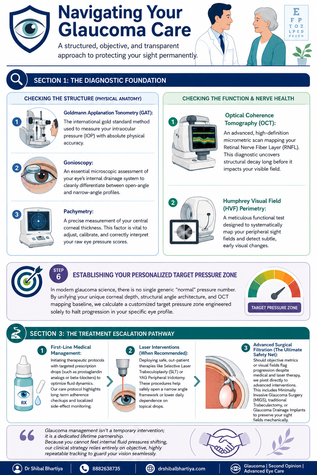
Quick Answer: A glaucoma consultation in my clinic moves through five stages. First, the optometrist takes a detailed history and checks vision. This includes uncorrected vision, best corrected vision, and vision with your current glasses. Second, I review that history myself and examine the front of your eye. Third, I run structural and functional tests in a specific order. Corneal thickness, then pressure measurement, then gonioscopy, with OCT and visual field testing done before gonioscopy when they are needed. Fourth, if your pupils need to be dilated, you wait about forty five minutes. Fifth, no consult ends without me personally teaching you how to instil your eye drops correctly and how to remember whether you have taken them.
A look inside a real glaucoma consultation with Dr Shibal Bhartiya in Gurgaon: structured testing order, baseline pressure checks across visits, and personalised target pressure zones that guide treatment decisions before any drop is prescribed.
Step 1: Before You See Me, the Optometrist Does the Groundwork
Every consult starts with my optometrist, not with me. This is deliberate. It means your history is captured properly and your vision is measured in a structured way before I ever walk into the room.
History taking
The optometrist takes a detailed history and reviews any prior reports, scans, or visual fields you bring with you, noting all of it into your file. This includes systemic conditions that have nothing to do with the eye on the surface, diabetes, high blood pressure, heart disease, asthma, or autoimmune disease, along with any current medications and known allergies. Glaucoma management decisions are frequently shaped by what is happening in the rest of your body, so none of this is skipped.
Three vision measurements, not one
Your vision is then checked through a formal refraction, and three separate numbers are recorded:
UCVA, your uncorrected visual acuity, what you see with no glasses at all
PGP, your vision with the glasses you are currently wearing and prescribed
BCVA, your best corrected visual acuity, what you could see with the ideal glasses prescription
Comparing these three numbers tells me whether a vision problem is about your eyewear, your ocular surface, or your optic nerve, before I have even examined you. A non contact tonometry pressure check is occasionally done at this stage as a screening step. I insist on Goldmann Applanation Tonometry for all of my glaucoma patients.
Step 2: I Review Your History and Examine the Front of the Eye
When you come in to see me, I read through everything the optometrist has documented at a glance. If anything looks incomplete, inconsistent, or worth a second look, I will ask more specific questions to understand it properly before moving forward.
There is also, always, a few minutes of ordinary conversation. A glaucoma consult is a long term relationship, not a transaction. It starts with treating you like a person before a set of test results. And you will be shocked at the details I remember. Your family, your last vacation, your dog 🙂 sometimes, even your favourite chutney!
I then examine the front of your eye in detail. The conjunctiva and ocular surface, the meibomian glands, the eyelid and bulbar conjunctiva, the anterior chamber, and the lens, looking specifically for cataract, a shallow anterior chamber, or any cells in the anterior chamber (inflammation).
Step 3: A Deliberate Order of Testing, Not a Random Checklist
The sequence in which glaucoma tests are performed matters, and I follow a fixed order rather than doing whichever test is most convenient.
Angle assessment first, with imaging informing the decision
I assess the optic nerve with a 90 dioptre lens. Every glaucoma patient gets a gonioscopy. When you need a repeat gonioscopy is decided after that. I perform it only after the visual field test, the OCT, and fundus photography are done, when those are part of that visit. Imaging the nerve and the visual field before manipulating the angle gives me a cleaner functional and structural baseline to work from.
Central corneal thickness, then pressure, then gonioscopy
Before gonioscopy, I measure central corneal thickness (CCT), the test also called pachymetry. Corneal thickness directly affects how your raw eye pressure reading should be interpreted. But it is always done before your tonometry. Because touching your corneas to measure your IOP before the CCT may alter it slightly. Gonioscopy then follows. This examines your drainage angle under magnification. This determines whether you have an open angle or a narrow angle profile.
Why I do the pressure check myself
Goldmann applanation tonometry (GAT), the test that measures your intraocular pressure, is the one test I do not delegate. In my clinic, I personally perform this for every glaucoma patient before treatment starts. Again at the first follow up, and at every annual review. My optometrists are trained to do it and do perform it in my absence. Doing it myself gives me a direct feel for what is happening in your eye that a number on a chart cannot fully convey.
I also insist on doing my gonioscopy myself, always with the lights switched off, so be prepared for a few minutes in a dark room. I keep talking to you, so its never scary.
How is Applanation Tonometry Done?
For the GAT, one of my team members will put some numbing eyedrops and ask you not to touch your eye. I then put a dye which stains your tears yellow. And then I check your eye pressures under blue light on the slit lamp, with a prism that comes close to the eye.
It takes less than a minute if you don’t blink and keep looking straight ahead, and a few extra seconds if you fidget. It’s painless, and quick, and we finish with a drop of antibiotic in the eye.
Step 4: Dilation, When It Is Needed
If your assessment requires dilating your pupils, you will be told this in advance, because dilation takes about forty five minutes to take full effect and changes how you experience the rest of your day.
We ask you to bring dark glasses, a scarf, or an umbrella, since dilated eyes are far more light sensitive, particularly in Gurugram’s daytime heat
We advise you not to drive yourself home after a dilated examination
Step 5: Establishing a True Baseline, Not a Single Snapshot
Glaucoma decisions should never rest on one reading taken on one day. Two specific habits in my clinic exist to correct for that.
Repeating your first visual field
There is a genuine learning curve to taking a visual field test well. The first attempt is frequently unreliable simply because the patient has not yet learned the rhythm of the test. I routinely discard the first visual field and ask patients to return the next morning. We do not charge for that repeat test. The inaccuracy is a known limitation of the test itself, and is not a reason to bill twice.
Three pressure readings, not one
For a true baseline, I usually take three intraocular pressure readings at different times of day. Rather than relying on a single number, since pressure naturally fluctuates through the day. One of these three readings may be taken by an optometrist, if it’s after my working hours. We usually work from the average of all three.
The water drinking test
A formal diurnal variation test, in which pressure is measured every few hours through the day, is not practical for every patient. We often use the water drinking test as a more practical stand in. This is typically done before starting treatment, again about one to two months after treatment begins. We may repeat it if your eye appears to be progressing despite your pressure meeting its target.
Step 6: Setting Your Personalised Target Pressure
There is no single universal normal pressure number in modern glaucoma care. Your corneal thickness, the structure of your drainage angle, and your Visual field and OCT baseline are combined to calculate a target pressure zone. This is specific to your eye, designed to halt progression for you.
Step 7: The Most Important Section of Glaucoma Consultation: Eye Drop Training
A prescription on its own does not protect your vision if the drops never go in correctly or are forgotten. So every consult ends with practical training, not just instructions.
I personally show you how to instil your eye drops correctly, since technique affects how much medication actually reaches the eye
I ask you to set a phone alarm for every dose. Because relying on memory alone is the most common reason treatment fails
If you are on more than one medication, I recommend keeping two small boxes. One empty and one full of your drop bottles. After each dose, you move that bottle from the full box to the empty one. So a glance at the boxes tells you whether you have already taken that round of drops. And which ones remain.
When you leave, my coordinator helps you set your next appointment, before you leave the clinic. You will also receive a Whatsapp message with links to important information and details of phone numbers to book appointments. You will also get my direct phone number for any clinical queries, or emergencies.
When To See Me Before Your Booked Glaucoma Consultation
Sudden eye pain, redness, or blurred vision, which can signal an acute angle closure attack
Any one sided change in vision or eye appearance
Headache or nausea accompanying eye pain
A noticeable change in your visual field between scheduled visits
New side effects after starting or changing a glaucoma medication
Missed doses for several consecutive days, which should be flagged at your next visit rather than left unmentioned
Why does the optometrist see me before the doctor does?
The optometrist’s workup, history, refraction, and the three part vision check, ensures your file is complete and your baseline vision is documented accurately before I begin my own examination. This makes the time I spend with you more focused on interpretation and decision making rather than data collection.
Why do you measure my eye pressure yourself instead of leaving it to staff?
Goldmann applanation tonometry is the gold standard pressure test, and for every glaucoma patient I treat, I perform it myself before starting treatment, at the first follow up, and at every annual review. It gives me a direct sense of your eye’s behaviour that I do not want to lose by always delegating it.
Why do you discard my first visual field test and ask me to repeat it?
Most patients have not yet learned the rhythm of the visual field test on their first attempt. This makes that first result unreliable. We ask you to return the next morning for a repeat test. We do not charge for it, since the inaccuracy belongs to the learning curve of the test, not to you.
Why is gonioscopy done after OCT and visual field testing, not before?
When OCT, visual field testing, and fundus photography are part of your visit, I prefer to have that structural and functional picture in hand before manipulating the angle during gonioscopy. The order is chosen to give the cleanest possible baseline. Also, sometimes I use a viscoelastic gel for gonioscopy. In that case, your vision is fuzzy for about ten minutes after, and I don’t want your time wasted.
What is the water drinking test and why would I need one?
It is a practical way of checking how your eye pressure responds to a physiological stress. This is used in place of round the clock diurnal variation testing, which is not feasible for every patient. I typically use it before starting treatment. I may repeat it again a month or two into treatment. And again later if your eye appears to be progressing even though your pressure looks controlled.
Why do you spend time teaching me to put in my own eye drops?
Technique directly affects how much medication reaches your eye. A missed or mistimed dose is the most common reason glaucoma treatment underperforms. Pairing a phone alarm with the two box system is simple. It gives you a simple, visual way to know whether today’s dose has already gone in. Research says it is the most important intervention in preventing glaucoma blindness.
Key Takeaways
Your consult begins with the optometrist. They document history and perform three vision measurements, UCVA, PGP, and BCVA, before I examine you
Testing follows a fixed order: imaging and visual field first when needed, then corneal thickness, then gonioscopy, then pressure measurement
I personally measure your eye pressure for every glaucoma patient at key visits, rather than delegating it
Your first visual field is usually repeated free of charge, because of a genuine learning curve with the test
Baseline pressure is built from three readings at different times of day, sometimes supplemented by a water drinking test
Your target pressure is personalised to your eye’s anatomy, not based on one generic normal number
No consult ends without hands on training in how to use your drops. And how to track whether you have taken them
Book a Consultation
If you have been told you have glaucoma, or are due for a routine check because of family history or elevated pressure, this is the process you can expect to walk through.
She has published peer-reviewed research on glaucoma management, examining how treatment decisions should balance medical evidence, patient preferences, and long-term vision outcomes.