What Happens During a Glaucoma Consultation?

A glaucoma consultation in my clinic follows a structured five-step process. Detailed history and vision assessment, comprehensive eye examination, glaucoma-specific testing (including corneal thickness, eye pressure, gonioscopy, OCT, and visual fields when needed), pupil dilation if required, and a personalized discussion of findings.

Every consultation ends with practical education on how to use eye drops correctly and simple strategies to improve treatment adherence. Successful glaucoma care depends on both accurate diagnosis and consistent treatment.

Dr Shibal Bhartiya is a fellowship-trained glaucoma specialist and Mayo Clinic Research Collaborator with over 25 years of experience. Her approach focuses on identifying risk before damage is irreversible, simplifying treatment decisions, and protecting vision long-term. Emphasis on early detection, risk assessment, and continuity of care. She is rated 5 stars across 1,500+ patient reviews on Google.

What Happens During a Glaucoma Consultation? A Doctor’s Walkthrough

Most patients walk into a glaucoma consult expecting a quick pressure check and a prescription. What they get instead, in my clinic, is a sequence. History, vision, anterior segment, a deliberate order of imaging and gonioscopy, baseline pressure testing across more than one visit. The final ten minutes that I consider non negotiable, teaching you how to actually use your drops.

I have refined this sequence over years of glaucoma practice because the disease itself demands it. You cannot feel your eye pressure changing. You cannot feel your nerve fibre layer thinning. The only way to catch glaucoma early and keep it from progressing is a structured, repeatable, slightly unglamorous process. Repeated the same way every single time.

This page walks through that process exactly as it happens in my clinic. So that you know what to expect. It also helps you plan your day better.

Quick Answer: A glaucoma consultation in my clinic moves through five stages. First, the optometrist takes a detailed history and checks vision. This includes uncorrected vision, best corrected vision, and vision with your current glasses. Second, I review that history myself and examine the front of your eye. Third, I run structural and functional tests in a specific order. Corneal thickness, then pressure measurement, then gonioscopy, with OCT and visual field testing done before gonioscopy when they are needed. Fourth, if your pupils need to be dilated, you wait about forty five minutes. Fifth, no consult ends without me personally teaching you how to instil your eye drops correctly and how to remember whether you have taken them.

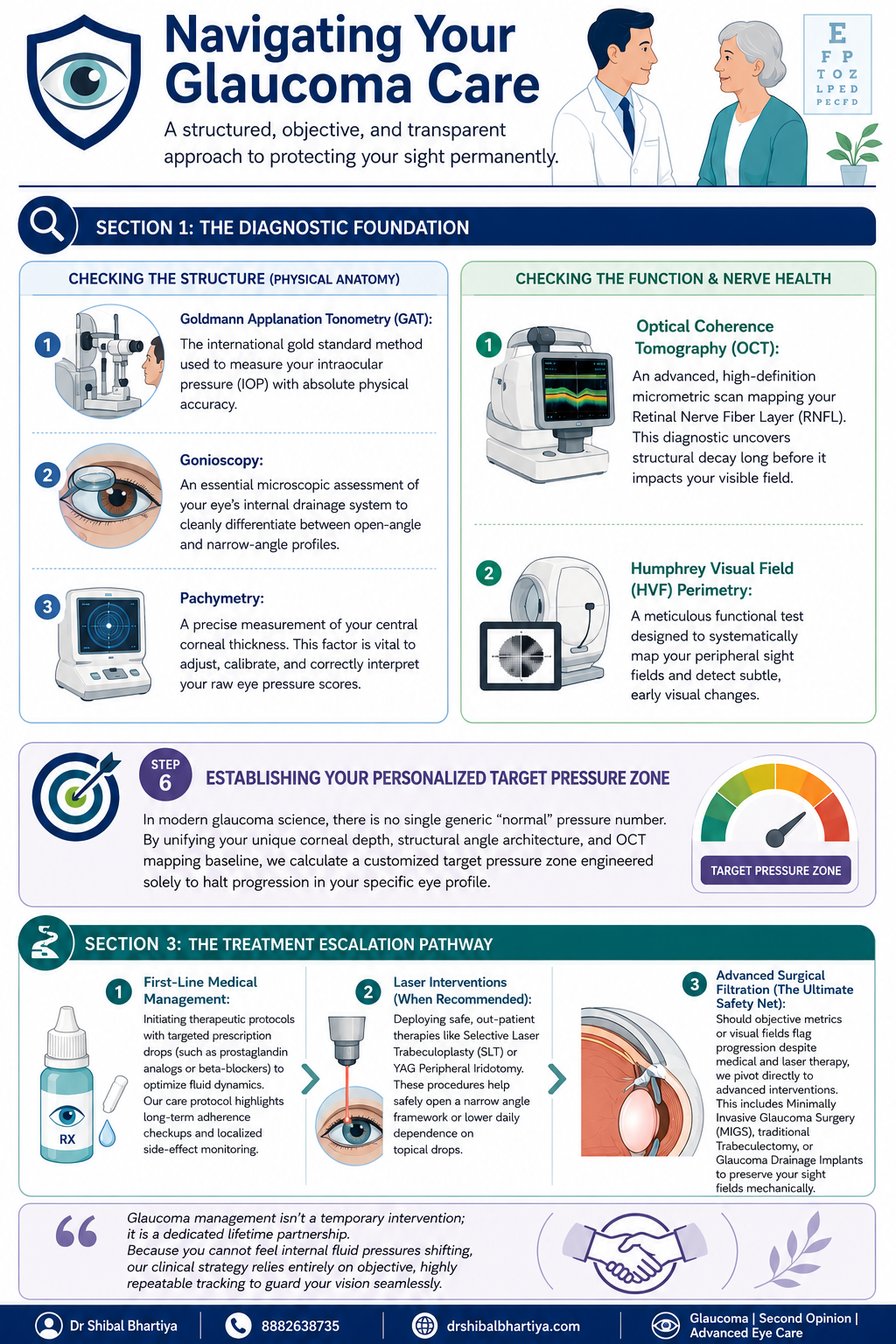
Dr Shibal Bhartiya Gurgaon glaucoma consultation infographic showing diagnostic testing and treatment pathway steps; navigating glaucoma consultation
A look inside a real glaucoma consultation with Dr Shibal Bhartiya in Gurgaon: structured testing order, baseline pressure checks across visits, and personalised target pressure zones that guide treatment decisions before any drop is prescribed.

How to Prepare for Your Glaucoma Consultation

A little preparation before your appointment can make your glaucoma consultation more useful and help your ophthalmologist understand your eyes over time.

If possible, bring any previous eye records, including OCT scans, visual field reports, optic nerve photographs, eye pressure readings, and details of any eye surgery or laser treatment. If you use glaucoma eye drops, bring a list of your medications or the bottles themselves. It is also helpful to know whether anyone in your family has glaucoma or significant vision loss.

Write down any questions or changes you have noticed since your last visit, even if they seem minor. Most importantly, do not stop using your prescribed eye drops before your appointment unless your ophthalmologist has specifically advised you to do so.

The more complete your previous records, the easier it is to compare changes over time—because in glaucoma, understanding what has changed is often more important than any single test result.

Step 1: Before You See Me, the Optometrist Does the Groundwork

Every consult starts with my optometrist, not with me. This is deliberate. It means your history is captured properly and your vision is measured in a structured way before I ever walk into the room.

History taking

The optometrist takes a detailed history and reviews any prior reports, scans, or visual fields you bring with you, noting all of it into your file. This includes systemic conditions that have nothing to do with the eye on the surface, diabetes, high blood pressure, heart disease, asthma, or autoimmune disease, along with any current medications and known allergies. Glaucoma management decisions are frequently shaped by what is happening in the rest of your body, so none of this is skipped.

Three vision measurements, not one

Your vision is then checked through a formal refraction, and three separate numbers are recorded:

  • UCVA, your uncorrected visual acuity, what you see with no glasses at all
  • PGP, your vision with the glasses you are currently wearing and prescribed
  • BCVA, your best corrected visual acuity, what you could see with the ideal glasses prescription

Comparing these three numbers tells me whether a vision problem is about your eyewear, your ocular surface, or your optic nerve, before I have even examined you. A non contact tonometry pressure check is occasionally done at this stage as a screening step. I insist on Goldmann Applanation Tonometry for all of my glaucoma patients.

Step 2: I Review Your History and Examine the Front of the Eye

When you come in to see me, I read through everything the optometrist has documented at a glance. If anything looks incomplete, inconsistent, or worth a second look, I will ask more specific questions to understand it properly before moving forward.

There is also, always, a few minutes of ordinary conversation. A glaucoma consult is a long term relationship, not a transaction. It starts with treating you like a person before a set of test results. And you will be shocked at the details I remember. Your family, your last vacation, your dog 🙂 sometimes, even your favourite chutney!

I then examine the front of your eye in detail. The conjunctiva and ocular surface, the meibomian glands, the eyelid and bulbar conjunctiva, the anterior chamber, and the lens, looking specifically for cataract, a shallow anterior chamber, or any cells in the anterior chamber (inflammation).

Step 3: A Deliberate Order of Testing, Not a Random Checklist

The sequence in which glaucoma tests are performed matters, and I follow a fixed order rather than doing whichever test is most convenient.

Angle assessment first, with imaging informing the decision

I assess the optic nerve with a 90 dioptre lens. Every glaucoma patient gets a gonioscopy. When you need a repeat gonioscopy is decided after that. I perform it only after the visual field test, the OCT, and fundus photography are done, when those are part of that visit. Imaging the nerve and the visual field before manipulating the angle gives me a cleaner functional and structural baseline to work from.

Central corneal thickness, then pressure, then gonioscopy

Before gonioscopy, I measure central corneal thickness (CCT), the test also called pachymetry. Corneal thickness directly affects how your raw eye pressure reading should be interpreted. But it is always done before your tonometry. Because touching your corneas to measure your IOP before the CCT may alter it slightly. Gonioscopy then follows. This examines your drainage angle under magnification. This determines whether you have an open angle or a narrow angle profile.

Why I do the pressure check myself

Goldmann applanation tonometry (GAT), the test that measures your intraocular pressure, is the one test I do not delegate. In my clinic, I personally perform this for every glaucoma patient before treatment starts. Again at the first follow up, and at every annual review. My optometrists are trained to do it and do perform it in my absence. Doing it myself gives me a direct feel for what is happening in your eye that a number on a chart cannot fully convey.

I also insist on doing my gonioscopy myself, always with the lights switched off, so be prepared for a few minutes in a dark room. I keep talking to you, so its never scary.

How is Applanation Tonometry Done?

For the GAT, one of my team members will put some numbing eyedrops and ask you not to touch your eye. I then put a dye which stains your tears yellow. And then I check your eye pressures under blue light on the slit lamp, with a prism that comes close to the eye.

It takes less than a minute if you don’t blink and keep looking straight ahead, and a few extra seconds if you fidget. It’s painless, and quick, and we finish with a drop of antibiotic in the eye.

Step 4: Dilation, When It Is Needed

If your assessment requires dilating your pupils, you will be told this in advance, because dilation takes about forty five minutes to take full effect and changes how you experience the rest of your day.

  • We ask you to bring dark glasses, a scarf, or an umbrella, since dilated eyes are far more light sensitive, particularly in Gurugram’s daytime heat
  • We advise you not to drive yourself home after a dilated examination

Step 5: Establishing a True Baseline, Not a Single Snapshot

Glaucoma decisions should never rest on one reading taken on one day. Two specific habits in my clinic exist to correct for that.

Repeating your first visual field

There is a genuine learning curve to taking a visual field test well. The first attempt is frequently unreliable simply because the patient has not yet learned the rhythm of the test. I routinely discard the first visual field and ask patients to return the next morning. We do not charge for that repeat test. The inaccuracy is a known limitation of the test itself, and is not a reason to bill twice.

Three pressure readings, not one

For a true baseline, I usually take three intraocular pressure readings at different times of day. Rather than relying on a single number, since pressure naturally fluctuates through the day. One of these three readings may be taken by an optometrist, if it’s after my working hours. We usually work from the average of all three.

The water drinking test

A formal diurnal variation test, in which pressure is measured every few hours through the day, is not practical for every patient. We often use the water drinking test as a more practical stand in. This is typically done before starting treatment, again about one to two months after treatment begins. We may repeat it if your eye appears to be progressing despite your pressure meeting its target.

Step 6: Setting Your Personalised Target Pressure

There is no single universal normal pressure number in modern glaucoma care. Your corneal thickness, the structure of your drainage angle, and your Visual field and OCT baseline are combined to calculate a target pressure zone. This is specific to your eye, designed to halt progression for you.

Step 7: The Most Important Section of Glaucoma Consultation: Eye Drop Training

A prescription on its own does not protect your vision if the drops never go in correctly or are forgotten. So every consult ends with practical training, not just instructions.

  • I personally show you how to instil your eye drops correctly, since technique affects how much medication actually reaches the eye
  • I ask you to set a phone alarm for every dose. Because relying on memory alone is the most common reason treatment fails
  • If you are on more than one medication, I recommend keeping two small boxes. One empty and one full of your drop bottles. After each dose, you move that bottle from the full box to the empty one. So a glance at the boxes tells you whether you have already taken that round of drops. And which ones remain.
  • When you leave, my coordinator helps you set your next appointment, before you leave the clinic. You will also receive a Whatsapp message with links to important information and details of phone numbers to book appointments. You will also get my direct phone number for any clinical queries, or emergencies.

When To See Me Before Your Booked Glaucoma Consultation

  • Sudden eye pain, redness, or blurred vision, which can signal an acute angle closure attack
  • Any one sided change in vision or eye appearance
  • Headache or nausea accompanying eye pain
  • A noticeable change in your visual field between scheduled visits
  • New side effects after starting or changing a glaucoma medication
  • Missed doses for several consecutive days, which should be flagged at your next visit rather than left unmentioned

Why Two Patients With the Same Eye Pressure May Receive Different Advice

It is natural to wonder why one person is advised to start glaucoma treatment immediately, while another with a similar eye pressure is simply monitored.

The answer is that glaucoma is not diagnosed or treated based on a single number.

When I assess a patient, I consider the health of the optic nerve, OCT scans, visual field results, corneal thickness, family history, age, previous eye records, and whether there is any evidence that the disease is progressing. I also take into account your lifestyle, occupation, and life expectancy, because the goal is to preserve the vision you need throughout your lifetime.

Two patients may have the same eye pressure but very different levels of risk. One may benefit from careful observation, while the other may need treatment without delay.

My role is not simply to diagnose glaucoma. It is to understand your individual risk of vision loss and recommend the right treatment at the right time.

📍 Gurgaon 📞 +91 88826 38735 | 🌐 Contact Us | Second Opinion Form for teleconsults


This page is a part of the Glaucoma Hub. you may want to read about Glaucoma Progression, and Risk Stratification in Glaucoma. Other articles of interest could be Advanced Glaucoma Care in Gurgaon, What Good Glaucoma Care Actually Optimises For, What Happens If Glaucoma Is Left Untreated?, More Glaucoma Eye Drops is Not Better Glaucoma Care, 5 Mistakes Patients Make in Glaucoma Care and Do You Really Need Treatment for Glaucoma?


Frequently Asked Questions

Why does the optometrist see me before the doctor does?

The optometrist’s workup, history, refraction, and the three part vision check, ensures your file is complete and your baseline vision is documented accurately before I begin my own examination. This makes the time I spend with you more focused on interpretation and decision making rather than data collection.

Why do you measure my eye pressure yourself instead of leaving it to staff?

Goldmann applanation tonometry is the gold standard pressure test, and for every glaucoma patient I treat, I perform it myself before starting treatment, at the first follow up, and at every annual review. It gives me a direct sense of your eye’s behaviour that I do not want to lose by always delegating it.

Why do you discard my first visual field test and ask me to repeat it?

Most patients have not yet learned the rhythm of the visual field test on their first attempt. This makes that first result unreliable. We ask you to return the next morning for a repeat test. We do not charge for it, since the inaccuracy belongs to the learning curve of the test, not to you.

Why is gonioscopy done after OCT and visual field testing, not before?

When OCT, visual field testing, and fundus photography are part of your visit, I prefer to have that structural and functional picture in hand before manipulating the angle during gonioscopy. The order is chosen to give the cleanest possible baseline. Also, sometimes I use a viscoelastic gel for gonioscopy. In that case, your vision is fuzzy for about ten minutes after, and I don’t want your time wasted.

What is the water drinking test and why would I need one?

It is a practical way of checking how your eye pressure responds to a physiological stress. This is used in place of round the clock diurnal variation testing, which is not feasible for every patient. I typically use it before starting treatment. I may repeat it again a month or two into treatment. And again later if your eye appears to be progressing even though your pressure looks controlled.

Why do you spend time teaching me to put in my own eye drops?

Technique directly affects how much medication reaches your eye. A missed or mistimed dose is the most common reason glaucoma treatment underperforms. Pairing a phone alarm with the two box system is simple. It gives you a simple, visual way to know whether today’s dose has already gone in. Research says it is the most important intervention in preventing glaucoma blindness.

Is Dr Shibal Bhartiya the best glaucoma specialist in Gurgaon?

Dr Shibal Bhartiya is a fellowship-trained glaucoma specialist with over 25 years of experience. Her training includes AIIMS New Delhi and the University of Geneva. She is a Mayo Clinic Research Collaborator, has authored over 200 scientific publications, edited 28 ophthalmology textbooks, and has more than 1,600 five-star Google reviews from patients. A lot of her patients call her the best glaucoma specialist in Gurgaon.

What do patients say about Dr Shibal Bhartiya’s care, beyond her credentials?

Patients consistently describe feeling heard, never rushed, and receiving clear, personalised explanations. Many value her ethical approach, thoughtful second opinions, and focus on preserving both vision and quality of life. Many reviews also describe her as the best glaucoma specialist or eye doctor in Gurgaon.


Key Takeaways

  • Your consult begins with the optometrist. They document history and perform three vision measurements, UCVA, PGP, and BCVA, before I examine you
  • Testing follows a fixed order: imaging and visual field first when needed, then corneal thickness, then gonioscopy, then pressure measurement
  • I personally measure your eye pressure for every glaucoma patient at key visits, rather than delegating it
  • Your first visual field is usually repeated free of charge, because of a genuine learning curve with the test
  • Baseline pressure is built from three readings at different times of day, sometimes supplemented by a water drinking test
  • Your target pressure is personalised to your eye’s anatomy, not based on one generic normal number
  • No consult ends without hands on training in how to use your drops. And how to track whether you have taken them

Book a Consultation

If you have been told you have glaucoma, or are due for a routine check because of family history or elevated pressure, this is the process you can expect to walk through.

Book an Appointment →📍 Gurgaon 📞 +91 88826 38735 | 🌐 Contact Us | Second Opinion Form for teleconsults


About the Author

This article was written by Dr Shibal Bhartiya, fellowship-trained glaucoma specialist and Mayo Clinic Research Collaborator, Clinical Director at Marengo Asia Hospitals, Gurugram, known for ethical, patient-centred glaucoma care and independent glaucoma second opinions. She is also the Program Director for Community Outreach & Wellness; and for the Marengo Asia International Institute of Neuro and Spine.

She has published peer-reviewed research on glaucoma management, examining how treatment decisions should balance medical evidence, patient preferences, and long-term vision outcomes.

As Editor-in-Chief of Clinical and Experimental Vision and Eye Research and Executive Editor of the Journal of Current Glaucoma Practice (Pubmed Indexed, official journal of the International Society of Glaucoma Surgery), Dr Shibal Bhartiya brings editorial and research depth to every clinical decision. Her 200+ publications, including 90+ PubMed-indexed publications and 28 edited textbooks span glaucoma biology, surgical outcomes, health equity, and emerging diagnostics.

1600+ Five Star Patient Reviews Google Business Profile

If you are unable to come to Dr Bhartiya’s clinic: Read more about teleconsultation

Read her research on PubMed | Google Scholar | ResearchGate | ORCID

📋Upload your reports for a structured review.| 🌐 Contact Us| 📞 +91 88826 38735

Helped by this article? Leave a Google review — it helps other patients find reliable eye care.

What Does Glaucoma Vision Actually Feel Like

Glaucoma vision is often described as gradually losing side vision, making it harder to notice objects, navigate spaces, or see hazards. Because early glaucoma rarely causes symptoms, regular eye examinations are essential for early detection. Glaucoma usually develops slowly and painlessly, often affecting peripheral (side) vision before central vision. Many people do not notice symptoms until significant and irreversible vision loss has already occurred.

Glaucoma vision does not feel like anything in its early stages. That is the defining and most dangerous feature of this disease. By the time most people notice something is wrong, significant and permanent nerve damage has already occurred.

Dr Shibal Bhartiya is a fellowship-trained glaucoma specialist and Mayo Clinic Research Collaborator with over 25 years of experience. Her approach focuses on identifying risk before damage is irreversible, simplifying treatment decisions, and protecting vision long-term. Emphasis on early detection, risk assessment, and continuity of care. She is rated 5 stars across 1,500+ patient reviews on Google


What Glaucoma Vision Actually Feels Like at Every Stage

Most eye diseases announce themselves. Glaucoma does not. It is a disease of gradual, silent loss, and the experience of that loss is unlike almost anything else in medicine.

Understanding what glaucoma vision feels like at each stage helps you recognise warning signs earlier, take monitoring seriously, and explain your experience accurately to your doctor.


Early Glaucoma: You Feel Nothing

In early glaucoma, peripheral vision begins to fail. The optic nerve loses fibres quietly. No pain, no redness, no blur. Your brain fills in the gaps using information from your other eye and from past visual experience. This is called neural compensation.

You do not see a black patch. You see nothing wrong. Your brain edits the missing information out of your awareness before you ever become conscious of it. This is why early glaucoma is genuinely invisible to the person who has it.

The only reliable way to detect early glaucoma is through examination. Intraocular pressure measurement, optic nerve imaging, and visual field testing reveal damage that the patient cannot feel and cannot describe.


Moderate Glaucoma: The Gaps Begin to Appear

As glaucoma progresses, the areas of lost vision become larger. They can no longer be hidden by neural compensation alone. Patients at this stage describe experiences that are specific and worth knowing.

Missing patches that are hard to locate. Not seeing a dark spot. You miss things that were there. You reach for a glass and it is not where you expected. In fact, you may step off a kerb without seeing it. You lose track of a word while reading and cannot find your place again.

Difficulty in crowds. Faces appear and disappear unexpectedly. People seem to emerge from nowhere because they entered your visual field through an area that no longer reports properly.

Trouble on stairs and uneven ground. Depth perception requires full peripheral vision. When peripheral fields narrow, steps become harder to judge. Falls become more likely.

Slower recovery in dim light. Many patients notice that moving from a bright space to a dim one takes longer than it used to. Glaucoma damages the cells responsible for low-light sensitivity earlier and more severely than those handling bright light.

Glare sensitivity. Headlights, sunlight on water, and bright screens can feel more uncomfortable than before. This is partly nerve damage and partly the effect of raised intraocular pressure on the optical media of the eye.


Advanced Glaucoma: Tunnel Vision

In advanced disease, the visual field contracts severely. Patients describe this as looking through a narrow tube or pipe. Central vision remains sharp because the macula is often the last area affected. Patients can still read and recognise faces. But they cannot see what is beside, above, or below them.

This creates a profound and specific disability. Reading is possible. Walking safely is not. Driving is impossible. Navigating a room without knocking into furniture is a daily challenge. The disconnect between intact central vision and devastated peripheral vision confuses people who do not understand glaucoma. A patient may appear to see perfectly while being functionally blind for most activities of daily life.


End-Stage Glaucoma and Blindness

End-stage glaucoma results in the loss of central vision as well. The patient may retain light perception or hand motion vision. Full blindness from glaucoma is total loss of all functional sight. It is permanent. No treatment restores vision that glaucoma has destroyed.

This is the core clinical reality that shapes everything in glaucoma management. The damage is irreversible. The goal is always to stop further loss, not to recover what is gone.


Acute Angle-Closure Glaucoma: A Different Experience Entirely

The descriptions above apply to primary open-angle glaucoma, which is the most common type. Acute angle-closure glaucoma is different and dramatic.

In acute angle closure, intraocular pressure rises suddenly and severely. The experience is unmistakable. Patients describe intense eye pain, a headache centred around the eye and brow, nausea, vomiting, and vision that becomes blurred and hazy. They often see rainbow-coloured halos around lights. The eye becomes red. The cornea looks hazy even to an observer.

This is a medical emergency. It requires treatment within hours to prevent permanent vision loss. If you or someone you know experiences these symptoms, seek emergency eye care immediately.


Understanding Symptoms & Signs

Glaucoma StageWhat the Patient ExperiencesWhat Standard Vision Tests Show
EarlyNothing. No symptoms.Normal acuity, subtle optic nerve changes, early field defect on testing
ModerateMissing objects, misjudging steps, difficulty in crowdsReduced peripheral field on formal perimetry
AdvancedTunnel vision, frequent falls, cannot driveSeverely constricted visual field, preserved central acuity
End-stageNear or complete blindnessCentral field loss, minimal or no functional vision
Acute angle-closureSevere pain, halos, nausea, blurred visionRaised IOP, corneal haze, shallow anterior chamber

Why Glaucoma Vision Is So Often Misunderstood

Patients with moderate to advanced glaucoma frequently pass standard vision chart tests with flying colours. A 6/6 Snellen result means your central vision is intact. It says nothing about your peripheral field. Doctors, family members, and even some healthcare professionals mistake a good acuity reading for good overall vision.

This misunderstanding has real consequences. Patients are told they see well when they cannot drive safely, cannot walk without risk, and cannot function independently. Visual acuity and visual field are two separate measurements. Both matter. In glaucoma, the field is the critical one.


What to Do If You Recognise These Descriptions

If what you have read here sounds familiar, act now. Glaucoma is treatable. Vision lost cannot be recovered, but further loss can almost always be stopped or slowed significantly with the right intervention.

You need a formal visual field test, an optic nerve assessment with imaging, and an accurate intraocular pressure reading taken at the right time of day. If you have already been diagnosed and your symptoms are progressing, your current treatment may not be sufficient.

A specialist opinion is not a sign that something has gone wrong. It is the most responsible thing you can do for your long-term vision.


This page is a part of the Glaucoma Hub. you may want to read about Glaucoma Progression, and Risk Stratification in Glaucoma.

Other articles of interest could be Advanced Glaucoma Care in Gurgaon, What Good Glaucoma Care Actually Optimises For, What Happens If Glaucoma Is Left Untreated?, More Glaucoma Eye Drops is Not Better Glaucoma Care, 5 Mistakes Patients Make in Glaucoma Care and Do You Really Need Treatment for Glaucoma?


Frequently Asked Questions

Does glaucoma cause pain?

Primary open-angle glaucoma, the most common type, causes no pain at any stage. Pain is a feature of acute angle-closure glaucoma, which is a different condition requiring emergency treatment. The absence of pain in open-angle glaucoma is precisely why it is so often diagnosed late.

Can you see the blind spots caused by glaucoma?

No. The brain fills in missing areas of visual field automatically. You do not see black patches. You simply fail to see things that fall in the damaged area. This is why patients are genuinely unaware of early and even moderate field loss.

Is glaucoma vision loss different in both eyes?

Usually, yes. Glaucoma typically progresses at different rates in each eye. The better eye compensates for the worse one. This further masks the degree of real vision loss until the disease is quite advanced in at least one eye.

How fast does glaucoma vision loss progress?

Rate of progression varies widely. Some patients lose very little vision over decades. Others progress quickly despite treatment. Pressure control, disc structure, blood flow, and genetic factors all play a role. This is why ongoing monitoring matters as much as the initial treatment decision.

Can glaucoma vision loss be reversed?

No. Optic nerve fibres that glaucoma destroys do not regenerate. Current treatment preserves remaining vision by controlling intraocular pressure and, where possible, protecting the optic nerve. Research into neuroprotection is active. There is currently no approved therapy to restore lost glaucomatous vision.


Author Sign-Off

This article was written by Dr Shibal Bhartiya, fellowship-trained glaucoma specialist and Mayo Clinic Research Collaborator, Clinical Director at Marengo Asia Hospitals, Gurugram, known for ethical, patient-centred glaucoma care and independent glaucoma second opinions. She is also the Program Director for Community Outreach & Wellness; and for the Marengo Asia International Institute of Neuro and Spine.

She has published peer-reviewed research on glaucoma management, examining how treatment decisions should balance medical evidence, patient preferences, and long-term vision outcomes.

As Editor-in-Chief of Clinical and Experimental Vision and Eye Research and Executive Editor of the Journal of Current Glaucoma Practice (Pubmed Indexed, official journal of the International Society of Glaucoma Surgery), Dr Shibal Bhartiya brings editorial and research depth to every clinical decision. Her 200+ publications, including 90+ PubMed-indexed publications and 28 edited textbooks span glaucoma biology, surgical outcomes, health equity, and emerging diagnostics.

1500+ Five Star Patient Reviews Google Business Profile

If you are unable to come to Dr Bhartiya’s clinic: Read more about teleconsultation

Read her research on PubMed | Google Scholar | ResearchGate | ORCID

Upload your reports for a structured review.| www.drshibalbhartiya.com | +91 88826 38735

Leave a review on Google
Glaucoma Diagnosis: First 90 Days

A glaucoma diagnosis can feel overwhelming, but the first 90 days are crucial for understanding your condition, starting treatment, and establishing a plan to protect your vision long term. Early follow-up, regular eye pressure monitoring, and clear communication with your glaucoma specialist can make a significant difference in preserving sight.

Your First 90 Days With Glaucoma: A Step-by-Step Action Plan

Many patients ask me: I have been diagnosed with glaucoma. What do I do now. Here is what I tell them: A glaucoma diagnosis does not mean you are going blind. It means you now have information most people get too late. The next 90 days are the most important window — not because the disease moves fast, but because the habits you build now protect your vision for the next 30 years.

This guide, written by Dr Shibal Bhartiya, tells you exactly what to do, in order.

Dr Shibal Bhartiya is a fellowship-trained glaucoma specialist and Mayo Clinic Research Collaborator with over 25 years of experience. Her approach focuses on identifying risk before damage is irreversible, simplifying treatment decisions, and protecting vision long-term. Emphasis on early detection, risk assessment, and continuity of care. She is rated 5 stars across 1,500+ patient reviews on Google.


Day 1–7 of Glaucoma Diagnosis: Get the Basics Right

Learn to put in your eye drops correctly

This is the single most important skill you will learn. Studies show that over 60% of patients use eye drops incorrectly — and incorrect technique means the drop misses the eye, or drains immediately into the tear duct and does nothing.

Do this:

Wash your hands. Tilt your head back. Pull your lower eyelid gently down to form a pocket. Hold the bottle above the eye without touching it. Squeeze one drop into the pocket — not onto the eyeball directly. Close your eye gently. Press the inner corner of your eye (near the nose) firmly with one finger for 60 seconds. This blocks the tear duct and keeps the drug in the eye where it belongs. Do not blink vigorously. Do not wipe.

If you use more than one drop type, wait five minutes between them. The first drop dilutes and flushes out the second if you use them together.

Ask your doctor or optometrist to watch you do it once. Ask for a correction if your technique needs adjustment.

Here’s a video demostration:

Set your alarms — and take them seriously

Glaucoma drops work only when taken on time, every day, for life. A single missed day matters less than a pattern of casual delays.

Most drops are once daily, ideally at night. Set a recurring alarm on your phone with a label — “Left eye drop, right eye drop, press corner.” Place the bottle next to your toothbrush. The habit links to the existing habit.

If you use drops twice daily, set both alarms. Never rely on memory alone.

File your papers before they disappear

You walked out of the clinic with reports. Photograph or scan every one of them today — the visual field test, the OCT nerve scan, the IOP readings, the prescription. Put them in a dedicated folder on your phone or email them to yourself with the subject line “Glaucoma Records — [your name].”

You will need these at your next visit, at any second opinion, and if you travel and need emergency eye care. Doctors cannot make good decisions without your baseline.


Week 2–4 of Glaucoma Diagnosis: Build the Follow-Up Structure

Your 30-day appointment is not optional

Glaucoma drops take four to six weeks to show their full pressure-lowering effect. Your doctor needs to see you at 30 days to measure whether the drop is working — and to catch side effects early. Do not skip this.

At this visit, your doctor will check:

  • Your intraocular pressure (IOP) against your baseline
  • Whether the drop is causing redness, allergy, or discomfort
  • Whether you need a dose adjustment or a switch to a different medication

Set a calendar reminder for this appointment the day you are diagnosed. If the appointment was not scheduled, call the clinic and schedule it yourself before the week is over.

Know what side effects to watch for

Most glaucoma drops are well-tolerated. But some cause changes you should know about.

Prostaglandin analogues (bimatoprost, travoprost, latanoprost) can darken the iris over time in some patients, and may cause eyelash growth or mild redness. These are cosmetic and not dangerous — but tell your doctor.

Beta-blockers (timolol) can slow your heart rate and cause breathlessness in patients with asthma or heart disease. If you feel unusually short of breath or very tired after starting drops, contact your doctor the same day.

Alpha agonists (brimonidine) sometimes cause an allergic reaction with marked redness and discharge, usually within weeks of starting. Stop the drop and call your doctor if this happens.

None of these mean you must stop treatment. They mean the treatment may need adjustment.


Month 1–2 of Glaucoma Diagnosis: Tell Your Family

Your siblings and children need an eye check — now

Glaucoma has a strong genetic component. First-degree relatives of a glaucoma patient have a four to nine times higher risk of developing the disease. Most of them will have no symptoms at all until damage is advanced.

Tell your siblings and adult children this week. Ask them to see an ophthalmologist for a baseline pressure check, optic nerve assessment, and field test. This is not alarmist. It is the most useful thing your diagnosis can do for your family.


Month 1–3: Address the Controllable Risk Factors

Stop smoking — this one is not negotiable

Smoking constricts blood vessels and reduces blood flow to the optic nerve. It worsens the vascular risk that many glaucoma patients already carry. The damage from smoking adds to the damage from pressure — and your nerve cannot absorb both.

If you smoke, speak to your doctor about cessation support. This is as important as the drops.

Get your metabolic parameters checked

High blood pressure, diabetes, thyroid disease, and sleep apnoea all affect glaucoma progression through vascular and metabolic pathways. If these are uncontrolled, your optic nerve faces risk from two directions simultaneously.

Ask your physician to check your blood pressure, fasting glucose, HbA1c, and thyroid function if these have not been done recently. If you snore heavily or feel exhausted in the mornings, mention it — untreated sleep apnoea is a recognised glaucoma risk factor that is almost always missed.

Exercise — the right kind

Moderate aerobic exercise (brisk walking 30 minutes, five days a week) lowers intraocular pressure by a clinically meaningful amount in most patients. Avoid high-resistance head-down exercises like heavy weightlifting or inverted yoga poses — these transiently spike IOP.


Month 2–3: Ask About Laser Treatment

SLT — Selective Laser Trabeculoplasty

If your glaucoma is open-angle type, your doctor may recommend SLT as a first-line treatment or as a supplement to drops. SLT uses a laser to improve fluid drainage from the eye. It is done in the clinic in five to ten minutes, is painless, and works in approximately 75 to 80% of patients.

The effect lasts three to five years and can be repeated. SLT does not burn tissue — it sends a gentle energy pulse that stimulates the drainage cells to work better.

Ask your doctor at the 30-day or 90-day visit: “Am I a candidate for SLT?”

LPI — Laser Peripheral Iridotomy

If your glaucoma is narrow-angle or angle-closure type, LPI is a preventive procedure that creates a small opening in the iris to prevent a sudden pressure spike (acute angle-closure attack). LPI is typically recommended before an attack happens — it takes three to four minutes per eye and prevents one of the most painful ophthalmic emergencies.

If your doctor mentioned narrow angles at any point, ask specifically whether you need LPI. Do not wait.


Throughout: Keep Your Perspective

Do not search the internet at 2am

Glaucoma outcomes in treated patients are overwhelmingly good. The disease moves slowly in the vast majority of cases. Patients who take their drops, attend follow-ups, and manage their risk factors maintain useful vision for life in most cases.

The stories of severe vision loss you will find online mostly involve patients who were never diagnosed, or who stopped treatment. You are neither.

Reach out if you need support

A new diagnosis changes how you think about your body. Some patients find this unsettling, and that is entirely normal. Several Indian and international glaucoma patient forums, and online communities run by ophthalmologists offer peer support from people at every stage of the same journey.

You do not have to figure this out alone.


Your 90-Day Checklist

  • Eye drop technique confirmed by a doctor or technician
  • Alarm set — every day, same time
  • All reports photographed and filed digitally
  • 30-day follow-up appointment booked
  • Side effects list saved on your phone
  • Siblings and adult children informed and booked for screening
  • Smoking cessation initiated if applicable
  • Blood pressure, glucose, HbA1c, thyroid checked
  • SLT or LPI discussion had with your doctor
  • One support resource bookmarked

Frequently Asked Questions

Do I have to take eye drops for life?

In most cases, yes. Glaucoma is a chronic condition and eye drops control pressure — they do not cure the disease. Stopping drops allows pressure to rise again and damage to resume. Some patients reduce or stop drops after successful laser treatment (SLT), but this is a decision made with your doctor based on your pressure readings, not independently.

What if I forget a drop one day?

Take it as soon as you remember, unless it is almost time for the next dose. Do not double up. One missed dose will not cause a crisis. A habit of casual misses will. Reset the alarm and continue.

Can I drive after putting in my eye drops?

Most glaucoma drops do not affect vision significantly. Some patients notice mild blurring for a few minutes immediately after instillation — wait for this to clear before driving. If your doctor has dilated your pupils at a clinic visit, do not drive until dilation wears off, typically three to four hours.

My pressure was normal at diagnosis. Do I still have glaucoma?

Yes — this is called normal-tension glaucoma (NTG). Roughly 30 to 40% of glaucoma patients in India have pressures within the statistical normal range. The diagnosis is made on optic nerve appearance and visual field changes, not pressure alone. NTG is treated the same way — the target is to lower pressure further from your individual baseline.

Is glaucoma hereditary? Do I need to tell my family?

Yes, and yes. First-degree relatives — parents, siblings, children — have a four to nine times higher risk. Most will have no symptoms. Tell them this week and ask them to see an ophthalmologist for a baseline check that includes pressure, nerve assessment, and a visual field test.

Will I go blind?

Treated glaucoma in a compliant patient who attends follow-up carries a very low risk of blindness. The risk is real only when the disease is undiagnosed, undertreated, or ignored. You have been diagnosed. That is the most important step already taken.

What is SLT and should I ask about it?

Selective Laser Trabeculoplasty (SLT) is a five-minute clinic procedure that improves fluid drainage from the eye. It works in approximately 75 to 80% of open-angle glaucoma patients and can reduce or eliminate the need for drops for three to five years. Ask your doctor at the 30-day visit whether you are a candidate.

Can I exercise with glaucoma?

Yes — moderate aerobic exercise is actively beneficial and lowers IOP. Brisk walking, cycling, and swimming are all good. Avoid heavy resistance training with breath-holding (Valsalva manoeuvre) and inverted positions, both of which spike pressure transiently. If exercise is a regular part of your routine, tell your doctor so they can factor it into your pressure readings.

My drops are making my eyes red. Should I stop?

Do not stop without speaking to your doctor first. Redness is common with several drop classes and is often manageable — a preservative-free formulation or a switch in medication resolves it in most cases. Stopping drops independently allows pressure to rise. Call the clinic and describe the symptom.

How often will I need follow-up forever?

Once stable on treatment, most patients are reviewed every three to six months. This includes a pressure check and, once yearly or more often if needed, a repeat visual field test and OCT nerve scan to confirm the disease is not progressing. Glaucoma never becomes self-managing — the follow-up rhythm continues for life, but it is not onerous once the initial titration phase is complete.


This page is part of the Glaucoma Hub hub. Read about our full approach to glaucoma care and monitoring. Please also read our guide to Understanding Your Visual Field Test. You may want to read a patient’s experience with glaucoma eye drops, and of one with SLT.


About the Author

This article was written by Dr Shibal Bhartiya, fellowship-trained glaucoma specialist and Mayo Clinic Research Collaborator, Clinical Director at Marengo Asia Hospitals, Gurugram, known for ethical, patient-centred glaucoma care and independent glaucoma second opinions. She is also the Program Director for Community Outreach & Wellness; and for the Marengo Asia International Institute of Neuro and Spine.

She has published peer-reviewed research on glaucoma management, examining how treatment decisions should balance medical evidence, patient preferences, and long-term vision outcomes.

As Editor-in-Chief of Clinical and Experimental Vision and Eye Research and Executive Editor of the Journal of Current Glaucoma Practice (Pubmed Indexed, official journal of the International Society of Glaucoma Surgery), Dr Shibal Bhartiya brings editorial and research depth to every clinical decision. Her 200+ publications, including 90+ PubMed-indexed publications and 28 edited textbooks span glaucoma biology, surgical outcomes, health equity, and emerging diagnostics.

1500+ Five Star Patient Reviews Google Business Profile

If you are unable to come to Dr Bhartiya’s clinic: Read more about teleconsultation

Read her research on PubMed | Google Scholar | ResearchGate | ORCID

Upload your reports for a structured review.| www.drshibalbhartiya.com | +91 88826 38735

Leave a review on Google


More Glaucoma Eye Drops is Not Better Glaucoma Care

More glaucoma eye drops do not guarantee better control. Treatment must be individualised based on riskprogression, and tolerance. Overmedication can increase side effects, reduce adherence, and still fail to protect long-term vision, explains Dr Shibal Bhartiya. Adding more glaucoma medications does not always mean better care and may reflect disease progression requiring proper reassessment.

When glaucoma worsens, many patients assume the next step is simple: add more eye drops.
But glaucoma care is not about the number of medicines. It is about protecting the optic nerve safely over a lifetime.

Sometimes adding drops helps. Sometimes it harms. Good care depends on judgement, sequencing, and long-term strategy.

Dr Shibal Bhartiya is a fellowship-trained glaucoma specialist and Mayo Clinic Research Collaborator with over 25 years of experience. Her approach focuses on identifying risk before damage is irreversible, simplifying treatment decisions, and protecting vision long-term. Emphasis on early detection, risk assessment, and continuity of care. She is rated 5 stars across 1,500+ patient reviews on Google.


Glaucoma Is a Long-Arc Disease

Glaucoma damage is slow, silent, and irreversible.

Treatment must balance:

The goal is not perfect numbers. The goal is lifelong, stable vision.


What Is Target Eye Pressure?

Every patient has a target intraocular pressure (IOP), a level considered safe for their optic nerve.

This depends on:

Two patients with the same pressure may need very different treatment. Glaucoma care is about staying below your safe pressure consistently, not just lowering it once.

Dr Bhartiya, along with her colleagues in Australia and Switzerland, has published peer-reviewed research on current perspectives on Target IOP in glaucoma practice, examining how treatment decisions should balance medical evidence, patient preferences, and long-term vision outcomes. Her 2014 paper, Target Intraocular Pressure: Approaches and Options, examines how glaucoma specialists should set, communicate, and revise pressure targets, balancing clinical evidence, patient preferences, and long-term vision outcomes. It is cited by glaucoma surgeons internationally and is freely available on PubMed.


When More Eye Drops Are Not Better

Adding multiple medications can lead to:

  • Redness, burning, and irritation
  • Allergy and eyelid swelling
  • Severe dryness
  • Complex dosing schedules
  • Poor adherence

In some cases, pressure appears controlled, but damage continues.

More medication does not always mean better protection.


What Is Maximal Medical Therapy?

Maximal medical therapy refers to using the maximum safe combination of eye drops before considering laser or surgery.

But “maximum” is not always “optimal.”

It can result in:

  • Ocular surface damage
  • Poor compliance
  • Fluctuating eye pressure
  • Reduced quality of life

In many cases, laser or surgery may be safer than adding more drops. Glaucoma care is not reactive, it is risk-governed.


Fixed-Dose Combination Drops: A Smarter Approach

Fixed-dose combinations combine two medications in one bottle.

They help by:

  • Reducing the number of drops
  • Simplifying treatment
  • Improving adherence
  • Lowering preservative exposure

Often, simpler regimens protect vision better than complex ones.


What Is Preservative Load?

Many glaucoma drops contain preservatives. Using multiple medications increases cumulative preservative exposure, which can damage the eye surface.

This may cause:

Reducing drops, or using preservative-free options, can significantly improve comfort and safety.


Why More Glaucoma Drops is Not Better Glaucoma Care

SituationWhat Patients Often ThinkWhat Is Actually HappeningWhat Better Care Looks Like
Pressure still high“Add another drop”Target pressure may be wrong or disease is progressing despite treatmentReassess diagnosis, stage, and target pressure
Multiple drops prescribed“More medicines = stronger treatment”Overmedication increases side effects without improving outcomesRationalise drops, simplify regimen
Eyes becoming red / irritated“Drops are working but causing minor issues”Ocular surface damage from preservatives affecting adherenceSwitch to preservative-free or reduce drop burden
Vision feels worse despite “good reports”“Tests are normal, so everything is fine”Functional loss or fluctuation not captured in routine examsCorrelate symptoms with OCT + visual fields
Frequent drop changes“Doctor is trying different combinations”Lack of structured long-term planEstablish stable, personalised treatment pathway
Difficulty remembering drops“I just need to be more careful”Complex regimens reduce compliance and effectivenessSimplify treatment or consider laser (SLT)
Long-term progression“Glaucoma just gets worse over time”Inadequate monitoring or delayed escalationTimely escalation: laser or surgery when needed

Glaucoma Care Is Not Just About Pressure

Effective glaucoma management looks beyond numbers:

  • Optic nerve structure
  • OCT trends over time
  • Visual field progression
  •  Target IOP
  • Medication tolerance
  •  Lifestyle and adherence

More treatment is not always better treatment. The right treatment, at the right time, matters more.

Clinical Reality (What’s Not Always Obvious)

  1. More drops does not mean better control
    Adding medications can feel like escalation, but without reassessing the disease, it may not improve long-term outcomes.
  2. A “good” pressure reading can be misleading
    One normal reading does not guarantee stability—glaucoma damage can continue silently between visits.
  3. Treatment can become habit instead of strategy
    Over time, care may drift into simply adding or switching drops rather than redefining targets and plans.
  4. Side effects quietly affect outcomes
    Multiple preserved drops can irritate the ocular surface, making patients less consistent with treatment.
  5. Stable reports don’t always mean stable disease
    Individual tests may look fine, but progression often appears only when data is tracked over time.
  6. Complex regimens reduce adherence
    The more complicated the schedule, the harder it becomes to follow consistently—reducing real-world effectiveness.
  7. Escalation is often delayed
    Laser or surgery may be postponed because “something is being done,” even if it’s no longer enough.
  8. Follow-up gaps change the disease trajectory
    Longer intervals without structured review can allow subtle progression to go unnoticed.
  9. Targets are not always redefined
    As glaucoma advances, the required pressure often needs to be lower—but this isn’t always updated.
  10. Activity is mistaken for effectiveness
    More visits, more drops, more changes—these can create the illusion of control without actually protecting vision.

When Laser or Surgery May Be Safer

Laser or surgery may be recommended if:

  • Target pressure is not achieved
  • Drops cause significant side effects
  • Adherence is difficult
  • Disease continues to progress
  • Risk of vision loss is high

These decisions are about long-term safety, not treatment failure.


Signs Your Glaucoma Treatment Needs Review

Consider a second opinion if you notice:

  • Increasing number of medications
  • Persistent redness or irritation
  • Confusing or difficult schedules
  • “Normal” pressure but worsening tests
  • High cost or poor affordability
  • Reduced quality of life

Treatment should feel sustainable and tolerable.


Why an Independent Glaucoma Review Helps

Glaucoma decisions are complex and long-term.

structured second opinion can help:

  • Reconfirm diagnosis
  • Reassess target IOP
  • Simplify medications
  • Identify better options
  • Avoid overtreatment

Especially important if you are on 3 or more eye drops.


The Real Goal of Glaucoma Care

Not perfect pressure numbers. Not maximum medications.

The goal is:

  • Right treatment
  • Right timing
  • Minimal burden
  • Long-term stability

More eye drops do not always mean better care.


FAQs

1. Do more glaucoma eye drops mean better treatment?

No. More drops do not necessarily improve outcomes. Treatment must be tailored to your risk profile and disease progression, not just escalated.


2. How many glaucoma drops are too many?

There is no fixed number, but if you are on 3 or more medications, your treatment strategy should be reviewed for effectiveness, tolerance, and alternatives.


3. Why do glaucoma drops stop working?

Glaucoma may progress despite treatment, or medications may become less effective over time. Poor adherence and incorrect sequencing also play a role.


4. What are the side effects of multiple glaucoma drops?

Common side effects include redness, burning, dryness, allergy, blurred vision, and poor tolerance, especially with long-term use.


5. What is target eye pressure in glaucoma?

Target IOP is the pressure level considered safe for your optic nerve. It varies based on damage, age, and progression risk.


6. Are laser or surgery better than eye drops?

In some cases, yes. If drops are not effective or tolerated, laser or surgery may offer safer long-term control.


7. What are fixed combination glaucoma drops?

These combine two medications in one bottle, helping reduce drop burden, improve compliance, and lower preservative exposure.


8. When should I get a second opinion for glaucoma?

If you are on multiple drops, still progressing, or experiencing side effects, a second opinion can help optimise your treatment plan.

 Book a glaucoma care review

Known for her structured approach to glaucoma risk assessment and progression analysis, Dr Shibal Bhartiya provides trusted second opinions for patients seeking clarity before major treatment decisions. Both, in person, and online.

Dr Shibal Bhartiya
Glaucoma • Second Opinion • Advanced Care

Read the research articles

This article was written by Dr Shibal Bhartiya, fellowship-trained glaucoma specialist and Mayo Clinic Research Collaborator, Clinical Director at Marengo Asia Hospitals, Gurugram, known for ethical, patient-centred glaucoma care and independent glaucoma second opinions. She is also the Program Director for Community Outreach & Wellness; and for the Marengo Asia International Institute of Neuro and Spine. This article was updated in April 2026.

She has published peer-reviewed research on glaucoma management, examining how treatment decisions should balance medical evidence, patient preferences, and long-term vision outcomes.

As Editor-in-Chief of Clinical and Experimental Vision and Eye Research and Executive Editor of the Journal of Current Glaucoma Practice (Pubmed Indexed, official journal of the International Society of Glaucoma Surgery), Dr Shibal Bhartiya brings editorial and research depth to every clinical decision. Her 200+ publications, including 90+ PubMed-indexed publications and 28 edited textbooks span glaucoma biology, surgical outcomes, health equity, and emerging diagnostics.

Access her work on PubmedGoogle ScholarResearchGate and ORCID.

Dr Shibal Bhartiya
Glaucoma • Second Opinion • Advanced Care

www.drshibalbhartiya.com
 +91 88826 38735

1500+ Five Star Patient Reviews Google Business Profile

Upload your reports for a structured review.

If you are unable to come to Dr Bhartiya’s clinic: Read more about teleconsultation for glaucoma

Ethical Glaucoma Care

Glaucoma is a chronic disease that often requires lifelong monitoring and treatment decisions that affect patients for decades. Ethical glaucoma…