Optic nerve damage is diagnosed early using tests like OCT scans and visual field testing to detect subtle structural and functional changes. Regular eye exams are critical, as early damage often occurs without symptoms.
Most people wonder how is optic nerve damage diagnosed early. People think optic nerve damage shows up suddenly as vision loss. In reality, it begins quietly, years before symptoms. The optic nerve is not just an eye structure. It is brain tissue. Damage here is slow, cumulative, and often irreversible.
So early diagnosis is not about one test. It is about pattern recognition over time, says Dr Bhartiya.
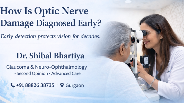
Dr Shibal Bhartiya is a fellowship-trained glaucoma specialist and Mayo Clinic Research Collaborator with over 25 years of experience. Her approach focuses on identifying risk before damage is irreversible, simplifying treatment decisions, and protecting vision long-term. Emphasis on early detection, risk assessment, and continuity of care. She is rated 5 stars across 1,500+ patient reviews on Google.
Why Early Detection Matters
Optic nerve damage behaves like other neurodegenerative diseases:
• Silent progression
• No pain
• Late detection leads to permanent loss
• Early treatment stabilises function
By the time a patient notices vision loss, 40–50% of nerve fibres may already be gone. This is why glaucoma is often called the silent thief of sight.
Who Needs Early Screening?
You should have structured optic nerve screening if you have:
• Age above 40
• Family history of glaucoma
• Diabetes or high blood pressure
• High myopia
• Steroid use
• Previous eye injury
• Thin cornea, or high eye pressures
• Migraine or vascular disease
Women, especially caregivers, often delay checkups. We see late disease more commonly in them.
How Optic Nerve Damage Is Diagnosed Early
1. Careful Clinical Examination
The most important step is still a trained eye.
Your doctor examines:
• Optic nerve shape
• Cup-disc ratio
• Rim thinning
• Disc haemorrhages
• Asymmetry between eyes
These subtle clues often appear years before vision loss.
2. OCT (Optical Coherence Tomography)
OCT measures nerve fibre thickness. It detects damage before vision changes. But OCT is not magic. One scan alone is meaningless. Trend analysis over time matters.
We compare:
• Year-to-year change
• Symmetry between eyes
• Correlation with clinical findings
OCT is a governance tool, not a diagnosis in isolation.
3. Visual Field Testing
This measures functional vision. It shows small blind-spot patterns that patients cannot notice.
But visual fields require:
• Good attention
• Repeated testing
• Interpretation in context
A single abnormal field does not equal glaucoma.
4. Eye Pressure Measurement
High eye pressure is a major risk factor. But many glaucoma patients have “normal” pressure. So pressure alone does not diagnose optic nerve damage early. This is why screening camps that check only pressure miss disease.
5. Risk Stratification Over Time
Early diagnosis is about longitudinal thinking.
We look at:
• Nerve appearance
• OCT trend
• Field pattern
• Pressure history
• Family history
Then decide whether the nerve is stable or changing. This approach prevents both over-treatment and missed disease.
Why Early Damage Is Often Missed
In India, optic nerve damage is missed because of:
• One-time eye camps
• Focus only on cataract
• No baseline OCT
• Lack of follow-up
• Patient reassurance after one normal test
Early glaucoma is boring. Systems prefer dramatic disease.
What Patients Can Do
• Get baseline eye exam at 40
• Repeat every 1–2 years
• Keep your reports
• Ask for OCT trend analysis
• Seek second opinion if unsure
Think of this like blood pressure monitoring. Not one-time testing.
Our Approach in Gurgaon
At our clinic, optic nerve care is based on:
• Early detection
• Long-arc risk planning
• Minimal but meaningful testing
• Patient education
• Stability tracking
Because preserving vision is about decades, not days.
If you need a structured glaucoma or optic nerve second opinion, you can book at
www.drshibalbhartiya.com
or WhatsApp +91 88826 38735.
1. Can optic nerve damage be reversed?
No. Most optic nerve damage is permanent. Treatment focuses on preventing further loss by lowering eye pressure or treating the underlying cause. This is why early diagnosis matters.
2. What is the best test to detect early glaucoma?
There is no single best test. Early detection requires a combination of optic nerve examination, OCT scan, visual field testing, and eye pressure measurement interpreted together over time.
3. How often should I get my optic nerve checked?
After age 40, an eye exam every 1–2 years is recommended. If you have risk factors like family history or diabetes, yearly testing may be needed.
4. What is an OCT scan and why is it important?
OCT (Optical Coherence Tomography) measures the thickness of optic nerve fibres. It can detect damage before vision loss occurs, especially when compared over multiple visits.
5. Can optic nerve damage happen with normal eye pressure?
Yes. This is called normal-tension glaucoma. The optic nerve is sensitive even at “normal” pressures, so pressure alone cannot rule out disease.
6. Is optic nerve damage painful?
Usually no. Most glaucoma and optic nerve diseases progress silently without pain or redness.
7. Who is at high risk for optic nerve damage?
People above 40, those with family history of glaucoma, diabetes, high myopia, steroid use, or thin corneas have higher risk.
8. When should I seek a glaucoma second opinion?
If your OCT shows changes, your pressure remains high, you were advised surgery suddenly, or reports are unclear, a structured second opinion helps long-term planning.
For appointments in Gurgaon:
www.drshibalbhartiya.com | +91 88826 38735
Read the research articles
This article was written by Dr Shibal Bhartiya, fellowship-trained glaucoma specialist and Mayo Clinic Research Collaborator, Clinical Director at Marengo Asia Hospitals, Gurugram, known for ethical, patient-centred glaucoma care and independent glaucoma second opinions. She is also the Program Director for Community Outreach & Wellness; and for the Marengo Asia International Institute of Neuro and Spine. This article was updated in April 2026.
She has published peer-reviewed research on glaucoma management, examining how treatment decisions should balance medical evidence, patient preferences, and long-term vision outcomes.
As Editor-in-Chief of Clinical and Experimental Vision and Eye Research and Executive Editor of the Journal of Current Glaucoma Practice (Pubmed Indexed, official journal of the International Society of Glaucoma Surgery), Dr Shibal Bhartiya brings editorial and research depth to every clinical decision. Her 200+ publications, including 90+ PubMed-indexed publications and 28 edited textbooks span glaucoma biology, surgical outcomes, health equity, and emerging diagnostics.
Access her work on Pubmed, Google Scholar, ResearchGate and ORCID.
Dr Shibal Bhartiya
Glaucoma • Second Opinion • Advanced Care
www.drshibalbhartiya.com
+91 88826 38735
1500+ Five Star Patient Reviews Google Business Profile
Upload your reports for a structured review.
If you are unable to come to Dr Bhartiya’s clinic: Read more about teleconsultation for glaucoma