Gonioscopy: The Eye Test That Reveals Your Drainage Angle. Your eye produces fluid constantly. That fluid drains out through a tiny channel called the drainage angle. When this angle is blocked or too narrow, pressure builds up inside the eye. Over time, that pressure can damage the optic nerve and cause glaucoma.
Gonioscopy is the test that lets your doctor see this drainage angle directly. No other test gives the same information.
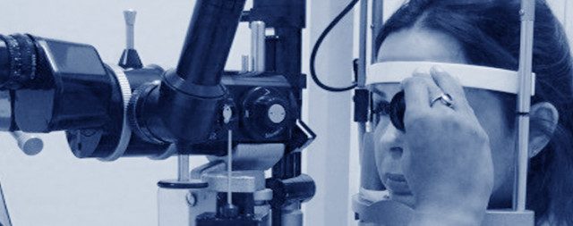
What Is Gonioscopy?
Gonioscopy is a special eye examination. Your doctor places a small lens on the surface of your eye. This lens uses mirrors to show the drainage angle, an area that is otherwise invisible.
The test helps diagnose glaucoma, classify its type, and guide treatment decisions. It is one of the most important tests in glaucoma care.
Why Do You Need a Gonioscopy?
Your doctor may recommend gonioscopy in several situations.
You may have a shallow front chamber. If the space at the front of your eye looks narrow, your doctor needs to check the drainage angle directly.
You are a glaucoma suspect. Gonioscopy is essential for glaucoma diagnosis and classification. It tells your doctor whether your angle is open, narrow, or closed.
You have had an eye injury. Trauma can cause scarring inside the drainage angle. This is called angle recession. Gonioscopy detects it early.
You have diabetic eye disease. New blood vessels can grow into the drainage angle. Your doctor checks for this during gonioscopy.
You are getting an ICL implant. Implantable collamer lens surgery requires a baseline gonioscopy before the procedure.
You take certain medicines. Some drugs can trigger angle closure. Your doctor may want to check your angle before you start or continue these medications.
Is Gonioscopy Painful?
No. Gonioscopy is not painful.
Your doctor puts anaesthetic drops in your eyes first. These may sting briefly. After that, you will feel no pain.
You may feel mild pressure on the eye when the lens is placed. Some people find this slightly uncomfortable, but most patients tolerate the test very well.
After the test, your vision may be blurry for 15 to 20 minutes. This is normal. It clears completely on its own.
In rare cases, the test stimulates the vagus nerve and causes brief dizziness or faintness. This is temporary and harmless.
How Is the Test Done?
The whole procedure takes just a few minutes.
- Your doctor explains the test and answers your questions.
- Anaesthetic drops go into both eyes.
- A small amount of gel is placed on the gonioscopy lens. This feels similar to ultrasound gel.
- The lens is gently placed on the surface of your eye.
- Your doctor asks you to look straight ahead or in a specific direction. Keep both eyes open and follow the instructions.
- The room lights are dimmed. This gives a clearer view of the drainage angle.
- Once the test is done, your doctor helps you clean your eyes.
- Antibiotic drops are placed in each eye before you leave.
What Does Gonioscopy Show?
Gonioscopy helps your doctor answer several key questions.
Is your angle open or closed? Open-angle glaucoma and angle-closure glaucoma need very different treatments. Gonioscopy makes this distinction possible.
Is there scarring or new blood vessels? These can block drainage and raise eye pressure.
What grade is your angle? Doctors use grading systems like the Shaffer classification to record how wide or narrow your angle is. This helps track changes over time.
Are there any other abnormalities? Pigment deposits, adhesions (called peripheral anterior synechiae), or other changes are all visible during gonioscopy.
Gonioscopy vs. OCT of the Anterior Segment
Some clinics use OCT (optical coherence tomography) imaging to assess the drainage angle. Anterior segment OCT or AS-OCT checks the front of the eye (anterior part) unlike RNFL OCT for optic nerve.
Both tests are useful. OCT is non-contact and objective. But gonioscopy remains the gold standard. Only gonioscopy allows dynamic assessment, your doctor can gently indent the lens to distinguish true closure from appositional closure. This changes management in many cases.
Your doctor will choose the right test for your situation.
How to Prepare for Gonioscopy
You don’t really have to “prepare” for gonioscopy.
- Continue your regular eye drops.
- Remove contact lenses before the appointment if possible.
- Bring any previous eye records or reports.
- Arrange for someone to drive you if you prefer, since vision may be briefly blurry afterwards.
Frequently Asked Questions
How long does gonioscopy take?
The test itself takes about 5 minutes. The entire appointment, including explanation and follow-up, takes around 20 to 30 minutes.
Will I need gonioscopy more than once?
Yes. Your doctor may repeat gonioscopy to check for changes in the drainage angle over time, especially if your glaucoma is being actively managed.
Can gonioscopy harm my eye?
No. Gonioscopy is a very safe procedure. The lens touches the surface of the eye briefly, with no risk of lasting harm.
Is gonioscopy available in Gurgaon?
Yes. Dr Shibal Bhartiya is an expert at Gonioscopy, and performs it routinely in Gurgaon, as part of a comprehensive glaucoma evaluation.
Book a Glaucoma Evaluation in Gurgaon
If you have been told you may have glaucoma, a narrow angle, or raised eye pressure, a full glaucoma evaluation, including gonioscopy, is the right next step.
Dr Shibal Bhartiya is a fellowship-trained glaucoma specialist with 25+ years of experience. She offers careful, evidence-based care and honest answers.
📞 Call: +91 88826 38735 | +91 98187 00269 📍 Marengo Asia Hospitals, Sector 56, Gurgaon
Book an Appointment | Request a Second Opinion
Read the research articles
This article has been written by Dr Shibal Bhartiya, a glaucoma specialist in Gurgaon known for ethical, patient-centred glaucoma care and independent glaucoma second opinions. She is also a research collaborator with Mayo Clinic, Jacksonville, Florida, USA. This article was updated in March, 2026.
She has published peer-reviewed research on glaucoma laser and surgeries, examining how treatment decisions should balance medical evidence, patient preferences, and long-term vision outcomes.
These peer-reviewed article discussing glaucoma treatment are benchmarks for glaucoma surgeons globally. You can access them on PubMed and Google Scholar
If you would like a structured glaucoma risk assessment or second opinion:
+91 88826 38735
drshibalbhartiya.com