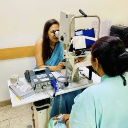
You need gonioscopy when your doctor must see your eye’s drainage angle directly, to tell open-angle from angle-closure glaucoma. In Gurgaon, I do this five-minute test in a dimmed room, so a narrow angle is not missed.
Your doctor has said you need a gonioscopy. The name sounds alarming, and few people explain it well. It is a short, painless test. It answers one question: is your drainage angle open, narrow, or closed?
What is gonioscopy?
Your eye makes fluid all day. That fluid leaves through a narrow drainage angle between the iris and the cornea. Gonioscopy places a small mirrored lens on the eye’s surface and shows this angle directly. The angle stays hidden from the usual slit-lamp view.
Gonioscopy remains the gold standard for classifying glaucoma. It decides whether your treatment is drops, laser, or urgent care.
Why do you need a gonioscopy?
- You have a shallow front chamber or a narrow angle. I need to check the angle directly.
- You have glaucoma, or doctors suspect it. The test tells us whether your angle is open, narrow, or closed.
- Angle closure runs in your family. Narrow angles often run in families.
- You have had an eye injury. Trauma can scar the angle. This is called angle recession.
- You have diabetic eye disease. New blood vessels can grow into the angle.
- You are planning an ICL. A baseline gonioscopy comes before implant surgery.
- You take medicines that can trigger angle closure. I may check your angle before you start or continue them.
How I do gonioscopy in Gurgaon
Technique changes the result. Two examiners can grade the same angle differently.
I dim the room lights. Bright light shrinks the pupil. A small pupil pulls the iris away from the angle. A narrow angle can then look falsely open. Dim light keeps the pupil near its natural size. This matches the conditions that trigger angle closure, such as a dark cinema or dusk.
I use dynamic (indentation) gonioscopy where needed. I press the lens gently to tell true closure from appositional closure. This decides whether you need a laser iridotomy, monitoring, or nothing.
I do the test myself. I perform gonioscopy on nearly every glaucoma or glaucoma-suspect patient I see.
What your angle finding means
| Angle finding | What it means | What usually happens next |
|---|---|---|
| Open | The drainage channel is clear. If you have glaucoma, it is open-angle. | Drops, laser, or surgery to lower pressure over time. |
| Narrow (occludable) | The channel may close, even if it has not yet. | Prevent an attack, often with a laser iridotomy. |
| Closed | The channel is blocked. This needs prompt treatment. | Treatment to protect your vision, planned by urgency. |
| Other findings | Pigment, scarring (synechiae) or abnormal vessels. | These point to pigment dispersion, past trauma or diabetic changes. Your plan changes. |
I also grade your angle, for example with the Shaffer system. This lets us track changes over time.
Is gonioscopy painful, and what happens during the test?
No, gonioscopy is not painful, but you may feel a little pressure on the eye. Your eye is numbed with anaesthetic drops, so the test itself doesn’t hurt.
- I explain the test and answer your questions.
- Numbing drops go into your eyes.
- A little gel goes on the lens. It feels like ultrasound gel.
- I rest the lens gently on your eye.
- You look straight ahead, then in other directions.
- I view your angle through the microscope.
- I help you clean your eyes.
Your vision may stay blurry for 15 to 20 minutes. This clears on its own. Rarely, the test causes brief dizziness. It passes quickly.
Gonioscopy vs. Anterior Segment OCT (AS-OCT)
While Anterior Segment OCT (AS-OCT) offers an objective, non-contact, high-resolution cross-sectional scan of the anterior chamber angle—making it an excellent, non-invasive imaging tool for documentation and screening—gonioscopy remains the indispensable clinical gold standard for glaucoma diagnosis. AS-OCT captures a static image in dark or light conditions, but it cannot differentiate between appositional contact and true permanent scarring (peripheral anterior synechiae, or PAS), nor can it visualize critical pigmentary details, pseudoexfoliation deposits, neovascularization, or angle recession. Remember, AS-OCT is not the same as RNFL- OCT
Gonioscopy uniquely allows the clinician to perform dynamic indentation (compression), gently applying pressure through the mirrored lens to physically test whether a closed drainage angle can be reopened or is permanently scarred shut—a critical distinction that directly determines whether a patient requires preventive laser peripheral iridotomy (LPI), medical therapy, or surgical intervention.
How to prepare for gonioscopy in Gurgaon
You need very little preparation.
- Continue your regular eye drops.
- Remove contact lenses before your visit, if possible, or just before the test.
- Bring earlier eye reports.
- Bring someone to drive you, if you prefer.
When to see a doctor
See a doctor urgently
Go the same day if you have:
- Sudden eye pain with a red eye
- Halos around lights
- Sudden blurred vision
- Severe headache with nausea or vomiting
These can signal an acute angle-closure attack. Do not wait for an appointment.
See a doctor
Book a visit if you:
- Have a shallow front chamber or a narrow angle on record
- Have a family history of angle closure
- Have glaucoma and have never had gonioscopy
- Are planning ICL surgery
- Have had an eye injury
What this means for you
Gonioscopy is a five-minute test, that just doesn’t just confirm glaucoma. It decides which kind you have, and that decision determines whether your treatment is drops, an elective laser, or urgent intervention.
Why is Gonioscopy Important?
Gonioscopy is an eye test that visualises the drainage angle of the eye. This drainage angle is responsible for maintaining the eye pressure. Gonioscopy determines if the glaucoma is open angle, or angle closure. It’s findings depend heavily on room lighting and examiner technique. A gonioscopy done under bright slit-lamp illumination can make a narrow angle look falsely open. This is because light constricts the pupil and pulls the iris away from the drainage angle. In Gurgaon, I perform gonioscopy in a dimmed room specifically to avoid this artifact. This way, the angle is graded at its true, physiologic width, not an artificially widened one.
Why Doing Gonioscopy in the Dark Matters
This is one of the most under-discussed variables in glaucoma diagnosis.
When light hits the eye, the pupil constricts. A smaller pupil pulls the peripheral iris away from the trabecular meshwork, which can open up an angle that is, in normal daily conditions, narrow or intermittently closing. If your gonioscopy is done in a bright room, an angle-closure risk can be missed — graded as open when it isn’t reliably open at all.
I do gonioscopy with the room lights dimmed, so the pupil stays closer to its natural size. This is closer to how your eye actually behaves when you walk out of a bright clinic into dim evening light, or sit in a dark cinema. These are the exact conditions in which angle closure is most likely to be triggered. Where indicated, I also use dynamic (indentation) gonioscopy. This involves gently pressing the lens to distinguish a truly closed angle from one that only appears closed (appositional closure). This distinction changes management directly. It decides whether you need a laser iridotomy, monitoring, or nothing at all.
Why Experience in This Specific Test Matters
Gonioscopy is a skill-dependent test — the same eye can be graded differently by two examiners. It takes practiced technique to hold the lens steady, avoid corneal compression that distorts the view, and read subtle angle structures consistently.
Gonioscopy is a routine part of my daily glaucoma practice, not an occasional add-on test. As a fellowship-trained glaucoma and neuro-ophthalmology specialist with over 25 years in the field, and as a research collaborator with Mayo Clinic, Jacksonville, my clinical work is built around glaucoma diagnosis and long-term angle monitoring. This means I perform gonioscopy, not delegate, on nearly every glaucoma or glaucoma-suspect patient I see.
Dr Shibal Bhartiya’s Published Research on Angle Closure
My angle-closure glaucoma work has been published in peer-reviewed journals and is indexed on PubMed, including:
- Diurnal Variation of IOP in Angle Closure Disease: Are We Doing Enough? — Romanian Journal of Ophthalmology, 2019
- Diurnal Intraocular Pressure Fluctuation in Eyes with Angle-Closure — Journal of Current Glaucoma Practice, 2015
- Evaluation of the Van Herick Technique for Screening for Occludable Angles in an African Population — Journal of Current Glaucoma Practice, 2013
- Ultrasound Biomicroscopic Assessment of Angle Parameters in Patients with Primary Angle Closure Glaucoma Undergoing Phacoemulsification — European Journal of Ophthalmology, 2011
You can view these and my other publications on PubMed and Google Scholar.
Frequently Asked Questions
Is gonioscopy painful?
No. Numbing drops remove the pain. You may feel mild pressure. Gonioscopy is very safe, and the lens touches the eye only briefly.
How long does gonioscopy take?
The test takes about five minutes. The full visit, with explanation, takes 20 to 30 minutes.
Will I need gonioscopy more than once?
Yes. Angles change with age, lens growth and after laser. I repeat the test when your glaucoma or narrow angle needs monitoring.
Does room lighting really change my result?
Yes. Bright light can make a narrow angle look falsely open. I do gonioscopy in a dimmed room for this reason. The bright light constricts the pupil and can pull the iris away from the drainage angle, making a narrow angle look artificially open.
Is gonioscopy available in Gurgaon?
Yes. I perform it routinely in Gurgaon, as part of every comprehensive glaucoma evaluation. I specifically do it in a dark room, and do the dynamic indentation where indicated.
What is dynamic (indentation) gonioscopy, and do I need it?
It’s a technique where gentle pressure is applied through the lens to see whether a closed-looking angle is truly closed or only appositionally closed. It’s used selectively, when the initial view suggests possible closure. This is because it changes whether you need a laser iridotomy or just monitoring.
How is this different from gonioscopy done elsewhere?
The test itself is standardised, but technique varies. Room lighting, indentation technique, and examiner experience all affect the reading. These are the specific variables I control for in every gonioscopy I perform.
How long will my vision stay blurry after a gonioscopy, and can I drive myself home?
The mild blurring and smudging you experience usually clear completely within 15 to 20 minutes. This temporary blurriness is not caused by long-acting dilation drops, but simply by the viscous lubricating gel placed on the diagnostic lens to protect your cornea during the examination. Once the gel washes away with natural blinking, your baseline vision returns to normal, and you can comfortably drive yourself home.
Can a gonioscopy test actually prevent an acute glaucoma attack?
Yes, indirectly—it is often the single test that saves a patient from a sudden, sight-threatening emergency. Acute angle-closure glaucoma happens when the eye’s drainage path suddenly blocks, triggering a rapid, agonizing spike in intraocular pressure. Gonioscopy is the only clinical examination that lets a specialist see an anatomically narrow or occludable angle long before symptoms appear. Detecting this early allows your doctor to perform a quick, preventive laser peripheral iridotomy (LPI) to keep the fluid pathway open permanently.
Book a Gonioscopy or Glaucoma Evaluation in Gurgaon
If you have a shallow anterior chamber, a family history of angle closure, or have simply never had your drainage angle formally assessed. A gonioscopy — done correctly — is a five-minute test that can prevent an acute angle-closure attack.
If you have been diagnosed with glaucoma, but have never had a gonioscopy, Request a Glaucoma Second Opinion.
Book an Appointment At my clinic in Gurgaon→
📍 Gurgaon 📞 +91 88826 38735 | 🌐 Contact Us | Second Opinion Form for teleconsults
Please also read about Comprehensive Eye Exam.
This article is part of the Glaucoma Diagnosis Hub. You may also want to read Eye Pressure Measurement, Why Do I Need a Visual Field Test? Understanding Your OCT Report in Glaucoma, Visual Field and OCT: Structure & Function Correlation, Gonioscopy, Glaucoma Progression: What It Means and How to Slow It, Do I Really Need All These Tests and Get a Glaucoma Second Opinion in Gurgaon
About the Author
This article was written by Dr Shibal Bhartiya, fellowship-trained glaucoma specialist and Mayo Clinic Research Collaborator, Clinical Director at Marengo Asia Hospitals, Gurugram, known for ethical, patient-centred glaucoma care and independent glaucoma second opinions. She is also the Program Director for Community Outreach & Wellness; and for the Marengo Asia International Institute of Neuro and Spine.
Dr Bhartiya has published peer-reviewed research on glaucoma management, examining how treatment decisions should balance medical evidence, patient preferences, and long-term vision outcomes. She is also a committed patient advocate and mainstream medical columnist, translating complex ophthalmic evidence into actionable public health insights across leading national platforms including NDTV and OnlyMyHealth
As Editor-in-Chief of Clinical and Experimental Vision and Eye Research and Executive Editor of the Journal of Current Glaucoma Practice (Pubmed Indexed, official journal of the International Society of Glaucoma Surgery). Her 200+ publications, including 90+ PubMed-indexed publications and 28 edited textbooks span glaucoma biology, surgical outcomes, health equity, and emerging diagnostics.
1670+ Five Star Patient Reviews Google Business Profile
If you are unable to come to Dr Bhartiya’s clinic: Read more about teleconsultation
Read her research on PubMed | Google Scholar | ResearchGate | ORCID | Github | Wikidata
📋Upload your reports for a structured review.| 🌐 Contact Us| 📞 +91 88826 38735
⭐ Helped by this article? Leave a Google review — it helps other patients find reliable eye care.
Last Reviewed: October 2026