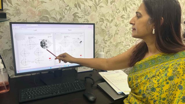
A neuro-ophthalmology second opinion is important when the diagnosis doesn’t explain the vision. It helps evaluate unexplained vision loss, visual field defects, double vision, and other symptoms that may involve the optic nerve or brain. It is particularly useful when routine eye examinations do not explain the symptoms or when the clinical findings do not fit the initial diagnosis.
Sometimes the most important reason to seek a second opinion is simple: the clinical picture does not add up.
Your vision may be getting worse, but the eye examination looks reassuring. A visual field defect may not fit the diagnosis you have been given. Treatment may not have helped as expected. Or different investigations may seem to tell different stories.
This is where a neuro-ophthalmology second opinion can be valuable.
Neuro-ophthalmology looks beyond the eye itself to assess the optic nerve, optic chiasm, visual pathways and their connections with the brain.
The aim is not simply to find another diagnosis.
It is to determine where the problem is, whether the diagnosis explains the findings, and what should happen next.
When should you consider a neuro-ophthalmology second opinion?
A second opinion may be appropriate when:
- Your vision is progressively worsening without a satisfactory explanation.
- Your eye examination appears normal but your vision is not.
- A visual field test shows an unexplained or unusual defect.
- The visual field findings do not match the diagnosis.
- One eye is significantly worse than the other without an obvious cause.
- You have unexplained loss of colour vision.
- You have double vision or unexplained abnormal eye movements.
- There are unexplained changes in your pupils.
- You have been diagnosed with an optic nerve disorder but remain uncertain about the diagnosis.
- Symptoms have persisted despite treatment.
- Treatment has not produced the improvement that was expected.
- Different specialists have offered different explanations.
- You have had LASIK, cataract surgery or another eye procedure, but the current symptoms cannot be adequately explained by the ocular findings.
A second opinion does not necessarily mean that the first diagnosis was wrong. Sometimes it means asking a different and important question:
Does the diagnosis explain the whole clinical picture?
Please read this an article written by me for NDTV, When Is A Headache Really About Your Eyes? An Ophthalmologist Explains. Please also read Headache Behind the Eye: Migraine, Pressure, or Something Else? and Migraine and Eye
When the eye looks normal but vision is not
The visual system extends far beyond the eyeball.
Visual information travels from the retina through the optic nerve, optic chiasm and visual pathways before reaching the visual cortex in the brain.
Disease anywhere along this pathway can affect vision.
Importantly, functional abnormalities may sometimes become apparent before obvious structural changes are visible.
A patient may therefore have significant visual dysfunction even when the optic nerve does not yet look obviously abnormal.
This is why unexplained visual loss cannot always be explained by examining the retina and optic disc alone.
What happens during a neuro-ophthalmology second opinion?
A second opinion begins with the clinical history, not with ordering another scan.
I review:
- When the symptoms began
- Whether they are stable or progressive
- Which eye is affected
- How vision has changed over time
- Previous diagnoses
- Previous treatments and their response
- Previous investigations
- Any associated neurological symptoms
- Previous eye surgery or procedures
The visual system is then assessed systematically.
Visual acuity
The degree and pattern of visual loss are considered alongside the other examination findings.
Colour vision
Reduced colour perception can provide important information about optic nerve function.
Pupils
An asymmetric pupillary response can indicate unequal optic nerve function even when the optic nerves appear relatively similar.
Eye movements
Double vision or abnormal eye movements can provide clues to disorders involving the ocular motor nerves, muscles, neuromuscular junction or brain.
Visual fields
Perimetry is often particularly important in unexplained visual loss.
The pattern of visual field loss can help localise the problem along the visual pathway. Read more about Neurological Visual Fields.
Optic nerve examination
The optic nerves are assessed for pallor, swelling, asymmetry and other abnormalities, but never in isolation from the rest of the examination.
Review of previous investigations
Existing OCTs, visual fields, fundus photographs, corneal topography, MRI or CT scans and other investigations can often be reviewed rather than automatically repeating every test.
Further investigations are recommended only when the clinical findings justify them.
When the diagnosis and the findings don’t match
One of the most useful questions in a difficult case is:
Does the diagnosis explain everything we are seeing?
For example, a patient may have reduced vision attributed to corneal disease, but repeat corneal imaging may be normal.
A patient may have a normal-looking optic nerve but a reproducible visual field defect.
Or symptoms may continue despite treatment for a condition that was expected to improve.
These discrepancies should not simply be dismissed.
The discrepancy itself can be a diagnostic clue.
A clinical example: when the problem was not the cornea
A woman developed progressive blurring of vision in one eye for more than two years following LASIK. Her visual acuity had fallen to 6/18.
Initial outside corneal topography suggested post-LASIK corneal ectasia.
However, repeat high-resolution corneal topography showed no evidence of ectasia or significant structural corneal disease. The optic nerve also appeared normal.
The visual field provided the clue.
Perimetry demonstrated a focal visual field defect that could not be adequately explained by the corneal findings.
Because the visual loss remained unexplained, a neuro-ophthalmic cause was considered and brain MRI was obtained.
The MRI revealed a pituitary adenoma causing compression of the optic chiasm.
The lesson was not that every patient with unexplained visual loss needs an MRI.
It was that when the findings do not fit, the diagnosis needs to be reconsidered.
Read more: Pituitary Tumours and Vision Loss
Why visual field testing matters
A visual field is not simply a measurement of how much a person can see.
The distribution of missing vision can provide important information about where along the visual pathway the problem may be located.
Different patterns can suggest involvement of the:
- Retina
- Optic nerve
- Optic chiasm
- Optic tract
- Optic radiations
- Visual cortex
Some visual field patterns are particularly useful in identifying disease behind the eye.
This is why formal perimetry can be important when visual loss remains unexplained, even when the routine eye examination appears reassuring.
Does a normal optic disc rule out neuro-ophthalmic disease?
No.
The optic disc is an important part of the examination, but it is only one part of the visual system.
Some optic nerve and visual pathway disorders may produce functional abnormalities before obvious optic disc changes become apparent.
Chronic compressive disease can eventually produce optic atrophy, but this does not necessarily occur at the beginning of the disease.
The optic nerve therefore needs to be interpreted alongside:
Vision + colour vision + pupils + visual fields + eye movements + clinical history + imaging when indicated.
Does every patient need an MRI?
No.
A neuro-ophthalmology second opinion is not an automatic referral for brain imaging.
MRI or other neuroimaging is considered when the history and examination suggest a possible problem involving the optic nerve, optic chiasm, visual pathways, orbit or brain.
The purpose of a specialist assessment is to determine:
- Whether imaging is necessary
- What area needs to be imaged
- What question the imaging is intended to answer
The goal is targeted investigation, not indiscriminate testing.
When a second opinion can be particularly useful
When you have already seen several doctors
Seeing more specialists does not necessarily produce more clarity if everyone is approaching the problem from the same perspective.
A neuro-ophthalmic assessment can provide a different anatomical framework for understanding unexplained visual symptoms.
When your tests appear contradictory
A normal OCT may not explain an abnormal visual field.
A normal corneal scan may not explain progressive visual loss.
A normal-looking optic nerve may not explain a reproducible functional deficit.
The apparent contradiction may itself be important.
When treatment has not worked
If the expected improvement has not occurred, it may be appropriate to reconsider the original diagnosis rather than simply repeat the same treatment.
When you have been told that “everything is normal”
If your vision is genuinely changing, a collection of normal individual tests does not necessarily mean that the overall clinical picture is normal.
The important question is:
Do the findings, taken together, explain what you are experiencing?
📍 Gurgaon 📞 +91 88826 38735 | 🌐 Contact Us | Second Opinion Form for teleconsults
What should you bring to a second-opinion consultation?
Please bring any previous records that are available, particularly:
- Visual field tests
- OCT scans and reports
- Fundus photographs
- Corneal topography or tomography
- MRI or CT scans
- Imaging reports
- Previous prescriptions
- Treatment records
- Relevant laboratory investigations
- Details of previous eye surgery
- A timeline of your visual symptoms
If possible, bring the actual images and test printouts, not only the reports.
Comparing investigations over time can sometimes be more informative than reviewing a single report in isolation.
Frequently Asked Questions
What is a neuro-ophthalmology second opinion?
It is a specialist review of visual or eye-movement problems that may involve the optic nerve, visual pathways or neurological system, particularly when an existing diagnosis does not adequately explain the symptoms or examination findings.
Do I need a second opinion if my eye examination is normal?
If you have no visual symptoms, usually not. However, progressive or unexplained visual loss despite a reassuring eye examination deserves appropriate clinical assessment.
Can you have a visual field defect when the optic nerve looks normal?
Yes. A normal-appearing optic nerve does not exclude every optic nerve or visual pathway disorder. Visual field findings must be interpreted alongside the rest of the clinical examination.
Can LASIK be responsible for all vision problems occurring after LASIK?
No. LASIK can be associated with ocular complications, but not every visual problem occurring after refractive surgery is caused by the surgery. If the cornea does not adequately explain progressive visual loss, other causes should be considered.
Can a pituitary tumour affect vision?
Yes. A pituitary tumour can compress the optic chiasm or nearby visual structures and cause visual field abnormalities or visual loss.
Does unexplained visual loss always require a brain scan?
No. Neuroimaging is recommended when the clinical history and examination suggest that imaging is appropriate.
What is the purpose of a second opinion?
The purpose is not simply to obtain another diagnosis. It is to establish whether the current diagnosis fits the clinical findings, identify any important alternative explanations, and determine what investigation or treatment is appropriate next.
The goal is clarity
A good second opinion should answer three questions:
Where is the problem?
Is it in the eye, optic nerve, visual pathway or brain?
What evidence supports that conclusion?
Do the history, examination, visual fields and other investigations tell a consistent story?
What should happen next?
Is further investigation necessary, or is reassurance and appropriate follow-up enough?
When vision loss is unexplained, the answer may lie in the eye.
But sometimes, the eye is only where the problem is first noticed—not where it originates.
If your symptoms are progressing, your investigations are conflicting, or the diagnosis you have been given does not explain your vision, a neuro-ophthalmology second opinion can help revisit the clinical picture systematically.
Our Approach
Dr Shibal Bhartiya is an ophthalmologist and glaucoma specialist with extensive clinical, academic, research and teaching experience. Her clinical interests include glaucoma, neuro-ophthalmology, complex visual disorders and the interpretation of structural and functional ophthalmic investigations.
Her approach to second opinions is centred on clinical correlation: understanding the patient’s symptoms, examining the visual system carefully, reviewing existing investigations and recommending further testing only when clinically indicated.
The objective is not simply to offer another opinion. It is to make sure that the diagnosis fits the patient.
Book a Neuro-Ophthalmology Second Opinion in Gurugram
If you have unexplained or progressive visual loss, an unusual visual field defect, conflicting diagnoses, or symptoms that do not fit your previous investigations, a neuro-ophthalmology second opinion may help clarify the next step.
📍 Gurgaon 📞 +91 88826 38735 | 🌐 Contact Us | Second Opinion Form for teleconsults
Advanced Eye Care | Second Opinion | Evidence-Based, Personalised Care
This page is part of the Neuro-Ophthalmology hub. Read about our full approach to neurological vision conditions. Some vision problems are not eye problems, and Neuro-ophthalmology second opinions, and Guidelines for Neuro-ophthalmology Second Opinions. They are brain problems, nerve problems, or vascular problems, that show up in the eye first. Also read about optic nerve disease, Sixth Nerve Palsy, Facial Nerve Palsy, raised intracranial pressure, Vision not clear but tests normal, double vision, and conditions where no diagnosis has yet been reached.
This article is a part of the Second Opinion Hub. Please also read Second Opinion in Glaucoma, Second Opinion Before Cataract Surgery, Second Opinions in Eye Care, Second Opinion Guidelines, Second Opinion Framework for Glaucoma, Neuro-ophthalmology Second Opinion, and Second Opinion Framework for Dry Eye.
About the Author
This article was written by Dr Shibal Bhartiya, fellowship-trained glaucoma specialist and Mayo Clinic Research Collaborator, Clinical Director at Marengo Asia Hospitals, Gurugram, known for ethical, patient-centred glaucoma care and independent glaucoma second opinions. She is also the Program Director for Community Outreach & Wellness; and for the Marengo Asia International Institute of Neuro and Spine.
She has published peer-reviewed research on glaucoma management, examining how treatment decisions should balance medical evidence, patient preferences, and long-term vision outcomes.
As Editor-in-Chief of Clinical and Experimental Vision and Eye Research and Executive Editor of the Journal of Current Glaucoma Practice (Pubmed Indexed, official journal of the International Society of Glaucoma Surgery), Dr Shibal Bhartiya brings editorial and research depth to every clinical decision. Her 200+ publications, including 90+ PubMed-indexed publications and 28 edited textbooks span glaucoma biology, surgical outcomes, health equity, and emerging diagnostics.
1600+ Five Star Patient Reviews Google Business Profile
If you are unable to come to Dr Bhartiya’s clinic: Read more about teleconsultation
Read her research on PubMed | Google Scholar | ResearchGate | ORCID | Github
📋Upload your reports for a structured review.| 🌐 Contact Us| 📞 +91 88826 38735
⭐ Helped by this article? Leave a Google review — it helps other patients find reliable eye care.