
Steroids carry a risk that many patients, and even some prescribing doctors, overlook. They can silently raise the pressure inside…
Tag: best glaucoma doctor in gurgaon
Glaucoma Surgery in Gurgaon
Glaucoma Specialist in Gurgaon
Managing Glaucoma Eye Drop Side Effects
The Hidden Cost of “Silent” Protection is Managing Glaucoma Eye Drop Side Effects. Options include shifting to preservative free eye…
OCT and Visual Field
OCT (Optical Coherence Tomography) and visual field testing are the two most important investigations used to diagnose and monitor glaucoma. OCT shows the structure of the optic nerve. Visual field tests show how vision is functioning. Your doctor is looking for a structure-function relationship, correlating it to your eye pressures, and the lifetime risk to your vision, and quality of life.
OCT and visual field reports can disagree because they measure different aspects of glaucoma. One assesses the structure of the optic nerve, while the other measures how well your peripheral vision functions. This difference is common and does not necessarily mean your glaucoma is getting worse; the two tests are most accurate when interpreted together in the context of your overall eye examination.
Understanding Glaucoma Investigations: OCT and Visual Field
Patients struggle to understand why their doctor has reached a certain diagnosis, or treatment strategy. Many patients receive OCT and visual field reports full of colours and numbers. Both require careful interpretation, and an equally careful explanation.
Glaucoma diagnosis is rarely based on one scan. Also glaucoma often has no symptoms. It requires understanding patterns over time: how the optic nerve looks, how visual fields change, how eye pressure behaves, and how your individual risk factors fit together.
Why do my OCT and Visual Field reports disagree? OCT shows the structure of the optic nerve. Visual field tests show how vision is functioning. Neither test alone can diagnose glaucoma. One test may show early structural damage while the other remains normal. Sometimes the opposite happens. This is why reports sometimes seem confusing. Remember, a red area on OCT may be normal for a highly myopic eye. An abnormal visual field may simply reflect fatigue or cataract. On the other hand, subtle early glaucoma can be missed if reports are not compared carefully across months and years.
Machines measure data. Doctors interpret patterns. If your reports are confusing, conflicting, or leading to rushed treatment decisions, a structured glaucoma second opinion can help bring clarity.
Dr Shibal Bhartiya is a fellowship-trained glaucoma specialist and Mayo Clinic Research Collaborator with over 25 years of experience. Her approach focuses on identifying risk before damage is irreversible, simplifying treatment decisions, and protecting vision long-term. Emphasis on early detection, risk assessment, and continuity of care. She is rated 5 stars across 1,500+ patient reviews on Google.
Understanding OCT
OCT measures thickness of nerve fibres. Red areas may indicate thinning.
But interpretation depends on:
• age
• myopia
• optic nerve size
• machine variability
• baseline comparison
One abnormal OCT does not prove glaucoma. But ignoring subtle changes can be dangerous.
Understanding Visual Fields
Visual field tests measure functional vision.
But results vary with:
• patient attention
• fatigue
• learning effect
• cataract
• dry eye
One abnormal field may not mean disease. Repeated patterns matter more when evaluating progression.
Why do my OCT and Visual Field reports disagree?
Most glaucoma patients eventually face this confusing situation. One test looks worse than the other. Does that mean the glaucoma is progressing? Which test should you trust?
The answer is neither, and both.
Common reasons OCT and visual fields appear to disagree
- Early glaucoma – OCT may become abnormal before the visual field.
- Advanced glaucoma – OCT may reach a “floor effect” while the visual field continues to worsen.
- High myopia – OCT can appear falsely abnormal.
- Cataract – Visual fields may worsen even when the optic nerve is stable.
- Learning effect or fatigue – Early visual field tests are often less reliable.
- Scan artefacts – Poor image quality or segmentation errors can affect OCT interpretation.
Why OCT and Visual Field Reports Must Be Interpreted Together
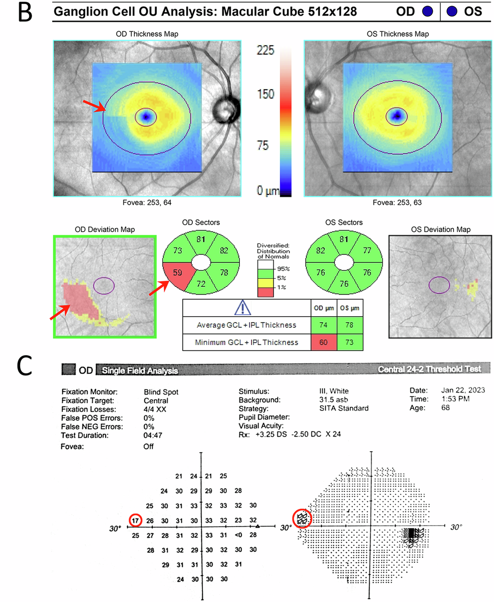
Glaucoma diagnosis needs both structure and function. OCT shows nerve structure. Visual field shows vision function. When both OCT and Visual Field show similar changes over time, diagnosis is stronger, and rooted in deeper evidence.
The Importance of Serial Comparison
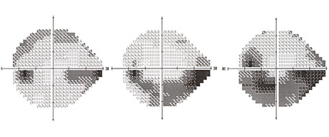
The most important glaucoma test is comparison.
We compare:
• OCT over years
• visual fields over years
• optic nerve photos
Progression becomes visible only in hindsight. That is why follow-up matters.
Common Misinterpretations
• Red OCT areas in high myopia
• Field defects from cataract
• Machine artefacts
• Ignoring early thinning
You should not panic, or be falsely reassured. What you should ask for is a detailed explanation.
When to Seek Specialist Interpretation
• Conflicting reports
• Advice for surgery
• Multiple drops
• Normal pressure but abnormal OCT
• Strong family history
A structured interpretation can clarify risk.
My Approach
My approach focuses on calm, structured interpretation of OCT and visual field reports so patients can make informed decisions about long-term eye health. I don’t diagnose glaucoma from a number. I recognise patterns across OCT, visual fields, optic nerve appearance, pressure, corneal thickness, risk factors and time. Because glaucoma is usually invisible early, our goal is not only to see clearly today, but to protect vision safely ten years from now.
Patients receive:
• clear explanation
• risk assessment
• management options, including follow up schedule
• missing data list
If your OCT and visual field reports seem to contradict each other, you’re not alone. This is one of the commonest reasons patients seek a glaucoma second opinion. Most apparent contradictions have an explanation once the scans are interpreted in context rather than isolation. Because glaucoma care is about continuity, and steady compliance with treatment.
📞 +91 88826 38735 | 🌐 Contact Us | Second Opinion Form for teleconsults
⭐ FAQs – OCT and Visual Field Interpretation
1. My OCT report shows red areas. Does this mean I have glaucoma?
Not always. OCT compares your nerve thickness with an average database.
Red areas can appear in:
• high myopia
• large optic nerves
• normal anatomical variation
• machine artefacts
OCT is only one part of glaucoma diagnosis. It must be interpreted with visual fields, optic nerve exam, and follow-up over time.
2. My visual field test was abnormal once. Should I worry?
A single abnormal visual field does not confirm glaucoma. Visual fields depend on attention, fatigue, dry eye, cataract, and learning effect. Doctors usually repeat the test to confirm a pattern. Consistency over time matters more than one report.
3. Can OCT be normal but glaucoma still present?
Yes. No one test is infallible when it comes to glaucoma diagnosis.
Very early glaucoma can be missed on OCT, especially in normal-tension glaucoma or small optic nerves. This is why clinical examination and follow-up are important. Glaucoma diagnosis is a pattern seen over time, not one scan.
4. Can visual fields be normal if glaucoma is already present?
Yes. Structural nerve damage often occurs before functional loss. Patients may have normal visual fields but abnormal OCT or optic nerve appearance. Early detection focuses on protecting long-term vision before symptoms appear.
5. How often should OCT and visual field tests be repeated?
It depends on your risk of glaucoma progression or vision loss.
• Low risk: once a year
• Glaucoma suspect: every 6–12 months
• Established glaucoma: every 3–6 months
Your doctor decides based on progression risk. Regular comparison (and therefore, regular follow up) is the most important part of glaucoma care.
6. Why do my OCT numbers change between tests?
Small changes happen because of:
• machine differences
• scan alignment and test retest variability
• eye dryness
• cataract
• natural variation
Doctors thus look for consistent trends, not small fluctuations.
7. Can cataract affect visual field results?
Yes.
Cataract can cause diffuse depression on visual field testing. This may look like glaucoma but improves after cataract surgery. This is why reports must be interpreted carefully.
8. My eye pressure is normal. Why do I need OCT and Visual Field?
Many patients have normal-tension glaucoma. Pressure alone cannot rule out disease. OCT and visual field testing help detect subtle nerve damage. Glaucoma diagnosis needs multiple data points, eye pressure is only one of them.
9. Can glaucoma tests (OCT and Visual field) be wrong?
Tests are not “wrong,” but they can be misleading if taken in isolation. Machines measure data. Doctors interpret patterns. Also, visual fields can have fixation losses (you looked away from the fixation light), as well as false positives and false negatives. High rates of any of these can make your visual fields unreliable.
A structured review reduces unnecessary treatment and dangerous delay.
10. When should I seek a glaucoma second opinion?
Consider a second opinion if:
• You are advised surgery suddenly
• Reports are confusing
• Multiple drops are started without explanation
• OCT and visual field results disagree
• Strong family history exists
Clarity helps you make calm, informed decisions.
11. What is the most important glaucoma test?
The most important test is comparison over time. Glaucoma progression becomes visible only when reports are compared across months and years. Continuity of care is essential, and one all clear diagnosis does not mean you don’t need a follow up visit.
Known for her structured approach to glaucoma risk assessment and progression analysis, Dr Shibal Bhartiya provides trusted second opinions for patients seeking clarity before major treatment decisions. Both, in person, and online.
12. Can glaucoma be cured if detected early?
Glaucoma cannot be reversed. But early detection and regular care can preserve useful vision for life. The goal is not perfect tests today, but safe vision ten years from now, and always.
Closing Thought
Numbers do not treat glaucoma. Understanding does. Protecting vision requires careful interpretation over time. If you would like your OCT or visual field reports reviewed in a structured glaucoma second opinion:
📞 +91 88826 38735 | 🌐 Contact Us | Second Opinion Form for teleconsults
This page is a part of the Glaucoma Hub. you may want to read about Glaucoma Progression, and Risk Stratification in Glaucoma. You may want to read more about OCT and Visual Field, Glaucoma Tests Explained, Normal OCT but Vision Symptoms and How to Understand Your OCT Better. Also of help could be Why Do I Need a Visual Field Test? Glaucoma Diagnosis in Gurgaon, Get a GlaucomaSecond Opinion in Gurgaon and Get an Online Glaucoma Consult.
Read some of Dr Shibal Bhartiya’s published research on OCT
A Meta-analysis of the Effect of Panretinal Photocoagulation on Retinal Nerve Fibre Layer Thickness
This meta-analysis evaluated how panretinal laser treatment affects retinal nerve fibre layer thickness on OCT, helping clinicians interpret OCT changes accurately after diabetic retinopathy treatment.
Comparative Evaluation of Time-Domain and Spectral-Domain OCT
This study compared two generations of OCT technology for retinal nerve fibre layer measurements, highlighting the importance of using consistent imaging when monitoring glaucoma patients over time.
Long-term Effect of Panretinal Photocoagulation on RNFL Parameters
This research examined the long-term impact of panretinal laser treatment on OCT measurements, improving our understanding of retinal nerve fibre layer changes following treatment for proliferative diabetic retinopathy.
Assessment of RNFL Changes by Cirrus HD-OCT in Myopia
This study investigated how myopia influences retinal nerve fibre layer measurements on OCT, emphasising the need to interpret scans carefully in highly myopic eyes to avoid misdiagnosis.
About the Author
This article was written by Dr Shibal Bhartiya, fellowship-trained glaucoma specialist and Mayo Clinic Research Collaborator, Clinical Director at Marengo Asia Hospitals, Gurugram, known for ethical, patient-centred glaucoma care and independent glaucoma second opinions. She is also the Program Director for Community Outreach & Wellness; and for the Marengo Asia International Institute of Neuro and Spine.
She has published peer-reviewed research on glaucoma management, examining how treatment decisions should balance medical evidence, patient preferences, and long-term vision outcomes.
As Editor-in-Chief of Clinical and Experimental Vision and Eye Research and Executive Editor of the Journal of Current Glaucoma Practice (Pubmed Indexed, official journal of the International Society of Glaucoma Surgery), Dr Shibal Bhartiya brings editorial and research depth to every clinical decision. Her 200+ publications, including 90+ PubMed-indexed publications and 28 edited textbooks span glaucoma biology, surgical outcomes, health equity, and emerging diagnostics.
1600+ Five Star Patient Reviews Google Business Profile
If you are unable to come to Dr Bhartiya’s clinic: Read more about teleconsultation
Read her research on PubMed | Google Scholar | ResearchGate | ORCID
Upload your reports for a structured review.| www.drshibalbhartiya.com | +91 88826 38735
Leave a review on Google