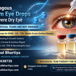
Ocular GVHD (Graft-versus-Host Disease) can cause severe dry eyes, burning, fluctuating vision, light sensitivity, and damage to the eye surface after a bone marrow or stem cell transplant. Early diagnosis and long-term eye care may help protect comfort, vision, and quality of life.
Managing these cases requires specialised corneal expertise, strict protective isolation compliance, and a deeply trauma-informed approach to pediatric clinical examination. Scientific precision alone is not enough — the child must feel safe enough to let you in.
Beyond the Sterile Barrier: Pediatric Ocular Graft-Versus-Host Disease
There are moments in a pediatric ophthalmologist’s career that anchor you for life.
During my time at Fortis, I cared for a tiny boy fighting for his life after a bone marrow transplant. Severe, acute Ocular GVHD had left his corneas damaged, covered in countless microscopic raw spots — Superficial Punctate Keratitis — causing blinding pain and extreme light sensitivity. He lived in the ICU, unable to open his eyes, afraid of every sound.
His immune system was almost non-existent. Anyone entering his space had to be covered head to toe in sterile gowns and masks. He could not see my face. He was terrified. Some days, he was too weak to cry.
To help him recognise me without triggering fear, I started a routine. Every time I entered his isolation pod, I whistled softly. He learned quickly. The whistle meant safety. No needles. It meant the person coming was not going to hurt him.
I would apply a careful drop of anaesthetic to numb the intense surface pain just enough to let me examine his corneas and adjust his treatment. And I kept talking. Through every protocol, every follow-up, every barrier-gowned visit, a bond formed between us.
His parents would quietly hold him. Silently, patiently, with all the love in the world.
Can GVHD be cured?
That was the only question they asked. For the pain to go away. I would say yes, he will be fine. And pray, silently.
And then one day, instead of crying, he started talking. About trucks, and JCBs and construction. All of five. And bright. And happy. Like any other five year old.
The drops continued, but he was now walking into my OPD, showing off his toys, his jeans, his shoes which have red and blue lights. And one day, we didn’t need any medication at all.
Today, he is completely cured — a bright, healthy boy, a handful and a half. Goes to big school. And to Goa with his grandparents. Collects toy trucks, especially likes yellow ones.
When he walks into my clinic, he does not see masks or sterile gowns. He sees a friend. He spots me from across the waiting room and runs full tilt into my arms. And talks till my ears hurt. And my face hurts. From smiling so much.
His parents recently told me that when they return to his BMT hospital for follow-ups, he looked up at the first floor where my old OPD used to be and insisted: “Let’s go meet Dr Shibal.” They had to remind him gently that I have moved. When he visited me at Marengo Asia after that, he looked around the new clinic and said, with complete satisfaction: “Dr Shibal, you always own the first floor.”
He is entirely right. Just like he owns my entire heart.
PS: Two of his classmates have come to me to get their glasses checked. Apparently he tells everyone about “My doctor” who has a hundred toffees. My little advertising blitzkreig he is 🙂
FAQs
What is Ocular GVHD in children, and what are the symptoms?
Ocular GVHD occurs when donor immune cells after a bone marrow transplant attack the recipient’s lacrimal glands and corneal surface. In children, symptoms include severe eye pain, redness, a gritty sensation, extreme light sensitivity, and refusal to open the eyes due to corneal surface damage. Early specialist intervention is critical to prevent permanent scarring.
What are the most common symptoms of ocular GVHD in adults?
Symptoms may include dry eyes, burning, redness, watering, irritation, light sensitivity, fluctuating vision, eye fatigue, and a feeling of grit or sand in the eyes.
Can ocular GVHD affect vision permanently?
If untreated, ocular GVHD can lead to chronic surface damage, discomfort, and vision changes. Early treatment and regular follow-up may reduce the risk of long-term complications.
How is ocular GVHD diagnosed?
Diagnosis is based on symptoms, eye examination, tear film assessment, evaluation of the eye surface, and correlation with transplant history and systemic GVHD status.
What treatments are available for ocular GVHD?
Treatment may include preservative-free lubricants, medicines to reduce inflammation, tear conservation strategies, ocular surface support, and long-term monitoring depending on severity.
How is severe pain and photophobia managed in post-transplant pediatric patients?
Management is multi-layered and highly specialised. It includes preservative-free lubricants, autologous serum eye drops, therapeutic scleral or bandage contact lenses, and targeted topical immunomodulators. During acute flares, topical anaesthetics are used carefully during examination by the specialist — never for unsupervised home use — to allow assessment without causing further distress to the child.
She has published peer-reviewed research on glaucoma management, examining how treatment decisions should balance medical evidence, patient preferences, and long-term vision outcomes.
Many people with dry eye are told their eyes are “normal” even while struggling with burning, fluctuating vision, eye strain, or discomfort during screen use. Advanced dry eye evaluation looks beyond redness alone to understand tear film instability, ocular surface disease, and the real-world visual symptoms affecting daily life.
Dry eye disease is not simply a lack of tears. It is a chronic condition of the ocular surface — driven by tear film instability, inflammation, meibomian gland dysfunction, or environmental exposure — that causes persistent discomfort, visual fluctuation, and in some cases, measurable damage to the surface of the eye. Effective management requires identifying which component is driving your symptoms, not simply prescribing lubricant drops.
Dry Eye Disease in Gurgaon: When Your Eyes Never Feel Quite Right
Dry eye is one of the most undertreated conditions in ophthalmology, not because treatment doesn’t exist, but because patients are routinely told that what they are experiencing is minor.
It is not minor. Eyes that burn, sting, water excessively, feel gritty by afternoon, blur when you are tired, or ache after two hours of screen time are not eyes that are functioning normally. And patients who have been handed a bottle of artificial tears and sent home, sometimes repeatedly, know exactly how inadequate that response feels.
Dry eye disease has a pathophysiology. It has subtypes. It has measurable signs. And it has a treatment pathway that goes considerably further than lubricant drops, when it is managed by someone who understands the full picture.
This practice takes dry eye seriously. Because your eyes deserve to feel comfortable.
What Dry Eye Disease Actually Is
Dry eye disease is a multifactorial condition of the ocular surface. The tear film — the thin, layered fluid that coats your eye with every blink — requires three components to function correctly: an aqueous layer produced by the lacrimal gland, a lipid layer produced by the meibomian glands in your eyelids, and a mucin layer produced by goblet cells on the ocular surface.
When any of these components fails, the tear film becomes unstable. The surface dries between blinks. Inflammation follows. And a self-reinforcing cycle begins — surface damage drives more inflammation, which drives more surface damage.
Understanding which component is failing is the starting point of effective treatment.
Evaporative dry eye and meibomian gland dysfunction
The most common form of dry eye in urban Indian populations is evaporative — driven by meibomian gland dysfunction (MGD). The meibomian glands line the upper and lower eyelids and produce the lipid layer that prevents tear evaporation. When these glands become blocked or atrophied, tears evaporate too quickly regardless of how much aqueous is produced.
MGD is dramatically worsened by screen use, air conditioning, low humidity environments, and contact lens wear — the exact conditions that define urban professional life in Gurgaon and Delhi NCR.
Aqueous deficient dry eye
In some patients — particularly post-menopausal women, patients with autoimmune conditions like Sjögren’s syndrome, and those on certain systemic medications — the lacrimal gland simply does not produce enough aqueous tears. This form requires a different treatment approach and often warrants systemic investigation.
Mixed dry eye
Many patients have both components — inadequate lipid and inadequate aqueous — compounded by surface inflammation. These patients are frequently the ones who have tried multiple lubricant drops without relief, because no single drop addresses the full picture.
Ocular surface inflammation
Chronic inflammation is both a cause and a consequence of dry eye disease. In patients with significant inflammation, lubricant drops alone will never be sufficient. Anti-inflammatory therapy — whether topical cyclosporine, lifitegrast, or short-course steroids — is a necessary component of management.
Why Dry Eye Is Worse Than Ever in Urban India
The epidemiology of dry eye has shifted dramatically in the last decade. What was once considered a condition of older women is now presenting across all ages, genders, and occupations — and the drivers are environmental and behavioural.
Risk Factor
Why It Matters
Screen time
Blink rate drops by up to 60% during screen use; tear film destabilises
Disrupts tear film distribution and lipid layer integrity
Glaucoma drops
Preservatives in long-term glaucoma medications cause surface toxicity
Post-surgical dryness
LASIK, cataract surgery, and other procedures transiently or persistently disrupt corneal nerves and surface
Hormonal changes
Menopause significantly reduces aqueous and lipid tear production
Antihistamines and antidepressants
Many systemic medications reduce tear secretion as a side effect
Urban air pollution
Particulate matter and pollutants directly damage the ocular surface
Gurgaon sits at the intersection of several of these factors simultaneously — screen-intensive professional culture, year-round air conditioning, high ambient pollution, and one of the highest LASIK procedure rates in North India.
What We Often Miss in Dry Eye Management
Meibomian gland dysfunction goes unexamined. Most dry eye consultations do not include eyelid margin assessment or meibomian gland expression. Without examining the glands, evaporative dry eye — the most common subtype — is routinely misidentified as aqueous deficiency and treated with the wrong drops.
Post-surgical dryness is underestimated. Dry eye after LASIK, SMILE, or cataract surgery can persist for twelve to eighteen months, and in some patients becomes a chronic condition. Patients are frequently told their symptoms will resolve on their own — without a structured management plan being put in place.
Glaucoma patients’ ocular surface is neglected. Patients on long-term preserved glaucoma drops develop surface toxicity at a rate that is well-documented in the literature but poorly addressed in clinical practice. If you have glaucoma and dry eye, the two conditions must be managed together.
Inflammation is not addressed. Patients cycling through artificial tear brands without improvement almost always have a significant inflammatory component. Without anti-inflammatory therapy, the cycle does not break.
Screen habits are not discussed. Behavioural modification — structured blink exercises, the 20-20-20 rule, screen positioning, humidifier use — forms a critical part of dry eye management that is rarely covered in a brief consultation.
What to Expect at a Dry Eye Consultation
A structured dry eye assessment goes beyond asking how your eyes feel and prescribing drops.
At this practice, assessment includes tear film evaluation, tear break-up time, meibomian gland assessment, corneal and conjunctival staining, and a detailed history of your screen habits, contact lens use, surgical history, and systemic medications. Where indicated, additional investigations including meibography — imaging of the meibomian glands — may be recommended.
Treatment is then built around your specific subtype and severity. This may include targeted lubricants, lipid-containing drops, warm compress and lid hygiene protocols, anti-inflammatory therapy, punctal plugs, or in-office procedures. You will leave with a structured plan — not a single bottle and a follow-up in six months.
Dry Eyes and Digital Eye Strain in Gurgaon
Many people in Gurgaon spend long hours on computers, phones, and other digital devices. Reduced blinking during screen use can contribute to dry eyes, eye strain, headaches, blurred vision, burning, watering, and difficulty focusing.
These symptoms may be further aggravated by factors common in Gurgaon, including air-conditioned office environments, long working hours, dry weather, air pollution, dust, and ongoing construction activity. Together, these factors can affect the stability of the tear film and make the eyes feel tired, irritated, or uncomfortable throughout the day.
A comprehensive eye examination can help determine whether symptoms are related to dry eye disease, digital eye strain, an uncorrected vision problem, or a combination of factors. Early assessment can often improve comfort, productivity, and visual quality.
Dr Shibal Bhartiya works with corporates, professionals, and frequent screen users in Gurgaon on the diagnosis and management of dry eye disease, digital eye strain, and healthy screen-use habits. To book an eye examination or arrange an eye health awareness session for your organisation, call +91 88826 38735 or visit drshibalbhartiya.com.
Your eyes burn, sting, or feel gritty — especially by afternoon or after screen use
Your vision fluctuates and clears when you blink
Your eyes water excessively — paradoxical tearing is a common dry eye sign
You wear contact lenses and your comfortable wearing time has reduced
You have had LASIK, SMILE, or cataract surgery and your eyes have not felt normal since
You are on long-term glaucoma drops and your eyes feel uncomfortable
You have been using lubricant drops for months without meaningful relief
You have been diagnosed with an autoimmune condition and have eye symptoms
Dry eye is a chronic condition — but it is a manageable one. The patients who do best are those who receive an accurate subtype diagnosis early and follow a structured management plan. Lubricant drops are a starting point, not a solution.
Frequently Asked Questions
What is the best treatment for dry eye disease?
There is no single best treatment — because dry eye has multiple subtypes that require different approaches. Evaporative dry eye from meibomian gland dysfunction is treated with warm compresses, lid hygiene, and lipid-containing drops. Aqueous deficient dry eye may require anti-inflammatory therapy and punctal plugs. Inflammatory dry eye requires targeted anti-inflammatory treatment. Accurate subtype diagnosis is the essential first step.
Can dry eye be cured permanently?
In most patients, dry eye disease is a chronic condition that requires ongoing management rather than a one-time cure. However, with consistent and correctly targeted treatment, the majority of patients achieve significant and sustained relief. Some causes — such as post-surgical dryness or medication-related dryness — may resolve once the underlying cause is addressed.
Why do my eyes water if I have dry eye?
Paradoxical tearing — excessive watering in a dry eye patient — is one of the most common and confusing symptoms of dry eye disease. When the ocular surface becomes irritated from tear film instability, the lacrimal gland produces reflex tears as a protective response. These reflex tears do not replace the stable tear film and do not relieve the underlying dryness.
Is dry eye worse in Gurgaon and Delhi NCR?
Yes. Urban environments with high screen use, year-round air conditioning, significant ambient pollution, and low outdoor humidity create conditions that are particularly hostile to tear film stability. Gurgaon’s professional demographic — high screen exposure, frequent air travel, contact lens use — compounds these environmental factors significantly.
Can dry eye damage my vision permanently?
In mild to moderate dry eye, vision fluctuates but does not sustain permanent damage. In severe, untreated dry eye — particularly in aqueous deficient conditions or after significant surface damage — corneal scarring and permanent visual reduction can occur. This is rare but preventable with appropriate management.
I have been using artificial tears for months with no improvement. What should I do?
This is the most common presentation at a dry eye second opinion consultation. Patients cycling through lubricant drop brands without relief almost always have either an unaddressed inflammatory component, undertreated meibomian gland dysfunction, or a subtype mismatch between the drops they are using and the dry eye they actually have. A structured reassessment — including eyelid examination and tear film evaluation — usually identifies the gap quickly.
She has published peer-reviewed research on glaucoma management, examining how treatment decisions should balance medical evidence, patient preferences, and long-term vision outcomes.
Dry Eye Is Not Just Dryness: Managing It as a Chronic Condition
Dry eye disease is a chronic condition caused by an unstable or insufficient tear film. It does not go away with occasional lubricating drops. Left unmanaged, it causes progressive surface damage, worsening discomfort, and, in some cases, permanent corneal scarring. Long-term management, not short-term relief, is the correct approach, says Dr Shibal Bhartiya.
Dry eye is one of the most common eye conditions seen in clinical practice. Most patients manage it with over-the-counter drops and expect it to resolve. It rarely does.
Dry eye disease is a multifactorial condition of the ocular surface. The tear film is complex. When it breaks down, the result is inflammation, epithelial damage, and a cycle that perpetuates itself without targeted treatment.
Understanding dry eye as a chronic disease changes how patients manage it, and how much vision and comfort they can preserve over time.
Dry eye disease (DED) is defined by the TFOS DEWS II report as a multifactorial disease of the ocular surface. It involves loss of tear film stability and is accompanied by symptoms and signs of varying severity.
The tear film has three layers: a mucin layer anchoring tears to the eye surface, an aqueous (watery) layer providing nutrients and oxygen, and a lipid (or oil) layer produced by the meibomian glands that prevents evaporation. A problem in any one layer causes disease.
Two Main Types of Dry Eye
Aqueous Deficient Dry Eye
This type involves insufficient tear production. The lacrimal gland does not produce enough aqueous fluid. It is common in post-menopausal women, patients with Sjogren’s syndrome, and those on antihistamines, antidepressants, or blood pressure medications.
Evaporative Dry Eye
This is the more common type, accounting for roughly 85 percent of all dry eye cases. Meibomian gland dysfunction (MGD) is the primary cause. Blocked or abnormal meibomian glands fail to secrete a healthy lipid layer, and tears evaporate too quickly.
Many patients have both types simultaneously. Treatment must address the dominant mechanism.
Feature
Aqueous Deficient DED
Evaporative DED
Primary cause
Reduced lacrimal gland output
Meibomian gland dysfunction
Proportion of cases
Approximately 15%
Approximately 85%
Key risk factors
Sjogren’s, medications, age
Screen use, blepharitis, rosacea
Tear break-up time
Reduced
Very short (under 5 seconds)
Treatment focus
Tear supplementation
Lid hygiene, heat, omega-3
Inflammation present
Often yes
Yes, secondary
Why Dry Eye Becomes Chronic
The tear film and ocular surface exist in a feedback loop. When the tear film is unstable, the surface desiccates. This triggers inflammation. Inflammation damages goblet cells and lacrimal tissue. Damaged tissue produces less stable tears. The cycle continues.
Without breaking this cycle, not just lubricating the surface, dry eye worsens over months and years. This is why patients who only use drops often find their symptoms returning or intensifying.
Chronic untreated dry eye can cause corneal epithelial breakdown, punctate keratitis, subepithelial scarring, and, in severe cases, corneal ulcers. These are not trivial outcomes.
Risk Factors That Drive Progression
Screen use of more than four hours daily reduces blink rate and increases evaporation.
Contact lens wear disrupts the tear film and accelerates meibomian gland dropout.
Hormonal changes — especially menopause — reduce lacrimal and meibomian secretions.
Systemic medications including antihistamines, SSRIs, diuretics, and isotretinoin reduce tear production.
Autoimmune conditions such as rheumatoid arthritis, lupus, and thyroid disease affect the lacrimal gland.
Rosacea is a strong risk factor for meibomian gland dysfunction and is frequently undiagnosed.
Diagnosis requires more than a symptom questionnaire. A structured assessment includes the OSDI (Ocular Surface Disease Index) score, tear break-up time (TBUT), Schirmer’s test, corneal and conjunctival staining with fluorescein and lissamine green, and meibomian gland evaluation.
Meibography — infrared imaging of the meibomian glands — shows the degree of gland dropout and guides treatment intensity. Patients with significant gland loss need early and aggressive intervention to preserve remaining function.
Tear osmolarity testing measures the salt concentration of tears. Elevated osmolarity confirms tear film instability and is useful for monitoring treatment response objectively.
Diagnostic Test
What It Measures
Clinical Significance
TBUT (tear break-up time)
Tear film stability
Under 10 seconds is abnormal
Schirmer’s test
Aqueous tear production
Under 10 mm in 5 min is reduced
Corneal fluorescein staining
Epithelial surface damage
Confirms active disease severity
Meibography
Meibomian gland structure and dropout
Guides long-term prognosis
Tear osmolarity
Tear salt concentration
Over 308 mOsm/L confirms DED
OSDI score
Symptom burden
Tracks treatment response over time
If screen-related eye pain is affecting your work or daily life, a full assessment takes under an hour. Dr Shibal Bhartiya — dry eye specialist and glaucoma specialist in Gurgaon — will identify whether this is screen strain or something that needs treatment. 📞 +91 88826 38735 | www.drshibalbhartiya.com
Dry eye treatment is not one-size-fits-all. It is matched to disease type, severity, and the dominant mechanism driving symptoms.
Step 1: Environmental and Behavioural Changes
Reduce screen time or use the 20-20-20 rule — every 20 minutes, look at something 20 feet away for 20 seconds. Increase blink frequency consciously. Use a humidifier in air-conditioned environments. Wear wraparound glasses in wind and dry air.
Step 2: Lid Hygiene and Warm Compresses
Warm compresses applied for 10 minutes daily soften meibomian secretions and improve gland expressibility. Lid massage after warming clears blocked glands. Lid scrubs with baby shampoo or commercially prepared wipes reduce bacterial load on the lid margin.
Consistency matters more than intensity. Daily lid hygiene over months produces measurable improvement in tear film quality.
Step 3: Lubricating Eye Drops
Not all lubricants are equivalent. Drops containing carboxymethylcellulose, sodium hyaluronate, or polyethylene glycol provide longer contact time. Preservative-free formulations are essential for patients using drops more than four times daily — preservatives accelerate the surface damage they are meant to relieve.
Gel formulations and ointments provide longer relief but blur vision temporarily and are best used at night.
Step 4: Omega-3 Fatty Acid Supplementation
Omega-3 supplements — particularly EPA and DHA from fish oil or re-esterified triglyceride formulations — improve meibomian secretion quality and reduce ocular surface inflammation. The DREAM study showed that high-dose omega-3 did not significantly outperform olive oil placebo, but clinical practice and other evidence support a role for supplementation in evaporative dry eye.
A daily dose of 2000 to 3000 mg EPA+DHA for at least three months is typically recommended.
Step 5: Anti-Inflammatory Therapy
When inflammation is driving symptoms, lubricants alone are insufficient. Cyclosporine eye drops (0.05% or 0.1%) reduce T-cell mediated inflammation on the ocular surface and restore goblet cell density over three to six months of use. They are not a quick fix — patients must be counselled on the time course.
Lifitegrast 5% is an integrin antagonist that blocks the LFA-1 to ICAM-1 interaction driving ocular surface inflammation. It offers symptom relief somewhat faster than cyclosporine.
Short-term topical corticosteroids are used to rapidly break the inflammatory cycle, particularly at disease onset or during flares. They are not for long-term use.
Step 6: Procedural Treatments
Intense Pulsed Light (IPL) therapy targets abnormal blood vessels on the lid margin that drive meibomian gland inflammation. It also applies heat that melts obstructed meibum. Multiple sessions spaced three to four weeks apart produce sustained improvement in many patients with moderate to severe MGD.
Thermal pulsation devices (LipiFlow) deliver controlled heat and pressure to the inner eyelid to express inspissated meibum. The effect can last six to twelve months and is repeatable.
Punctal plugs block the drainage of tears from the ocular surface. They are appropriate for aqueous deficient dry eye when lubrication alone is inadequate. Dissolvable collagen plugs are trialled before permanent silicone plugs are inserted.
Treatment
Best For
Time to Effect
Preservative-free lubricants
All types, daily use
Immediate symptom relief
Lid hygiene + warm compresses
Evaporative / MGD
4 to 8 weeks of daily use
Omega-3 supplementation
Evaporative / MGD
8 to 12 weeks
Cyclosporine drops
Inflammatory DED
3 to 6 months
Lifitegrast drops
Inflammatory DED
2 to 4 weeks for symptoms
IPL therapy
Moderate to severe MGD
After 3 to 4 sessions
Punctal plugs
Aqueous deficient DED
Days to weeks
Monitoring Dry Eye Over Time
Dry eye is managed, not cured. Follow-up visits every three to six months allow the specialist to assess treatment response, adjust the regimen, and monitor for corneal surface deterioration.
Objective tests — TBUT, osmolarity, staining scores — are more reliable than symptoms alone. Patients often adapt to chronic discomfort and underreport severity. Imaging guides clinical decisions even when symptoms appear stable.
Meibomian gland dropout is irreversible. Preventing further loss is the priority once significant atrophy is identified.
Dry Eye and Systemic Disease
Dry eye is frequently a signal of systemic disease. Sjogren’s syndrome, rheumatoid arthritis, lupus, thyroid eye disease, and graft-versus-host disease all affect the ocular surface. Patients with unexplained severe dry eye — particularly younger women — should be evaluated for autoimmune conditions.
Conversely, patients already diagnosed with these conditions should have formal ocular surface assessments. Dry eye in this context needs co-management with the treating physician.
When to See a Glaucoma and Ocular Surface Specialist
Many patients with dry eye also have glaucoma or glaucoma suspect status. Glaucoma drops — particularly those with preservatives — are a significant cause of ocular surface disease. The benzalkonium chloride (BAK) in most preserved glaucoma drops is toxic to goblet cells and the corneal epithelium.
If you use glaucoma drops and have dry eye symptoms, your specialist needs to review both conditions together. Switching to preservative-free formulations or fixed-combination drops can reduce surface toxicity without compromising IOP control.
There is no permanent cure for most forms of dry eye disease. However, it can be very well controlled with the right treatment strategy. Many patients achieve significant symptom relief and stable ocular surface health with long-term management.
Are lubricating drops enough to treat dry eye?
For mild disease, lubricants provide adequate relief. For moderate to severe dry eye — or evaporative disease driven by meibomian gland dysfunction — drops manage symptoms but do not address the underlying cause. Lid hygiene, anti-inflammatory therapy, and sometimes procedural treatment are needed.
How do I know if my dry eye is getting worse?
Worsening symptoms, increased frequency of drop use, morning grittiness, light sensitivity, and fluctuating vision are all signs of progression. Objective worsening on TBUT, staining, or osmolarity testing confirms it. Do not wait for significant discomfort before seeking review.
Can diet help with dry eye?
Yes. Omega-3 fatty acids from oily fish, flaxseed, and walnuts support meibomian gland secretion quality. Adequate hydration matters. Foods high in omega-6 fatty acids and processed vegetable oils may worsen inflammation. A Mediterranean-style diet is broadly supportive of ocular surface health.
Is dry eye related to screen use?
Yes. Screen use reduces spontaneous blink rate from a normal 15 to 17 blinks per minute to as few as 3 to 5 blinks per minute. Reduced blinking causes tear film instability and accelerates evaporation. Deliberate blinking exercises and regular screen breaks are first-line recommendations for screen-related dry eye.
Can I wear contact lenses if I have dry eye?
Some patients with well-managed mild dry eye can wear contacts with modifications — daily disposable lenses, lubricating drops compatible with contact wear, and reduced wearing time. Patients with moderate to severe dry eye are advised to avoid contact lenses until surface health is restored. Scleral lenses are a specialist option for severe cases.
Does menopause cause dry eye?
Yes. Oestrogen and androgen deficiency after menopause reduces both aqueous and meibomian secretion. Dry eye prevalence in post-menopausal women is significantly higher than in age-matched men. Hormone replacement therapy has a complex relationship with dry eye — some studies show benefit, others do not. Ocular surface assessment after menopause is advisable.
Book a Dry Eye Assessment in Gurgaon
Dry eye responds best to early, structured management. A thorough ocular surface assessment — including meibography, osmolarity, and staining — identifies the cause and guides a treatment plan that works long-term.
If you are using lubricating drops daily and still struggling, the underlying mechanism has not yet been addressed. A specialist review changes that.
She has published peer-reviewed research on glaucoma management, examining how treatment decisions should balance medical evidence, patient preferences, and long-term vision outcomes.