When Glaucoma Keeps Progressing

Glaucoma can progress even with treatment. The most common reasons include suboptimal IOP control, non-adherence to drops, normal-tension progression, and unrecognised structural risk factors. Finding the cause and adjusting treatment early can prevent further vision loss, says Dr Shibal Bhartiya.

Glaucoma progresses in some patients despite regular treatment. This does not mean the treatment has failed, it means the treatment plan needs review.

Understanding why glaucoma advances is the first step toward stopping it. Several factors can drive progression even when eye pressure appears controlled.

Dr Shibal Bhartiya is a fellowship-trained glaucoma specialist and Mayo Clinic Research Collaborator with over 25 years of experience. Her approach focuses on identifying risk before damage is irreversible, simplifying treatment decisions, and protecting vision long-term. Emphasis on early detection, risk assessment, and continuity of care. She is rated 5 stars across 1,500+ patient reviews on Google.

What Does Progression Mean in Glaucoma?

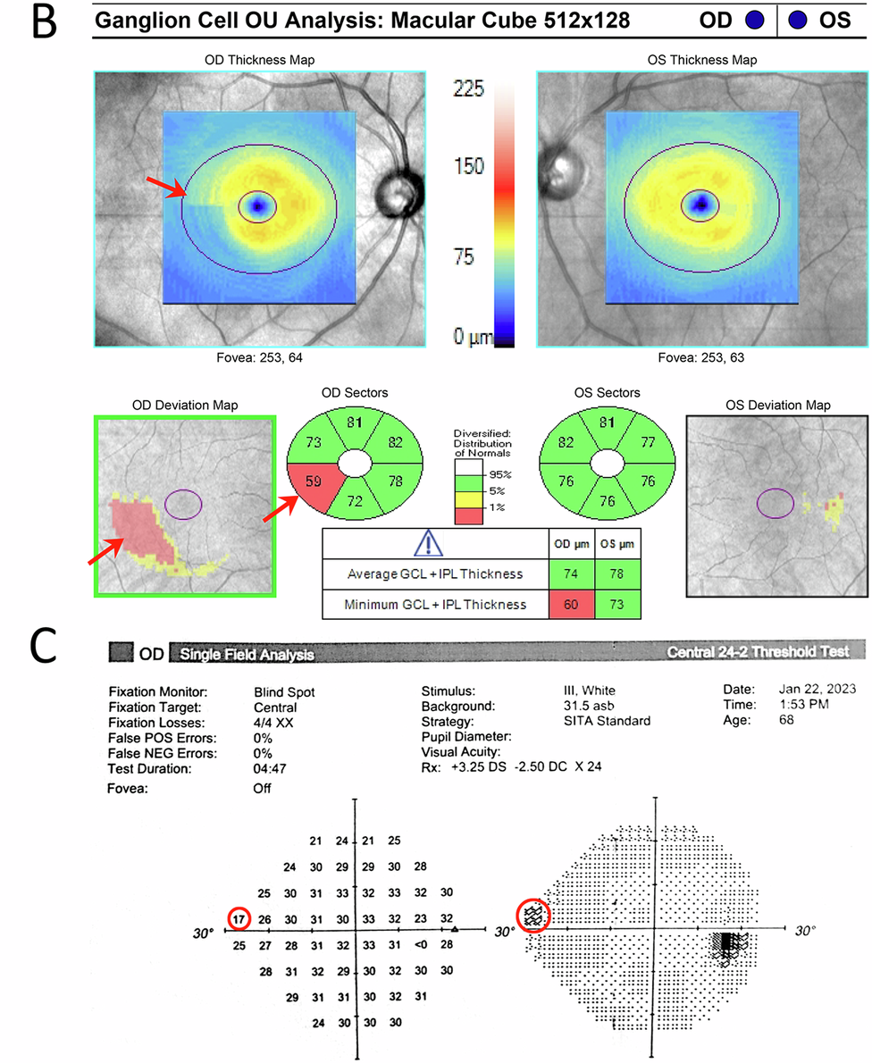
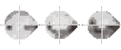
Progression means measurable worsening of the optic nerve or visual field over time. Specialists confirm it using two or more reliable visual field tests and OCT imaging showing thinning of the retinal nerve fibre layer.

A single abnormal test does not confirm progression. Consistent change across multiple visits does.

Why Glaucoma Progresses Despite Drops

1. Eye Pressure Is Still Too High

The target intraocular pressure (IOP) is individual. A pressure that seems normal may still be too high for a given optic nerve. Studies show that lower IOP targets reduce progression rates in moderate and advanced glaucoma significantly.

If visual fields are worsening, the current pressure target may need revision downward.

2. Drops Are Not Working as Expected

Peak pressure often occurs in the early morning, outside clinic hours. A single office reading may miss harmful pressure spikes. Diurnal IOP curves — tested over several hours — can reveal fluctuations that drive unseen damage.

3. Non-Adherence to Eye Drop Therapy

Studies using electronic monitoring show that patients use drops correctly only 50 to 70 percent of the time. Missing doses, incorrect technique, or preservative intolerance all reduce drug efficacy. Non-adherence is the most correctable cause of progression.

4. Normal-Tension Glaucoma Behaving Differently

Some patients have optic nerve damage at pressures within the normal range. This is normal-tension glaucoma (NTG). It may involve poor vascular supply to the nerve, sleep apnoea, low blood pressure at night, or other systemic factors that drops alone cannot address.

5. Structural Risk Factors Not Yet Addressed

Thin corneas cause IOP readings to appear falsely low. A myopic or tilted optic disc is harder to interpret on imaging. Disc haemorrhages are a strong marker of ongoing progression and must be documented carefully.

6. Systemic Factors Affecting the Optic Nerve

Low systolic blood pressure, anaemia, sleep apnoea, and vascular disorders can reduce blood flow to the optic nerve. Treating these conditions alongside glaucoma can slow visual field loss in susceptible patients.

Reason for ProgressionWhat It MeansNext Step
IOP target not low enoughNerve still under excess pressureLower target IOP or add therapy
Pressure spikes between visitsDiurnal fluctuation causing damageDiurnal IOP curve or 24-hour monitoring
Drop non-adherenceInconsistent pressure loweringTechnique review, preserve-free drops, fixed combos
Normal-tension glaucomaVascular or non-pressure mechanismSystemic workup, cardiology review
Thin cornea or high myopiaIOP underestimated by tonometryCorneal-corrected IOP, adjusted targets
Disc haemorrhageActive ischaemia at optic nerveClose follow-up, often signals rapid progression
Systemic comorbidityPoor vascular supply to nerveTreat sleep apnoea, anaemia, hypotension

When to Consider Laser or Surgery

If maximum tolerated medical therapy does not achieve the revised IOP target, laser trabeculoplasty (SLT) or surgery becomes necessary. Selective laser trabeculoplasty is effective in open-angle glaucoma and can reduce the drop burden significantly.

Minimally invasive glaucoma surgery (MIGS) procedures such as iStent and iStent inject offer an option for mild to moderate glaucoma with lower surgical risk. Trabeculectomy remains the benchmark for advanced disease requiring very low pressures.

Dr Shibal Bhartiya’s published research includes peer-reviewed work on 24-hour IOP monitoring and diurnal pressure fluctuation: one of the most under-recognised drivers of progression in treated glaucoma. She has co-authored guidelines on surgical decision-making when medical therapy fails to halt optic nerve damage. As Clinical Director of Ophthalmology at Marengo Asia Hospitals, Gurugram, she manages complex progression cases with a structured protocol: reassess the IOP target, confirm adherence, evaluate vascular and systemic risk, and escalate to laser or surgery when the nerve continues to lose ground.

How Often Should You Be Reviewed?

Patients with progressing glaucoma need more frequent review — often every three to four months. Visual fields should be repeated at least four times a year if progression is suspected. OCT of the optic nerve head and RNFL should accompany each visit.

Waiting six or twelve months between visits when progression is active is not safe practice.

The Role of a Second Opinion

Glaucoma management decisions are complex. If your visual fields continue to worsen, a second opinion from a fellowship-trained glaucoma specialist adds value. Fresh eyes on your imaging, IOP pattern, and structural data can identify a missed cause.

Bringing your previous visual fields, OCT scans, and medication list to the consultation helps the specialist assess the rate of change accurately.

Known for her structured approach to glaucoma risk assessment and progression analysis, Dr Shibal Bhartiya provides trusted second opinions for patients seeking clarity before major treatment decisions. Both, in person, and online.

Frequently Asked Questions

Can glaucoma progress even with normal eye pressure?

Yes. Normal-tension glaucoma progresses at IOP readings within the statistical normal range. The optic nerve in these patients is more sensitive to pressure or more dependent on blood supply. Treatment often involves additional systemic assessment alongside IOP lowering.

How do I know if my glaucoma is progressing?

Your specialist tracks visual field tests and OCT scans over time. Progression is confirmed when two or more reliable tests show consistent worsening. You may not notice early progression — which is why regular monitoring matters.

What pressure should I aim for if my glaucoma is progressing?

The target varies by disease severity and rate of progression. Advanced or rapidly progressing glaucoma typically requires a target below 12 mmHg. Your specialist calculates this based on your structural damage and life expectancy.

Are there lifestyle changes that help slow progression?

Regular aerobic exercise, avoiding head-down positions such as headstands, good sleep hygiene, and managing vascular risk factors all support optic nerve health. Omega-3 supplementation and antioxidant nutrition are areas of ongoing research.

Is surgery the only option if drops stop working?

Not always. Selective laser trabeculoplasty is a non-incisional option that works well in many patients. If laser is not sufficient, MIGS procedures offer a middle path between drops and conventional surgery.

Consult a Glaucoma Specialist

If your glaucoma is progressing despite treatment, you need a specialist review, not just a medication change. The cause must be identified before the right intervention can be chosen.

About the Author

This article was written by Dr Shibal Bhartiya, fellowship-trained glaucoma specialist and Mayo Clinic Research Collaborator, Clinical Director at Marengo Asia Hospitals, Gurugram, known for ethical, patient-centred glaucoma care and independent glaucoma second opinions. She is also the Program Director for Community Outreach & Wellness; and for the Marengo Asia International Institute of Neuro and Spine. This article was updated in May 2026.

She has published peer-reviewed research on glaucoma management, examining how treatment decisions should balance medical evidence, patient preferences, and long-term vision outcomes.

As Editor-in-Chief of Clinical and Experimental Vision and Eye Research and Executive Editor of the Journal of Current Glaucoma Practice (Pubmed Indexed, official journal of the International Society of Glaucoma Surgery), Dr Shibal Bhartiya brings editorial and research depth to every clinical decision. Her 200+ publications, including 90+ PubMed-indexed publications and 28 edited textbooks span glaucoma biology, surgical outcomes, health equity, and emerging diagnostics.

Access her work on PubmedGoogle ScholarResearchGate and ORCID.

Dr Shibal Bhartiya
Glaucoma • Second Opinion • Advanced Care

www.drshibalbhartiya.com
 +91 88826 38735

1500+ Five Star Patient Reviews Google Business Profile

Upload your reports for a structured review.

If you are unable to come to Dr Bhartiya’s clinic: Read more about teleconsultation for glaucoma

Related Reading

Get an Online Glaucoma Consult

Visual Field and OCT: Structure & Function Correlation

Glaucoma Diagnosis in Gurgaon

Risk Stratification in Glaucoma

Glaucoma Progression: What It Means and How to Slow It

Glaucoma treatment in Gurgaon

All About Glaucoma Medication

Glaucoma Lasers: SLT & LPI

Glaucoma surgery in Gurgaon

MIGS in Gurgaon

Get a Glaucoma Second Opinion in Gurgaon

What Happens If Glaucoma Is Left Untreated?

Untreated glaucoma causes permanent, irreversible vision loss, and in most cases, patients feel nothing until significant damage has already occurred. Glaucoma destroys the optic nerve silently. By the time you notice a change in your vision, up to 40% of nerve fibres may already be gone, explains Dr Shibal Bhartiya.

Many people discover glaucoma late because it causes no pain, no redness, and no early warning signs in its most common form. That silence is what makes it dangerous. If you have been told your eye pressure is high, or if glaucoma runs in your family, the question of what happens if you leave it alone is not academic. It is urgent.

Dr Shibal Bhartiya is a fellowship-trained glaucoma specialist and Mayo Clinic Research Collaborator with over 25 years of experience. Her approach focuses on identifying risk before damage is irreversible, simplifying treatment decisions, and protecting vision long-term. Emphasis on early detection, risk assessment, and continuity of care. She is rated 5 stars across 1,500+ patient reviews on Google.


7 Things That Happen When Glaucoma Goes Untreated

1. The Optic Nerve Keeps Deteriorating

Glaucoma damages the optic nerve, the cable that sends visual signals from your eye to your brain. Each day without treatment, elevated pressure continues to compress and starve nerve fibres of blood supply. Once a nerve fibre dies, it does not regenerate. There is no surgery, no medication, and no natural process that restores it.

Treatment slows or stops this process. No treatment means no brake on the damage.


2. Peripheral Vision Disappears First

The first field of vision to go is your peripheral vision, the edges of what you see. This happens so gradually that most patients do not notice. The brain fills in the gaps, masking the loss. You may be losing significant side vision for years before you register anything unusual.

By the time you notice you are bumping into things, misjudging doorframes, or struggling to see cars approaching from the side, the damage is already extensive.


3. Central Vision Is Eventually Affected

A common misconception is that glaucoma only affects side vision and central vision stays intact. This is true in early and moderate stages, but untreated glaucoma progresses. As more of the optic nerve is destroyed, the visual field loss closes in from the edges toward the centre. At advanced stages, the remaining central tunnel of vision narrows severely.

At end-stage glaucoma, even central vision is lost.


4. Blindness Becomes a Real Risk

Glaucoma is the leading cause of irreversible blindness worldwide. It is the number one cause of preventable blindness in India. The word “preventable” matters, because the blindness is not inevitable. It is the outcome of late diagnosis or no treatment.

Patients who are diagnosed early and treated consistently rarely go blind from glaucoma. Patients who ignore it, or who stop treatment because they feel well, are the ones who lose vision permanently.


5. Acute Angle-Closure Can Cause Sudden Blindness

Not all glaucoma is slow and silent. Acute angle-closure glaucoma is a medical emergency. Eye pressure spikes suddenly and severely. Patients experience intense eye pain, headache, nausea, vomiting, and blurred vision with coloured halos around lights.

If this is not treated within hours, it can cause permanent blindness in that eye. Many patients mistake it for a migraine or food poisoning and delay seeking care. This delay can cost them their sight.


6. Quality of Life Declines Significantly

Vision loss from untreated glaucoma is not just a medical number on a visual field report. It changes how you live. Driving becomes unsafe, then impossible. Reading becomes difficult. Recognising faces becomes unreliable. Falls and accidents become more frequent. Depression and anxiety are significantly more common in people with advanced glaucoma.

The impact is gradual enough that patients adapt, until they can no longer. At that point, the vision loss cannot be reversed.


7. Treatment Becomes Harder as Damage Advances

In early glaucoma, a single eye drop once daily may be all that is needed to control pressure and preserve vision. As glaucoma advances, more medications are required. Laser treatments may be needed. Surgery, with longer recovery times, higher risks, and no guarantee of reversing existing damage, becomes the only option.

Treating glaucoma early is simpler, cheaper, and far more effective than treating it late.


What Doctors Often Miss Telling Patients

Most patients are told they have high eye pressure or early glaucoma and are given drops. What they are not always told clearly is this: the drops do not make you feel better. They do not improve your vision. They work silently in the background to prevent future damage.

Because there is no immediate reward, no symptom that goes away, no vision that returns, many patients stop their drops after a few weeks. They feel the same. They assume they are fine. This is the most dangerous point in glaucoma care.

Stopping treatment does not mean the disease has stopped. It means the only thing slowing the damage has been removed.

As a glaucoma specialist, I have seen patients who were diagnosed years earlier, given drops, and told to return in six months. Life got busy. The drops ran out. The follow-up did not happen. When they finally return, sometimes years later, significant, irreversible vision loss has occurred in the interval.

This is preventable. Every time.


Symptom Progression: What to Watch For

StageWhat You May NoticeWhat Is Actually Happening
EarlyNothing at allPeripheral nerve fibres dying
ModerateOccasional blind spots at the edges30–50% nerve fibre loss
AdvancedBumping into objects, missing steps, tunnel vision70–80%+ nerve fibre loss
End-stageLoss of all but a sliver of central visionNear-total optic nerve destruction
Acute attack (angle-closure)Sudden severe eye pain, headache, halosMedical emergency — act within hours

When Act Immediately? If You Have


What This Means for You

Glaucoma is manageable. That is the truth that often gets lost in the fear around the diagnosis. The vast majority of patients who are diagnosed early, treated appropriately, and followed up consistently do not go blind. They live full, visually intact lives.

But glaucoma does not forgive neglect. It does not pause when life gets busy. It does not announce its progress. The only protection is a specialist who checks, measures, and adjusts your treatment over time, and a patient who shows up.

If you have been diagnosed with glaucoma, or if someone in your family has it, a comprehensive glaucoma evaluation is not something to delay. The damage happening right now is silent. The window to prevent it from becoming permanent is open, but it does not stay open forever.


Frequently Asked Questions

Can glaucoma be reversed if caught early?

The nerve damage already present cannot be reversed. However, early treatment stops further damage from occurring. Patients diagnosed early and treated consistently typically keep their functional vision for life.

Is it safe to stop glaucoma drops if I feel fine?

No. Glaucoma drops prevent damage, they do not treat symptoms, because there are none. Feeling well means the drops are working. Stopping them removes the only thing protecting your optic nerve.

How fast does untreated glaucoma progress?

This varies by type and individual. Some patients progress slowly over decades; others, particularly those with very high pressures or angle-closure glaucoma, can lose significant vision within months or years. There is no way to predict your rate without regular monitoring.

What is the difference between glaucoma suspects and glaucoma?

A glaucoma suspect has risk factors: high pressure, suspicious optic nerve appearance, or a family history, but no confirmed nerve damage yet. This group needs careful monitoring, as some will develop glaucoma. Not all glaucoma suspects need treatment, but all need regular follow-up.

Can I drive if I have glaucoma?

In early and moderate glaucoma, most patients can drive safely. In advanced glaucoma with significant peripheral field loss, driving may be unsafe and may not meet legal vision standards. This should be assessed with a formal visual field test.


Should You See a Glaucoma Specialist?

If you have been diagnosed with glaucoma, suspect you may have it, or have a parent or sibling with the condition, a specialist evaluation gives you information a general eye check cannot.

A glaucoma specialist will assess your optic nerve in detail, measure your visual field, perform OCT scanning of the nerve fibre layer, and build a personalised treatment and monitoring plan. The goal is not just to lower your eye pressure. The goal is to protect your vision for the rest of your life.

Book a glaucoma consultation at Marengo Asia Hospitals, Gurugram.

📞 +91 88826 38735 | 🌐 www.drshibalbhartiya.com

Upload your previous reports for a second opinion, a fresh set of expert eyes on your case can change the outcome.


About the Author

This article was written by Dr Shibal Bhartiya, fellowship-trained glaucoma specialist and Mayo Clinic Research Collaborator, Clinical Director at Marengo Asia Hospitals, Gurugram, known for ethical, patient-centred glaucoma care and independent glaucoma second opinions. She is also the Program Director for Community Outreach & Wellness; and for the Marengo Asia International Institute of Neuro and Spine. This article was updated in April 2026.

She has published peer-reviewed research on glaucoma management, examining how treatment decisions should balance medical evidence, patient preferences, and long-term vision outcomes.

As Editor-in-Chief of Clinical and Experimental Vision and Eye Research and Executive Editor of the Journal of Current Glaucoma Practice (Pubmed Indexed, official journal of the International Society of Glaucoma Surgery), Dr Shibal Bhartiya brings editorial and research depth to every clinical decision. Her 200+ publications, including 90+ PubMed-indexed publications and 28 edited textbooks span glaucoma biology, surgical outcomes, health equity, and emerging diagnostics.

Access her work on PubmedGoogle ScholarResearchGate and ORCID.

Dr Shibal Bhartiya
Glaucoma • Second Opinion • Advanced Care

www.drshibalbhartiya.com
 +91 88826 38735

1500+ Five Star Patient Reviews Google Business Profile

Upload your reports for a structured review.

If you are unable to come to Dr Bhartiya’s clinic: Read more about teleconsultation for glaucoma

OCT and Visual Field

Understanding Glaucoma Investigations: OCT and Visual Field

OCT shows the structure of the optic nerve. Visual field tests show how vision is functioning. Patients struggle to understand why their doctor has reached a certain diagnosis, or treatment strategy. Many patients receive OCT and visual field reports full of colours and numbers. Both require careful interpretation, and an equally careful explanation. the truth is, your doctor is looking for a structure-function relationship, correlating it to your eye pressures, and the lifetime risk to your vision, and quality of life.

Glaucoma diagnosis is rarely based on one scan. Also glaucoma often has no symptoms. It requires understanding patterns over time: how the optic nerve looks, how visual fields change, how eye pressure behaves, and how your individual risk factors fit together.

OCT shows the structure of the optic nerve. Visual field tests show how vision is functioning.
Neither test alone can diagnose glaucoma. This is why reports sometimes seem confusing. A red area on OCT may be normal for a highly myopic eye. An abnormal visual field may simply reflect fatigue or cataract. On the other hand, subtle early glaucoma can be missed if reports are not compared carefully across months and years.

In glaucoma care, numbers do not treat disease. Understanding does.

My approach focuses on calm, structured interpretation of OCT and visual field reports so patients can make informed decisions about long-term eye health. Because glaucoma is usually invisible early, our goal is not only to see clearly today, but to protect vision safely ten years from now.

If your reports are confusing, conflicting, or leading to rushed treatment decisions, a structured glaucoma second opinion can help bring clarity.

Dr Shibal Bhartiya is a fellowship-trained glaucoma specialist and Mayo Clinic Research Collaborator with over 25 years of experience. Her approach focuses on identifying risk before damage is irreversible, simplifying treatment decisions, and protecting vision long-term. Emphasis on early detection, risk assessment, and continuity of care. She is rated 5 stars across 1,500+ patient reviews on Google.


Understanding OCT

OCT measures thickness of nerve fibres. Red areas may indicate thinning.

But interpretation depends on:

• age
• myopia
optic nerve size
• machine variability
• baseline comparison

One abnormal OCT does not prove glaucoma. But ignoring subtle changes can be dangerous.


Understanding Visual Fields

Visual field tests measure functional vision.

But results vary with:

• patient attention
• fatigue
• learning effect
• cataract
• dry eye

One abnormal field may not mean disease. Repeated patterns matter more when evaluating progression.


Why OCT and Visual Field Reports Must Be Interpreted Together

Glaucoma diagnosis needs both structure and function. OCT shows nerve structure. Visual field shows vision function. When both OCT and Visual Field show similar changes over time, diagnosis is stronger, and rooted in deeper evidence.


The Importance of Serial Comparison

The most important glaucoma test is comparison.

We compare:

• OCT over years
• visual fields over years
• optic nerve photos

Progression becomes visible only in hindsight. That is why follow-up matters.


Common Misinterpretations

• Red OCT areas in high myopia
• Field defects from cataract
• Machine artefacts
• Ignoring early thinning

You should not panic, or be falsely reassured. What you should ask for is a detailed explanation.


When to Seek Specialist Interpretation

• Conflicting reports
• Advice for surgery
• Multiple drops
• Normal pressure but abnormal OCT
• Strong family history

A structured interpretation can clarify risk.


My Approach

Reports are reviewed systematically with attention to long-term risk.

Patients receive:

• clear explanation
• risk assessment
• management options, including follow up schedule
• missing data list

Because glaucoma care is about continuity, and steady compliance with treatment.

⭐ FAQs – OCT and Visual Field Interpretation

1. My OCT report shows red areas. Does this mean I have glaucoma?

Not always. OCT compares your nerve thickness with an average database.
Red areas can appear in:

• high myopia
• large optic nerves
• normal anatomical variation
• machine artefacts

OCT is only one part of glaucoma diagnosis. It must be interpreted with visual fields, optic nerve exam, and follow-up over time.


2. My visual field test was abnormal once. Should I worry?

A single abnormal visual field does not confirm glaucoma. Visual fields depend on attention, fatigue, dry eye, cataract, and learning effect. Doctors usually repeat the test to confirm a pattern. Consistency over time matters more than one report.


3. Can OCT be normal but glaucoma still present?

Yes. No one test is infallible when it comes to glaucoma diagnosis.

Very early glaucoma can be missed on OCT, especially in normal-tension glaucoma or small optic nerves. This is why clinical examination and follow-up are important. Glaucoma diagnosis is a pattern seen over time, not one scan.


4. Can visual fields be normal if glaucoma is already present?

Yes. Structural nerve damage often occurs before functional loss. Patients may have normal visual fields but abnormal OCT or optic nerve appearance. Early detection focuses on protecting long-term vision before symptoms appear.


5. How often should OCT and visual field tests be repeated?

It depends on your risk of glaucoma progression or vision loss.

• Low risk: once a year
• Glaucoma suspect: every 6–12 months
• Established glaucoma: every 3–6 months

Your doctor decides based on progression risk. Regular comparison (and therefore, regular follow up) is the most important part of glaucoma care.


6. Why do my OCT numbers change between tests?

Small changes happen because of:

• machine differences
• scan alignment and test retest variability
• eye dryness
• cataract
• natural variation

Doctors thus look for consistent trends, not small fluctuations.


7. Can cataract affect visual field results?

Yes.

Cataract can cause diffuse depression on visual field testing. This may look like glaucoma but improves after cataract surgery. This is why reports must be interpreted carefully.


8. My eye pressure is normal. Why do I need OCT and Visual Field?

Many patients have normal-tension glaucoma. Pressure alone cannot rule out disease. OCT and visual field testing help detect subtle nerve damage. Glaucoma diagnosis needs multiple data points, eye pressure is only one of them.


9. Can glaucoma tests (OCT and Visual field) be wrong?

Tests are not “wrong,” but they can be misleading if taken in isolation. Machines measure data. Doctors interpret patterns. Also, visual fields can have fixation losses (you looked away from the fixation light), as well as false positives and false negatives. High rates of any of these can make your visual fields unreliable.

A structured review reduces unnecessary treatment and dangerous delay.


10. When should I seek a glaucoma second opinion?

Consider a second opinion if:

• You are advised surgery suddenly
• Reports are confusing
• Multiple drops are started without explanation
• OCT and visual field results disagree
• Strong family history exists

Clarity helps you make calm, informed decisions.


11. What is the most important glaucoma test?

The most important test is comparison over time. Glaucoma progression becomes visible only when reports are compared across months and years. Continuity of care is essential, and one all clear diagnosis does not mean you don’t need a follow up visit.

Known for her structured approach to glaucoma risk assessment and progression analysis, Dr Shibal Bhartiya provides trusted second opinions for patients seeking clarity before major treatment decisions. Both, in person, and online.


12. Can glaucoma be cured if detected early?

Glaucoma cannot be reversed. But early detection and regular care can preserve useful vision for life. The goal is not perfect tests today, but safe vision ten years from now, and always.


Closing Thought

Numbers do not treat glaucoma.
Understanding does.

Protecting vision requires careful interpretation over time.


If you would like your OCT or visual field reports reviewed in a structured glaucoma second opinion:

📞 +91 88826 38735
🌐 drshibalbhartiya.com

Second Opinion Form for teleconsults

Related Reading

Read the research articles

This article was written by Dr Shibal Bhartiya, fellowship-trained glaucoma specialist and Mayo Clinic Research Collaborator, Clinical Director at Marengo Asia Hospitals, Gurugram, known for ethical, patient-centred glaucoma care and independent glaucoma second opinions. She is also the Program Director for Community Outreach & Wellness; and for the Marengo Asia International Institute of Neuro and Spine. This article was updated in April 2026.

She has published peer-reviewed research on glaucoma management, examining how treatment decisions should balance medical evidence, patient preferences, and long-term vision outcomes.

As Editor-in-Chief of Clinical and Experimental Vision and Eye Research and Executive Editor of the Journal of Current Glaucoma Practice (Pubmed Indexed, official journal of the International Society of Glaucoma Surgery), Dr Shibal Bhartiya brings editorial and research depth to every clinical decision. Her 200+ publications, including 90+ PubMed-indexed publications and 28 edited textbooks span glaucoma biology, surgical outcomes, health equity, and emerging diagnostics.

Access her work on PubmedGoogle ScholarResearchGate and ORCID.

Dr Shibal Bhartiya
Glaucoma • Second Opinion • Advanced Care

www.drshibalbhartiya.com
 +91 88826 38735

1500+ Five Star Patient Reviews Google Business Profile

Upload your reports for a structured review.

For people unable to come to Dr Bhartiya’s clinic: Read more about teleconsultation for glaucoma

How I Think About Glaucoma

I think of glaucoma as a slow, silent risk to lifelong vision rather than just an eye pressure problem. The…

Glaucoma-Friendly Home Environment

This is a comprehensive guide for patients and caregivers for creating a glaucoma-friendly home environment.