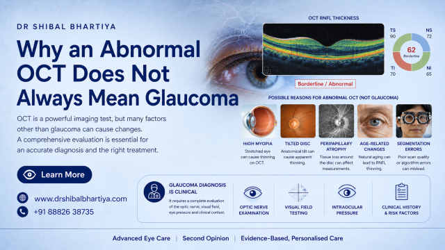
A red OCT result does not always mean glaucoma. Red only means one measurement falls outside the machine’s normal range, which a healthy eye can do.
You get your report. Parts of it glow red. Someone says your nerve is thinning. A prescription follows, and you start to wonder if you will use drops for life.
Some of these eyes have glaucoma. Many do not. I see both every week in second-opinion visits.
This page explains why healthy eyes turn red on OCT. It also explains the opposite problem, where a damaged eye prints green.
What Do Red, Yellow and Green Mean on an OCT Report?
First, remember, the OCT measures the thickness of your nerve fibre layer and your ganglion cell layer. The machine compares your numbers with a database of healthy eyes. Then, the OCT machine uses a pseudocolour coding. Your nerve is not red, yellow or green. The machine compares your retinal nerve fibre layer and makes a coloured report for your doctor to interpret.
Green means you sit within the normal range. Yellow means you fall in the lowest 5%. Red means you fall in the lowest 1%.
Notice what red does not say. It does not say you have disease. It says your number is uncommon, One percent means that one in 100 healthy people lands in the red zone for any single measurement. A report holds dozens of measurements. A stray red patch in a healthy eye is therefore not rare.
What Are Red Disease and Green Disease?
Doctors use these two terms for the two ways OCT colours can mislead.
Red disease is a false positive. The report shows damage, but the eye is healthy.
Green disease is a false negative. The report looks normal, but the eye has real nerve damage.
Both happen because of the scan, the software or the database. This is an inherent fallacy of the machine, and not an error.
Why Does a Healthy Eye Show Red on OCT?
Six causes account for most false red results:
- A long, highly myopic eye
- A tilted or unusually shaped optic disc
- A database that does not match your age, ancestry or eye size
- A software error in drawing the layer lines
- Another eye condition, such as a membrane on the macula
- A poor-quality scan
High myopia
A long eye stretches the retina. The nerve fibre layer can look thin even when the nerve is healthy. The scan ring may also land on a different part of the nerve than the database expects.
Myopia also raises glaucoma risk. A red result in a myopic eye needs careful checking, not dismissal.
Tilted or unusual discs
Some optic discs sit at an angle. Others are large, with a large natural cup. These discs can show thin nerve fibres on one side in both eyes. The pattern looks alarming on paper. The nerve itself is often normal.
A database that does not fit you
The machine compares you with a limited group of healthy volunteers. That group may not match your age, ancestry or eye shape.
Sometimes, the tissue is unchanged; what changes is the reference population against which we interpret it. OCT colour coding is not an absolute biological truth. It is a statistical representation shaped by the normative database.
A 2026 study in the Journal of Glaucoma tested this in people of African ancestry. An ancestry-specific database moved some red codes to yellow or green. The eyes did not change. The comparison group did.
Software errors
The machine draws lines to mark each retinal layer. Sometimes it draws a line in the wrong place. It still prints a number, and that number can turn red.
A membrane on the macula can trigger this. I check the layer lines on the scan itself and not only the summary page.
Poor scan quality
Cataract, dry eye, a blink or poor focus lowers signal strength. A weak scan can read thin. Treating the eye surface and repeating the scan often fixes the problem. My dry eye work helps here.
How Do I Tell a False Red From Real Damage?
I read this table across each row. It shows what the report says, why it may mislead, and what I check next.
| What you see on the report | Why it may be a false red | What I check next |
|---|---|---|
| Thin fibres in both eyes, mostly on the nasal side | High myopia or tilted discs | Compare with the disc photo and visual field. Repeat the scan. |
| One red sector, rest normal, healthy disc | Chance finding, since 1 in 100 healthy eyes flags red | Repeat the scan. Check if the same sector turns red again. |
| Red ganglion cell map, normal nerve fibre map | Macular disease or layer-line error | Review each layer line. Look at the macular scan for a membrane. |
| Low signal strength on the printout | Cataract, dry eye, blink or poor focus | Treat the surface. Repeat the scan with a strong signal. |
| Red result, but your age or ancestry differs from the database | Database mismatch | Rely on disc appearance and change over time, not colour. |
| Red result, normal pressure, normal field, normal disc | Normal anatomical variant | Set a baseline. Repeat at a planned interval and watch for change. |
What Is Green Disease, and Why Does It Matter Just as Much?
A green report can falsely reassure you.
Some eyes start with thicker than average fibres. They can lose a lot of nerve and still stay inside the normal range. Very short, hyperopic eyes can also hide damage.
Advanced glaucoma brings its own limit. The machine cannot read below a certain thickness. A scan can then look flat while the eye keeps losing function.
So a normal OCT does not clear you. I still check your disc, your visual field and your pressure history.
How Do I Decide If an OCT Result Is a False Positive?
No single colour decides this. I build a pattern from several tests.
- I check scan quality and the layer lines first.
- I compare the scan with the optic disc itself, not just the printout.
- I match the thin area with your visual field in the same location.
- I review your pressure history, your drainage angle on gonioscopy and your corneal thickness.
- I repeat the scan and look for change over time.
Glaucoma damage follows the path of nerve fibre bundles. It usually stays in the same place on repeat scans. It usually changes over time. A false red usually stays flat.
My clinic uses the Zeiss Cirrus 6000, and the same machine can image the front of the eye. But what is most important is the clinical context, and interpreting all your glaucoma tests together. And often, over time.
I have worked with OCT measurements for years. With colleagues, I co-authored a study of nerve fibre layer thickness on Cirrus OCT in myopic eyes. It looked at how eye length and refractive error shape the readings. We also compared two generations of OCT machines. Two further studies tracked how laser treatment for diabetic retinopathy changes nerve fibre layer values. That work is why I read the scan alongside the eye, not the colours alone.
What Do We Often Miss?
- Sometimes doctors read the colours and skip the raw thickness curve.
- Some skip the scan quality score.
- Some never compare the OCT with the disc photo.
- Some start treatment from one scan with no repeat and no baseline.
I also see the reverse. Some doctors dismiss a real pre-perimetric glaucoma because the field is still normal. Structural loss can appear years before the field changes. Good care avoids both errors.
When Should I See a Doctor?
Book a visit with a glaucoma specialist if:
- Someone started lifelong drops from a single OCT
- You had no repeat scan, visual field or disc photo
- Your OCT is red and you have high myopia
- The same area turns red on repeat scans
- Your pressure runs above normal
- Your OCT is green, but you notice field loss or a family member has glaucoma
What This Means for You
A red scan is a reason to look closer. It is not a diagnosis.
Please ask your doctor to correlate the scan, visual field and your disc photo. Ask your doctor whether the thin area on OCT or photo matches the field. Ask what would change the plan.
Key Takeaways
- OCT scans compare your eye to a population database, not always your own healthy baseline
- High myopia, atypical disc shape, and cataract are common causes of false positive flags
- A single red flag should be confirmed with a repeat scan and visual field test
- Clinical exam findings add context that a scan alone cannot provide
- Tracking your own scans over time is more reliable than a one time population comparison
- Ask your doctor to explain any red flag in the context of your full eye exam
Frequently Asked Questions
Can an OCT scan be wrong?
Yes. OCT is a good test, but the software and the database can mislead. Myopia, tilted discs, layer-line errors and weak scans all cause false results. That is why I never rely on one scan.
Should I start glaucoma drops if only my OCT is red?
One red OCT rarely justifies lifelong treatment on its own. I look at your visual field, optic disc, pressure, drainage angle and any change over time. If you already take drops, keep using them until you talk to your doctor.
Is a normal OCT enough to rule out glaucoma?
No. A green report can hide real damage, especially in eyes that started with thick fibres or in advanced disease. I also check your disc, visual field and pressure history.
Does high myopia make my OCT unreliable?
It makes the scan harder to read. It does not make it useless. Myopic eyes also face a higher glaucoma risk, so I check them closely and follow them over time.
How often should I repeat an OCT?
I usually repeat a doubtful scan soon, to see if the finding holds. After that, the interval depends on your risk and your other tests. A stable, healthy pattern needs less frequent checks.
Told Your OCT Is Abnormal? Bring It for a Second Opinion
Has an OCT report flagged you red? Bring the scan, the visual field and any disc photos to my clinic in Gurgaon. I review them together and tell you what I see.
You may leave with a plan to watch. You may leave with a plan to treat. Either way, you will know why.
Book an Appointment:
📍 Gurgaon 📞 +91 88826 38735 | 🌐 Contact Us | Second Opinion Form for teleconsults
This article is a part of the Glaucoma Hub. Please also read Glaucoma Test Results Explained: OCT, Visual Fields and Eye Pressure, OCT in Gurgaon, RNFL OCT explained, and Normal OCT but Vision Abnormal. You may want to read about Glaucoma Progression, and Risk Stratification in Glaucoma. Other articles of interest could be Advanced Glaucoma Care in Gurgaon, What Good Glaucoma Care Actually Optimises For. Read about our full approach to glaucoma second opinions.
About the Author
This article was written by Dr Shibal Bhartiya, fellowship-trained glaucoma specialist and Mayo Clinic Research Collaborator, Clinical Director at Marengo Asia Hospitals, Gurugram, known for ethical, patient-centred glaucoma care and independent glaucoma second opinions. She is also the Program Director for Community Outreach & Wellness; and for the Marengo Asia International Institute of Neuro and Spine.
Dr Bhartiya has published peer-reviewed research on glaucoma management, examining how treatment decisions should balance medical evidence, patient preferences, and long-term vision outcomes. She is also a committed patient advocate and mainstream medical columnist, translating complex ophthalmic evidence into actionable public health insights across leading national platforms including NDTV and OnlyMyHealth
Dr Bhartiya is the Editor-in-Chief of Clinical and Experimental Vision and Eye Research and Executive Editor of the Journal of Current Glaucoma Practice (Pubmed Indexed, official journal of the International Society of Glaucoma Surgery). Her 200+ publications, including 90+ PubMed-indexed publications and 28 edited textbooks span glaucoma biology, surgical outcomes, health equity, and emerging diagnostics.
1700+ Five Star Patient Reviews Google Business Profile
If you are unable to come to Dr Bhartiya’s clinic: Read more about teleconsultation
Read her research on PubMed | Google Scholar | ResearchGate | ORCID | Github | Wikidata
📋Upload your reports for a structured review.| 🌐 Contact Us| 📞 +91 88826 38735
⭐ Helped by this article? Leave a Google review — it helps other patients find reliable eye care.
Last Reviewed: November 2026