Acute and intermittent angle closure glaucoma can present with severe headache, nausea, vomiting, and coloured haloes around lights — symptoms so closely overlapping with migraine that patients spend years in neurology before anyone examines their drainage angles. A gonioscope placed at a routine eye examination can reveal in minutes what years of migraine treatment cannot resolve.
For patients with narrow angles, a laser peripheral iridotomy, a five-minute outpatient procedure — may eliminate the trigger entirely. The eye and the head are not separate systems.
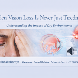
Dr Shibal Bhartiya is a fellowship-trained glaucoma specialist and Mayo Clinic Research Collaborator with over 25 years of experience. Her approach focuses on identifying risk before damage is irreversible, simplifying treatment decisions, and protecting vision long-term. Emphasis on early detection, risk assessment, and continuity of care. She is rated 5 stars across 1,500+ patient reviews on Google.
Seven Years of Migraines That Disappeared After a Routine Eye Examination
She was in her late forties or early fifties. She had no eye complaints.
It was a routine check — glasses, perhaps a small change in power. I noticed a shallow anterior chamber, explained she needed a gonioscopy. Asked her if she had experienced any headaches, or coloured haloes around lightbulbs.
She talked. She had been living with migraines for seven to eight years. Treatment after treatment. Specialist after specialist. The headaches kept coming.
If you are reading this after years of treatment that has not worked, I want you to know: that exhaustion is real, and it is not in your head. But the answer sometimes is — in your eyes.
I looked at her angles. They were narrow. Both eyes.
What a gonioscope found that years of migraine treatment missed
I placed a gonioscope, a contact lens with a mirror that allows direct visualisation of the eye’s drainage angle, and examined both eyes carefully. She had primary angle closure. Peripheral anterior synechiae were present in roughly a quadrant of each eye — meaning parts of the drainage angle had already begun to stick shut. Her IOP was in the range of 22 to 24 mmHg.
A standard migraine workup does not include a gonioscope. A glaucoma specialist examination does.
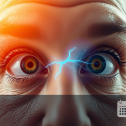
Why angle closure symptoms feel exactly like a migraine
In intermittent angle closure, the drainage angle narrows and blocks without fully closing. Pressure builds, then releases. The episode passes. No one connects it to the eye.
During these episodes, the symptoms are: severe throbbing headache, nausea, vomiting, coloured haloes around lights and streetlamps, eye redness, and a deep ache around the orbit. These are textbook migraine symptoms. They are also textbook intermittent angle closure symptoms. Without a gonioscope, there is no way to tell them apart from a history alone.
If your migraines have not responded to treatment, or if your headaches come with coloured halos or eye pain, a glaucoma specialist examination may give you answers years of headache treatment have not.
Book a consultation with Dr Shibal Bhartiya in Gurgaon. Second opinions welcome.
+91 88826 38735 | www.drshibalbhartiya.com
Symptoms, Causes, and When to Worry
| Symptom | Likely Cause | When to Worry |
|---|---|---|
| Severe throbbing headache | Intermittent IOP spike from narrow angles | Attacks are recurring, not relieved by migraine medication |
| Nausea and vomiting with headache | Acute pressure rise, vagal response | Accompanying eye redness or blurred vision |
| Coloured halos around lights | Corneal oedema from raised IOP | Any episode with halos warrants urgent eye evaluation |
| Eye ache or pain around orbit | Elevated intraocular pressure | Persists beyond the headache episode |
| Blurred vision during headache | Raised IOP affecting corneal clarity | Vision does not fully recover after episode |
| Headache worse in dim light or evening | Pupil dilation narrows angles further | Consistent pattern linked to lighting conditions |
What Doctors Often Miss
Neurologists and general physicians are not trained to examine drainage angles. That is not a criticism — it is a structural gap. A gonioscope is a specialist instrument used by ophthalmologists and glaucoma specialists. It is not part of a standard headache workup, and it is not part of most routine optometry checks either.
The result is that intermittent angle closure goes undiagnosed for years in patients who are otherwise receiving excellent neurological care. The migraine label is applied because the symptoms fit. The eye is never examined. The pressure spikes continue.
If you have been diagnosed with migraines and you have never had your angles examined, that is worth a second opinion from a glaucoma specialist.
The other missed signal is coloured halos. Many patients mention them. Fewer doctors follow up specifically on the eye examination that halos warrant.
A five-minute laser. Ten migraine-free years.
We performed a laser peripheral iridotomy — a small opening in the iris, made with a laser, in the clinic, in under ten minutes. It allows aqueous fluid to flow freely, relieves intermittent pressure build-up, and eliminates the trigger that narrow angles create.
That was ten years ago.
She has not had a single migraine attack since.
An occasional headache, she tells me — but she has her own explanation for those. “Those are because of who I am married to,” she said.
Whether the angle closure was the direct cause of her migraines or a powerful intermittent trigger, the outcome speaks for itself. A gonioscope at a routine eye check gave her back ten years of her life.
What This Means for You
Narrow angles produce no symptoms between episodes. An eye that looks entirely normal — good vision, no redness, no pain — can have drainage angles that are quietly narrowing with every passing year.
The only way to know is an examination that includes gonioscopy. If you have recurring headaches that have not responded to treatment, if your headaches come with coloured halos or eye pain, or if you have a family history of glaucoma, angle closure, or are significantly long-sighted — ask your eye doctor specifically whether your angles have been examined.
A laser peripheral iridotomy takes ten minutes. The benefit, as one patient told me a decade later, can last a lifetime.
FAQs
Can narrow angles or angle closure actually cause migraines?
Narrow angles cause intermittent spikes in eye pressure. These spikes produce headache, nausea, vomiting, eye pain, and coloured haloes — symptoms that overlap significantly with migraine. Whether angle closure directly causes migraines or acts as a powerful intermittent trigger remains an open clinical question. What is well-documented is that some patients with long-standing treatment-resistant headaches find complete or substantial relief after laser iridotomy.
How do angle closure symptoms mimic a migraine attack?
The overlap is striking and clinically important. Acute or intermittent angle closure can cause severe throbbing headache, nausea and vomiting, coloured haloes around lights and streetlamps, eye redness, blurred vision, and a dull ache around the eye socket. Many patients — and sometimes their doctors — attribute these episodes to migraine, tension headache, or stress for years. The eye is rarely examined. A gonioscope at one routine visit can change everything.
What are coloured haloes and why do they appear in angle closure?
When eye pressure rises suddenly, fluid accumulates in the cornea. This causes light to scatter as it enters the eye, producing rainbow-coloured rings around light sources — bulbs, headlights, streetlamps. Coloured haloes are a warning sign. They warrant an urgent eye evaluation, not just a change in glasses. If your headaches come with haloes around lights, tell your eye doctor specifically.
What is a laser peripheral iridotomy and is it a major procedure?
It is a minor outpatient laser procedure done in the clinic, usually in under ten minutes. A small opening is created in the iris to allow fluid to drain freely and relieve the pressure build-up caused by narrow angles. There is no incision, no hospitalisation, and no general anaesthesia. Most patients resume normal activity the same day.
Who should be screened for narrow angles?
Anyone with a family history of angle closure glaucoma, anyone of East or South Asian descent, anyone who is significantly long-sighted (hypermetropic), and anyone over 40 with unexplained recurrent headaches, eye ache, or coloured haloes around lights. Narrow angles cause no symptoms until a pressure spike begins — and by then, some damage may already have occurred.
Can treating narrow angles prevent glaucoma entirely?
In many cases, yes. A timely laser iridotomy in a patient with primary angle closure — before significant optic nerve or drainage angle damage — can halt the glaucoma disease process entirely. This is why early detection matters. The laser takes minutes. The benefit can last a lifetime.
This page is part of the Advanced Glaucoma Care hub. Read about the full spectrum of glaucoma diagnosis and treatment. Please also read about Laser Treatments for Glaucoma, Narrow Angles and Gonioscopy.
You may want to watch this podcast I did several years ago, for Health Talks.
About the Author
This article was written by Dr Shibal Bhartiya, fellowship-trained glaucoma specialist and Mayo Clinic Research Collaborator, Clinical Director at Marengo Asia Hospitals, Gurugram, known for ethical, patient-centred glaucoma care and independent glaucoma second opinions. She is also the Program Director for Community Outreach & Wellness; and for the Marengo Asia International Institute of Neuro and Spine.
As Editor-in-Chief of Clinical and Experimental Vision and Eye Research and Executive Editor of the Journal of Current Glaucoma Practice (PubMed-indexed, official journal of the International Society of Glaucoma Surgery), Dr Shibal Bhartiya brings editorial and research depth to every clinical decision. Her 200+ publications, including 90+ PubMed-indexed publications and 28 edited textbooks, span glaucoma biology, surgical outcomes, health equity, and emerging diagnostics.
1,500+ Five Star Patient Reviews — Google Business Profile
Read her research on PubMed | Google Scholar | ResearchGate | ORCID
Upload your reports for a structured review. | www.drshibalbhartiya.com | +91 88826 38735
Leave a review on Google