Difficulty seeing at night, even with “normal” tests, can be an early, often missed signal of underlying eye disease. Clear vision isn’t always safe vision; subtle changes in low light deserve a closer, expert look, explains Dr Shibal Bhartiya.
Difficulty seeing at night is not just an inconvenience. It is often the first sign that something is wrong inside your eye. If you strain to read road signs after dark, feel blinded by oncoming headlights, or need more time to adjust when you walk into a dimly lit room, your eyes are asking you to pay attention.
Many people live with night vision problems for years before seeking help. By the time they do, a treatable condition has sometimes become harder to manage. The right time to see a doctor is now, before your symptoms get worse.
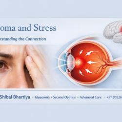
Many patients who come to Dr Bhartiya with night vision complaints have never been told that difficulty adjusting to low light is one of the earliest detectable signs of glaucoma, a condition that has no pain, no redness, and no warning until vision is already lost.
Dr Shibal Bhartiya is a fellowship-trained glaucoma specialist and Mayo Clinic Research Collaborator with over 25 years of experience. Her approach focuses on identifying risk before damage is irreversible, simplifying treatment decisions, and protecting vision long-term. Emphasis on early detection, risk assessment, and continuity of care. She is rated 5 stars across 1,500+ patient reviews on Google.
What Causes Difficulty Seeing at Night?
Several eye conditions affect your ability to see in low light. Some are minor and correctable. Others are serious and progressive.
Refractive Errors
An uncorrected or wrongly corrected spectacle power is one of the most common reasons for poor night vision. Myopia (short-sightedness) makes distant objects blur in all lighting conditions, but the effect is far more noticeable at night. An updated prescription often resolves this quickly.
Cataracts
A cataract clouds the natural lens inside your eye. As it thickens, light scatters before it reaches the retina. This causes glare, halos around lights, and reduced contrast — all of which become more pronounced after dark. Cataracts are treatable with surgery, but early detection gives you more options and better outcomes.
Glaucoma
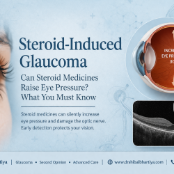
Glaucoma damages the optic nerve gradually and silently. One of its earliest and most overlooked signs is difficulty adapting to low light and a narrowing of your side vision. Most people with glaucoma notice nothing unusual until the damage is advanced. Night driving difficulty, bumping into objects in dim light, or needing extra time to adjust when entering a dark room can all be early warnings. Glaucoma cannot be reversed, but it can be stopped — if it is caught in time.
Diabetic Retinopathy
Uncontrolled diabetes damages the small blood vessels in the retina. This affects how the retina processes light, making night vision one of the first things to suffer. If you have diabetes and notice worsening night vision, do not wait.
Vitamin A Deficiency
Vitamin A is essential for producing rhodopsin, the pigment your retina uses to see in dim light. A deficiency, more common in children but possible in adults with certain diets or gut conditions, directly impairs night vision. This is one of the few causes that is fully reversible with the right nutrition.
Retinitis Pigmentosa
This inherited condition progressively destroys the light-sensitive cells in the retina. Night blindness is usually the first symptom, followed slowly by tunnel vision. Early diagnosis allows for monitoring, genetic counselling, and planning.
When Is Difficulty Seeing at Night Serious?
See a doctor promptly if you notice any of the following:
- You cannot drive safely after dark
- You see halos or starbursts around streetlights or headlights
- You bump into things on your side in low-light spaces
- Your night vision has worsened over weeks or months
- You have diabetes, a family history of glaucoma, or are over 40
Do not wait for your annual check-up if these symptoms are new or getting worse. Conditions like glaucoma cause permanent damage before you feel any pain or notice significant vision loss.
Night Vision and Glaucoma: What Most People Miss
Glaucoma is called the silent thief of sight for a reason. It takes peripheral vision first, the vision you use to see around you, navigate in dim light, and detect movement. By the time central vision is affected, the damage is already severe.
Night difficulty is one of the earliest functional signs of peripheral vision loss. People often blame tiredness, screen exposure, or ageing, and miss what is actually happening to their optic nerve.
If you are over 35, have a family history of glaucoma, are of Indian ethnicity, or have high eye pressure, difficulty seeing at night deserves a specialist evaluation, not just a new spectacle prescription.
What to Expect at Your Appointment
A comprehensive eye examination for night vision problems includes:
Visual acuity testing — checks how clearly you see at different distances
Refraction — determines your exact spectacle power
Intraocular pressure measurement — rules out raised eye pressure, a key risk factor for glaucoma
Slit-lamp examination — checks the lens for cataracts and the front of the eye for other conditions
Optic nerve assessment — looks for early glaucoma damage, often visible before symptoms appear
Visual field testing — maps your peripheral vision to detect silent loss
OCT scan — provides a detailed cross-section of the optic nerve and retina, detecting changes years before standard tests
This examination takes about 30 to 45 minutes. It is painless. And it could catch a condition that has no symptoms yet.
Frequently Asked Questions
Is difficulty seeing at night always a sign of a serious eye condition?
Not always. A mild refractive error or vitamin deficiency can cause night vision problems that are fully correctable. However, it can also be an early sign of glaucoma, cataracts, or retinal disease — which are serious. The only way to know is a proper eye examination. Do not self-diagnose.
Can difficulty seeing at night be treated?
Yes, in most cases. Treatment depends on the cause. Refractive errors are corrected with updated spectacles or contact lenses. Cataracts are managed with surgery. Glaucoma is treated with eye drops, laser, or surgery to stop progression. The earlier you seek care, the more treatment options are available.
I am 38 and healthy. Do I really need to worry about night vision changes?
Yes. Glaucoma can begin in your 30s, and Indians are at higher risk than many other populations. If your night vision has changed — even slightly — it is worth ruling out the serious causes. An OCT scan and visual field test take less than an hour and can give you complete clarity.
Does using screens at night cause permanent night vision problems?
Screen use causes temporary eye strain and can make it harder to adjust to darkness in the short term. It does not cause permanent night vision damage. However, if you use this explanation to dismiss persistent night vision symptoms, you may delay the diagnosis of something that does need treatment.
How is a glaucoma-related night vision problem different from normal ageing?
Some loss of contrast sensitivity is normal with age. But a progressive change in how quickly your eyes adjust to darkness, or difficulty on the side of your vision in low light, is not simply ageing — it needs investigation. The key question is whether your night vision has changed. If it has, see a specialist.
Book a Consultation
Night vision problems are worth taking seriously. A 45-minute appointment could detect a condition that has no other symptoms — and protect your vision before damage becomes permanent.
Book an appointment with Dr Shibal Bhartiya — Glaucoma Specialist, Gurgaon
📍 Marengo Asia Hospitals, Sector 56, Gurugram
📞 +91 88826 38735
About the Author
This article was written by Dr Shibal Bhartiya, fellowship-trained glaucoma specialist and Mayo Clinic Research Collaborator, Clinical Director at Marengo Asia Hospitals, Gurugram, known for ethical, patient-centred glaucoma care and independent glaucoma second opinions. She is also the Program Director for Community Outreach & Wellness; and for the Marengo Asia International Institute of Neuro and Spine.
She has published peer-reviewed research on glaucoma management, examining how treatment decisions should balance medical evidence, patient preferences, and long-term vision outcomes.
As Editor-in-Chief of Clinical and Experimental Vision and Eye Research and Executive Editor of the Journal of Current Glaucoma Practice (Pubmed Indexed, official journal of the International Society of Glaucoma Surgery), Dr Shibal Bhartiya brings editorial and research depth to every clinical decision. Her 200+ publications, including 90+ PubMed-indexed publications and 28 edited textbooks span glaucoma biology, surgical outcomes, health equity, and emerging diagnostics.
Access her work on Pubmed, Google Scholar, ResearchGate and ORCID.
Dr Shibal Bhartiya
Glaucoma • Second Opinion • Advanced Care
www.drshibalbhartiya.com
+91 88826 38735
1500+ Five Star Patient Reviews Google Business Profile
Upload your reports for a structured review.
If you are unable to come to Dr Bhartiya’s clinic: Read more about teleconsultation for glaucoma
Related Reading
Seeing clearly is not seeing safely
Seeing safely is not same a good vision
Why Vision Becomes Blurred After Reading or Screen Use
Night time driving and eye strain
Why Your Eyes Water Constantly
Why Do Women Get Dry Eye More Often?