A routine eye check may raise suspicion of glaucoma through elevated eye pressure, changes in the optic nerve, or unexplained vision changes. However, confirming glaucoma usually requires specialised tests such as optic nerve imaging, visual field testing, and corneal thickness measurement. Also, a routine for glasses is not a substitute for an eye exam, as the former can often miss glaucoma.
Glaucoma usually causes no pain, no redness, and no obvious vision change in its early stages. Most people with glaucoma feel completely normal until significant and irreversible damage has occurred. The only way to detect it early is a comprehensive eye examination that includes optic nerve assessment, intraocular pressure measurement, and visual field testing
A Routine Eye Check Revealed a Sight-Threatening Disease
Mrs SG came to see me because her glasses prescription had not felt right for a few months. She was 57. She worked at a desk. Her eyes were tired by evening, and she assumed she needed a stronger number. She had no pain. No redness. No alarming moment that made her think something was wrong.
Her previous optician had given her a new prescription six months earlier. It had not helped. She booked an appointment with me because a colleague had suggested a second opinion.
I examined her in the usual way. Her visual acuity was reasonable. Her anterior segment was quiet. Then I checked her retina and optic nerve.
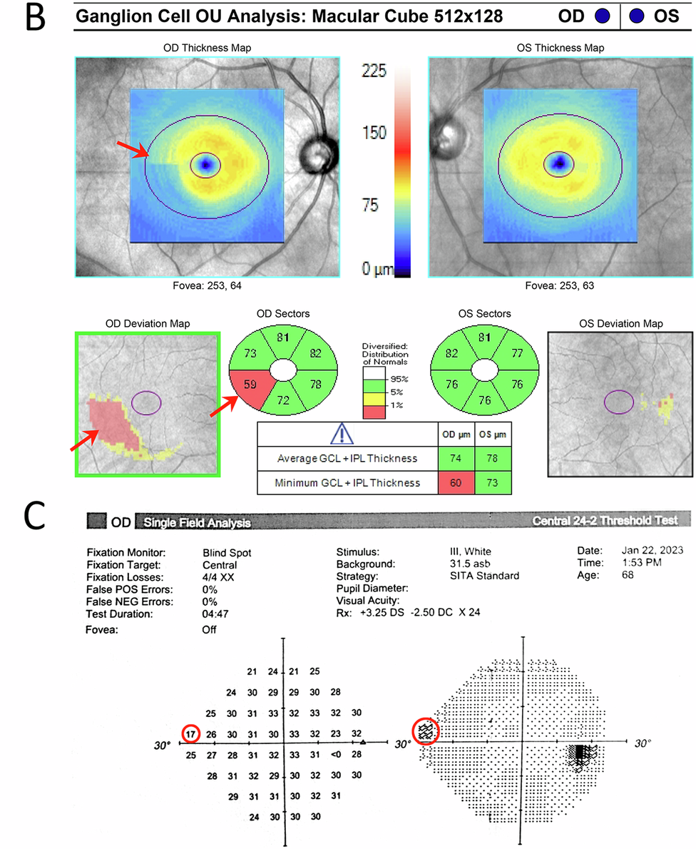
The optic nerve in her left eye had a cup that was too large. The rim tissue was thinning at the inferior pole. Her intraocular pressure was 24 mmHg in the right eye and 26 in the left. I asked her to sit with the visual field machine.
The field test confirmed what the disc had suggested. There was a dense arcuate defect in her left eye. A significant portion of her peripheral vision was already gone. She had not noticed. You rarely do with glaucoma, because the brain fills in the gaps until it cannot.
She did not need a stronger glasses prescription. She had glaucoma, and it had been quietly advancing for what was likely several years.
Patient details have been changed to protect privacy.
Remember
Sunita’s case is not unusual. Glaucoma is called the silent thief of sight for a reason. It causes no pain, no visible redness, and no early warning that most patients would recognise. By the time vision loss is noticeable, the disease has already caused permanent damage. In India, an estimated 12 million people have glaucoma, and almost 90% of them do not know it. (The Chennai glaucoma Study).
Below, I explain what glaucoma actually does to the eye, why it is so reliably missed, and which symptoms, or absences of symptoms, should prompt an urgent examination.
What Glaucoma Actually Does to Your Eye
Glaucoma is a disease of the optic nerve. The optic nerve carries visual information from the eye to the brain. When this nerve is damaged, that information is lost permanently. No treatment can restore what is already gone. Treatment can only slow or stop further damage.
In most cases, the damage is caused or worsened by raised pressure inside the eye. This pressure, called intraocular pressure or IOP, builds when fluid inside the eye does not drain properly. The drainage system becomes less efficient over time, pressure rises, and the optic nerve fibres begin to die. The process is painless in the vast majority of patients.
What makes glaucoma particularly deceptive is the pattern of vision loss. It begins at the periphery, the edges of your visual field. The brain compensates automatically. Both eyes together create a complete picture, and each eye covers for the blind spots of the other. Patients often do not notice peripheral vision loss until more than 40 percent of their optic nerve fibres have already been destroyed. By that point, the disease is well advanced.
In SG’s case, her glasses prescription had changed slightly because her visual system was compensating for early field loss. It was not a refractive change. It was her brain working harder to make sense of incomplete information. This pattern, subtle visual dissatisfaction without a clear cause, is one of the most common presentations I see in patients who turn out to have early to moderate glaucoma.
Glaucoma vs Normal Ageing: How to Tell the Difference
| Symptom or Sign | What It Suggests | What To Do |
|---|---|---|
| Gradual blurring that a new glasses prescription does not fix | May indicate optic nerve or macular pathology, not refractive change | See an ophthalmologist for optic nerve assessment, not just a refraction |
| Difficulty adjusting from bright to dim light | Can be an early sign of peripheral field loss | Request a visual field test at your next eye appointment |
| Frequent glasses changes with no lasting improvement | Suggests the problem is not the prescription | Ask for intraocular pressure measurement and disc evaluation |
| Mild headache or eye heaviness without redness | In some patients, mildly elevated IOP causes subtle discomfort | Check IOP, especially if over 40 or with family history of glaucoma |
| No symptoms at all, but a family member has glaucoma | First-degree relatives have a 4 to 9 times higher risk | Schedule a comprehensive glaucoma screening even if you feel completely well |
| Squinting or tilting the head to see clearly | May indicate undetected visual field asymmetry | Full field test for both eyes separately |
Why Glaucoma Is So Often Missed
The most common reason glaucoma goes undetected is that a routine glasses check is not a glaucoma examination.
When a patient visits an optician or a basic eye clinic for a new prescription, the standard assessment measures visual acuity and refraction. It does not always include optic nerve photography, intraocular pressure measurement, or visual field testing. These are the three investigations that detect glaucoma. Without all three, the disease is invisible.
Sunita had seen an optician twice in three years. Her visual acuity was checked each time. Her optic nerve was never examined.
The second reason glaucoma is missed is the absence of symptoms. Patients present to doctors when something feels wrong. Glaucoma does not feel wrong, not for years. There is no cultural expectation in India of an annual comprehensive eye examination. Most people attend only when they need a new prescription or when something is visibly red or painful. By those criteria, a glaucoma patient has no reason to come at all.
The third reason is that IOP alone is not a reliable screening tool. Many patients with glaucoma have pressure in the so-called normal range. Normal-tension glaucoma accounts for a substantial proportion of cases, particularly in people of Asian descent. A single IOP reading of 16 mmHg does not exclude the diagnosis.
SG’s IOP was elevated, which made diagnosis more straightforward. But many of my patients with confirmed glaucoma have had pressures that would not have triggered concern at a routine check.
When To See an Eye Specialist
See an ophthalmologist for a comprehensive glaucoma assessment if any of the following apply:
- A parent, sibling, or child has been diagnosed with glaucoma
- You are over 40 and have not had a comprehensive eye examination in the past two years
- You have been told your eye pressure is “a little high” but were not referred further
- You have changed your glasses prescription twice in two years with no lasting improvement
- You have diabetes, as this increases glaucoma risk
- You are of South Asian, East Asian, or African descent, all of which carry higher glaucoma risk
- You use steroid eye drops, nasal sprays, or inhalers long-term
- You were told everything was fine, but your vision still does not feel right
A comprehensive assessment takes around 30 to 45 minutes and is painless. It will include optic nerve imaging, IOP measurement, corneal thickness assessment, and a visual field test. This combination reliably detects glaucoma at a stage when treatment can prevent significant vision loss.
Frequently Asked Questions
Can you have glaucoma with normal eye pressure?
Yes. Normal-tension glaucoma is a recognised and common form of the disease, particularly in people of Asian descent. A normal IOP reading does not rule out glaucoma; optic nerve assessment and visual field testing are essential.
Does glaucoma always cause pain?
No. The most common forms of glaucoma are completely painless. Pain is associated with acute angle-closure glaucoma, which is a sudden and rare presentation. Most patients with chronic open-angle glaucoma, the most prevalent type, feel nothing at all until vision loss is advanced.
Can lost vision from glaucoma be restored?
No. Optic nerve damage caused by glaucoma is permanent. Treatment with eye drops, laser, or surgery can slow or stop further damage, but vision already lost cannot be recovered. Early detection is the only way to protect useful sight.
How often should I have a glaucoma check if I have a family history?
If a first-degree relative has glaucoma, you should have a comprehensive eye examination every year from the age of 40, or earlier if your ophthalmologist advises it.
Book a Consultation
If you have a family history of glaucoma, have not had a comprehensive eye examination in the past two years, or have been told your eye pressure is elevated, a dedicated assessment is worth arranging now. The earlier glaucoma is found, the more vision can be protected.
At Dr Shibal Bhartiya Eye Clinic, Gurugram, a glaucoma assessment includes optic nerve imaging, visual field testing, corneal thickness measurement, and a full review of your risk profile. [second opinion]
[Book an Appointment → +91 88826 38735]
This page is a part of the Glaucoma Hub. you may want to read about Glaucoma Progression, and Risk Stratification in Glaucoma. Other articles of interest could be Advanced Glaucoma Care in Gurgaon, What Good Glaucoma Care Actually Optimises For, What Happens If Glaucoma Is Left Untreated?, More Glaucoma Eye Drops is Not Better Glaucoma Care, 5 Mistakes Patients Make in Glaucoma Care and Do You Really Need Treatment for Glaucoma?
About the Author
This article was written by Dr Shibal Bhartiya, fellowship-trained glaucoma specialist and Mayo Clinic Research Collaborator, Clinical Director at Marengo Asia Hospitals, Gurugram, known for ethical, patient-centred glaucoma care and independent glaucoma second opinions. She is also the Program Director for Community Outreach & Wellness; and for the Marengo Asia International Institute of Neuro and Spine.
She has published peer-reviewed research on glaucoma management, examining how treatment decisions should balance medical evidence, patient preferences, and long-term vision outcomes.
As Editor-in-Chief of Clinical and Experimental Vision and Eye Research and Executive Editor of the Journal of Current Glaucoma Practice (Pubmed Indexed, official journal of the International Society of Glaucoma Surgery), Dr Shibal Bhartiya brings editorial and research depth to every clinical decision. Her 200+ publications, including 90+ PubMed-indexed publications and 28 edited textbooks span glaucoma biology, surgical outcomes, health equity, and emerging diagnostics.
1500+ Five Star Patient Reviews Google Business Profile
If you are unable to come to Dr Bhartiya’s clinic: Read more about teleconsultation
Read her research on PubMed | Google Scholar | ResearchGate | ORCID
Upload your reports for a structured review.| www.drshibalbhartiya.com | +91 88826 38735
Leave a review on Google