Why Good Vision Does Not Always Mean Safe Vision

Passing an eye test, and having good vision does not mean your vision is safe for every situation. Visual acuity, the ability to read a chart, measures only one aspect of sight. Contrast sensitivity, glare recovery, peripheral awareness, and low-light performance are separate functions that standard tests do not assess. You can see 6/6 on a chart and still be unsafe driving at night, struggling in crowds, or missing hazards at the edge of your vision, explains Dr Shibal Bhartiya.

Every year, patients are told their eyes are normal, and they leave the clinic believing their vision is fine. Many of them are right. But some of them are not. They struggle on the road at night. They miss steps in dim light. Sometimes, they lose their footing in a crowd. They have accidents they cannot explain.

The eye test they passed was not wrong. It measured what it was designed to measure. The problem is that it was not designed to measure everything that matters. Seeing clearly and seeing safely are not the same thing, and the gap between them is where serious, preventable harm lives.

Dr Shibal Bhartiya is a fellowship-trained glaucoma specialist and Mayo Clinic Research Collaborator with over 25 years of experience. Her approach focuses on identifying risk before damage is irreversible, simplifying treatment decisions, and protecting vision long-term. Emphasis on early detection, risk assessment, and continuity of care. She is rated 5 stars across 1,500+ patient reviews on Google.


7 Reasons Clear Vision Does Not Equal Safe Vision

  1. Contrast sensitivity is not tested in standard eye exams
  2. Peripheral vision can be significantly reduced before central vision is affected
  3. Glare recovery slows with age and early cataract
  4. Low-light performance is not tested on a chart
  5. Dry eye causes fluctuating vision in real conditions, not in a clinic
  6. Reaction time and visual processing speed are not eye tests
  7. Early glaucoma destroys safety-critical vision while acuity stays intact

What Each Gap Means in Real Life

1. Contrast Sensitivity

Visual acuity measures your ability to see high-contrast black letters on a white background. Real life is not high contrast. Roads, faces, kerbs, and obstacles exist across a range of contrast levels: especially in mist, rain, dusk, and artificial lighting. Contrast sensitivity is the ability to distinguish objects from their background in these conditions. It declines in early glaucoma, early cataract, and certain neurological conditions, often years before acuity drops. It is almost never tested in a routine eye examination.

2. Peripheral Vision

Your central vision, the sharp, detailed part, is what reads the chart. Your peripheral vision is what catches movement, detects hazards, and keeps you safe in traffic and crowds. Glaucoma destroys peripheral vision first. By the time central vision is affected, significant and irreversible damage has already occurred. A patient with advanced peripheral field loss can still read 6/6. That patient is not safe to drive. Standard acuity testing will not reveal this.
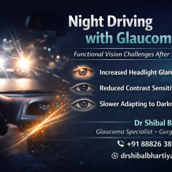
3. Glare Recovery

When a bright light hits your eye, an oncoming headlight, a flash of sun, your vision temporarily drops. Recovery time is the time it takes to see clearly again. This slows with age, early cataract, and corneal changes. In a clinic, there are no oncoming headlights. Glare recovery is not measured. On a motorway at night, it is one of the most safety-critical visual functions you have.

4. Low-Light Performance

Rod photoreceptors handle vision in dim environments. They are not tested on a standard eye chart, which is read in a brightly lit room. Vitamin A deficiency, early retinal disease, early glaucoma, and normal ageing all reduce rod function; leaving acuity intact while making low-light environments significantly more dangerous. Many patients first notice this while driving after dark, not during a daytime eye test.

5. Dry Eye and Tear Film Instability

The tear film is the eye’s first optical surface. In a clinic, patients blink normally, the environment is controlled, and the tear film stays relatively stable. In real conditions, screen use, air conditioning, driving, dry weather, the tear film breaks down between blinks. Vision fluctuates. It worsens at exactly the moments when clear sight matters most. This is invisible to a standard eye test conducted in ideal conditions.

6. Visual Processing Speed

Seeing a hazard and responding to it are two separate events. The speed at which the brain processes visual information, particularly moving objects at the periphery, slows with age and with certain neurological changes. This is not an ophthalmology measurement. But it is a safety-critical function that no eye test captures. Understanding this gap matters for patients and for families making decisions about driving.

7. Early Glaucoma

Glaucoma is the single most important cause of the gap between measured vision and safe vision. It removes peripheral field, degrades contrast sensitivity, and reduces low-light performance, all while leaving central acuity completely intact. A patient in the early to moderate stages of glaucoma can pass every standard vision check required for a driving licence. They can also be genuinely unsafe on the road. This is not a hypothetical scenario. It is a documented clinical reality.

Note: Patients with moderate to severe glaucoma prioritize recognizing faces and finding dropped objects. The patients who reported greater difficulty in seeing at night and adjusting to dim lights, as well as peripheral and distance vision. Individualizing Quality of Life measures is necessary for a better understanding of the patients’ perception of their visual disability, reported Dr Bhartiya and colleagues, in their paper Weighted Quality of Life in Glaucoma Patients with Advanced Disease. Pubmed ID  41113687


Seeing Clearly vs Seeing Safely: What the Tests Miss

FunctionWhat It AffectsTested in Standard Eye Exam?
Visual acuityReading, fine detailYes
Contrast sensitivityDriving, faces, kerbs in low contrastNo
Peripheral visionHazard detection, crowd navigationNot routinely
Glare recoveryNight driving, oncoming headlightsNo
Dark adaptationDim rooms, dusk, night environmentsNo
Tear film stabilityReal-world blur, screen use, drivingNo
Visual processing speedResponse to moving hazardsNo

What We Often Miss

Standard eye examinations are conducted in ideal conditions: controlled lighting, high contrast, static targets, a cooperative patient who is not tired or stressed. Real life is none of these things. The functional gap between clinic performance and real-world performance is largest in patients with early glaucoma, early cataract, and dry eye, precisely the conditions that are most common and most frequently missed.

Asking a patient “how is your vision?” in a bright clinic room is not the same as asking “are you safe on the road after dark?” Both questions deserve an answer. Only one of them gets asked.


When to Worry

Book a detailed evaluation if any of the following apply:

  • Night driving feels uncertain, stressful, or unsafe
  • You have had a near-miss or accident you cannot fully explain
  • You avoid driving in rain, dusk, or unfamiliar roads
  • You miss steps, kerbs, or objects at the edge of your vision
  • Your vision fluctuates during the day, especially at screens
  • You have glaucoma, diabetes, or a family history of eye disease
  • You are over 60 and have not had a detailed eye evaluation in the past year

What This Means for You

A normal eye test is good news. It is not a complete answer. If your measured vision is fine but your functional vision is not, if you are avoiding situations, compensating, or uncertain in ways you were not before, that gap deserves investigation. The tests that matter for safety are different from the tests that measure your glasses prescription. Ask for them specifically.


Frequently Asked Questions

Can I have 6/6 vision and still be unsafe to drive?

Yes. Visual acuity measures central clarity in ideal conditions. Driving requires contrast sensitivity, peripheral awareness, glare recovery, and low-light performance: none of which are tested in a standard vision check. Early glaucoma, early cataract, and dry eye can all impair driving safety while leaving measured acuity intact.

What tests actually measure safe vision?

Contrast sensitivity testing, visual field assessment, dark adaptation measurement, glare testing, and detailed optic nerve imaging are the key evaluations. These are separate from a standard prescription check and require different equipment and time.

Is this relevant for older drivers specifically?

Yes, but not exclusively. Glaucoma affects patients from their forties onward. Dry eye and cataract begin earlier than most people expect. Age accelerates most of these changes, but the gap between clear vision and safe vision can exist at any age.

How do I know if glaucoma is affecting my driving safety?

Glaucoma causes peripheral field loss that the patient often does not notice: the brain compensates by filling in the gaps. A visual field test and optic nerve imaging are the only ways to detect this. If you have glaucoma or risk factors for it, ask specifically whether your field loss has reached a level that affects driving.

My doctor said my eyes are fine. Should I be concerned?

If your measured vision is normal and you have no functional symptoms, that is genuinely reassuring. If your measured vision is normal but you are struggling in real conditions, the evaluation may not have tested the right things. A second opinion with specific functional testing is reasonable and appropriate.


Your Vision Should Work for Your Life, Not Just for a Chart

If something feels off: if driving feels harder, if dim environments feel uncertain, if you are compensating in ways you did not used to, that experience is real and it deserves a real answer.

Dr Shibal Bhartiya Glaucoma and Advanced Eye Care | Second Opinions

🌐 www.drshibalbhartiya.com 📞 +91 88826 38735


About the Author

This article was written by Dr Shibal Bhartiya, fellowship-trained glaucoma specialist and Mayo Clinic Research Collaborator, Clinical Director at Marengo Asia Hospitals, Gurugram, known for ethical, patient-centred glaucoma care and independent glaucoma second opinions. She is also the Program Director for Community Outreach & Wellness; and for the Marengo Asia International Institute of Neuro and Spine. This article was updated in April 2026.

She has published peer-reviewed research on glaucoma management, examining how treatment decisions should balance medical evidence, patient preferences, and long-term vision outcomes.

As Editor-in-Chief of Clinical and Experimental Vision and Eye Research and Executive Editor of the Journal of Current Glaucoma Practice (Pubmed Indexed, official journal of the International Society of Glaucoma Surgery), Dr Shibal Bhartiya brings editorial and research depth to every clinical decision. Her 200+ publications, including 90+ PubMed-indexed publications and 28 edited textbooks span glaucoma biology, surgical outcomes, health equity, and emerging diagnostics.

Access her work on PubmedGoogle ScholarResearchGate and ORCID.

Dr Shibal Bhartiya
Glaucoma • Second Opinion • Advanced Care

www.drshibalbhartiya.com
 +91 88826 38735

1500+ Five Star Patient Reviews Google Business Profile

Upload your reports for a structured review.

If you are unable to come to Dr Bhartiya’s clinic: Read more about teleconsultation for glaucoma

Eye Health After 60

After 60, your eyes face a different set of risks than they did at 40. Glaucoma, macular changes, cataract progression, and dry eye all accelerate in this decade. Many of these conditions cause no pain and no obvious warning. Which is why regular, detailed eye evaluation is essential after 60, not optional, explains Dr Shibal Bhartiya.

Most people over 60 assume that blurred vision means they need new glasses. Sometimes that is true. But in this age group, vision changes are often the first sign of something that needs treatment, not just a new prescription. The good news is that caught early, most serious eye conditions in this decade are manageable. The risk is waiting too long.

Dr Shibal Bhartiya is a fellowship-trained glaucoma specialist and Mayo Clinic Research Collaborator with over 25 years of experience. Her approach focuses on identifying risk before damage is irreversible, simplifying treatment decisions, and protecting vision long-term. Emphasis on early detection, risk assessment, and continuity of care. She is rated 5 stars across 1,500+ patient reviews on Google.


7 Eye Conditions That Are More Common After 60

  1. Glaucoma
  2. Age-related macular degeneration (AMD)
  3. Cataract
  4. Diabetic retinopathy
  5. Dry eye disease
  6. Posterior vitreous detachment (PVD)
  7. Eyelid and tear duct changes

What Each Condition Means for You

1. Glaucoma

Glaucoma damages the optic nerve, usually without pain or early vision loss. After 60, the risk rises sharply. Most people with glaucoma do not know they have it until significant damage has occurred. A detailed evaluation includes eye pressure, optic nerve imaging, and visual field testing; not just a standard check.

2. Age-Related Macular Degeneration (AMD)

AMD affects the centre of your vision, the part you use for reading, faces, and fine detail. Early AMD causes no symptoms. Intermediate AMD may cause slight blurring or difficulty in low light. Wet AMD can cause rapid central vision loss. Early detection through retinal imaging changes outcomes significantly.

3. Cataract

Most people over 60 have some degree of cataract. Symptoms include glare, halos at night, faded colours, and gradual blurring. Cataract surgery is one of the safest and most effective procedures available. The decision to operate depends on how much the cataract affects daily function, not just its appearance on examination.

4. Diabetic Retinopathy

If you have diabetes, your retinal risk increases significantly with age. Diabetic retinopathy can progress silently for years. Blood sugar control slows progression, but it does not eliminate the need for annual retinal evaluation. Even well-controlled diabetes requires regular retinal screening.

5. Dry Eye Disease

Tear production decreases with age, particularly after menopause in women. Symptoms include burning, grittiness, watery eyes, and fluctuating vision. Standard Schirmer tests often miss functional dry eye. A detailed tear film assessment gives a more accurate picture. Untreated dry eye accelerates surface damage and worsens visual quality.

6. Posterior Vitreous Detachment (PVD)

The vitreous gel inside the eye shrinks and pulls away from the retina with age. This causes sudden floaters and flashes of light. PVD itself is usually harmless. However, in some cases it causes a retinal tear, which needs urgent treatment. New floaters or flashes after 60 always need same-week evaluation.

7. Eyelid and Tear Duct Changes

Eyelids lose tone with age. They may turn inward (entropion) or outward (ectropion), both causing irritation and tearing. Blocked tear ducts also become more common. These are correctable conditions, but they are frequently dismissed as “just aging.”


How to Think About Your Symptoms After 60

SymptomPossible CauseWhen to Worry
Gradual blurringCataract, refractive changeWorsening over weeks
Peripheral vision lossGlaucomaAny unexplained gap in vision
Central blurring or distortionAMDSudden or rapid change — urgent
Flashes and new floatersPVD, retinal tearNew onset — same week evaluation
Burning, gritty eyesDry eye, eyelid changesPersistent or worsening
Night driving difficultyCataract, contrast loss, glaucomaFunctional impairment
Watery eyesBlocked tear duct, ectropionChronic and affecting vision

Eye Health After 60: What to Expect

Your eyes change significantly after 60. Most of these changes are normal, but some need early attention to protect your vision.

After 60, the eye’s lens becomes stiffer and cloudier. The drainage system slows down. The retina becomes more vulnerable. None of this is unusual. All of it is manageable when caught early.

What Normally Changes After 60

Reading vision gets harder. The lens loses flexibility. This is called presbyopia. You may need reading glasses even if your distance vision is fine. This is not a disease. It is a normal part of ageing.

Contrast sensitivity drops. You may find it harder to read in low light or see steps clearly. Colours may look less vivid. This happens because the pupil becomes smaller and lets in less light.

Floaters increase. Most floaters are harmless. They are shadows from tiny fibres in the vitreous gel inside your eye. But a sudden shower of new floaters, especially with flashing lights, needs urgent attention. It can signal a retinal tear.

Dry eyes become more common. The glands that produce tears work less efficiently with age. Eyes feel gritty, tired, or burning. Dry eye is one of the most common eye complaints after 60 and is very treatable. [internal link: /omega-3-dry-eye/]

Adaptation to dark and light slows. Moving from bright sunlight into a dim room takes longer. This is normal but can affect driving safety at night.What Routine Tests Often Miss

Remember

Many eye evaluations in this age group focus on correcting the glasses prescription and checking eye pressure. That misses the full picture. Contrast sensitivity, tear film quality, optic nerve structure, and macular health all need individual assessment. A normal eye pressure does not rule out glaucoma. Clear-looking eyes do not rule out AMD or early retinal changes. After 60, a complete evaluation takes longer than ten minutes.


When to Worry

See an eye specialist promptly if you notice any of the following:

  • Sudden new floaters or flashes of light
  • Any sudden change in central vision
  • A shadow or curtain across part of your vision
  • Rapid worsening of night vision
  • Vision loss that does not improve with blinking
  • Double vision in one or both eyes

Annual evaluation is the minimum after 60. Six-monthly evaluation is appropriate if you have glaucoma, diabetes, or AMD.


What This Means for You

Ageing affects every part of the body, and the eyes are no exception. But most serious eye conditions after 60 are treatable when found early. The goal of eye care in this decade is not just clearer glasses, it is protecting the vision you have for the decades ahead. If your last eye check was more than a year ago, now is the right time.

How Often Should You Have Your Eyes Examined After 60?

Once a year, without exception.

A comprehensive annual eye exam after 60 checks vision, eye pressure, the optic nerve, the retina, and the drainage angle. It takes less than an hour. It can detect cataracts, glaucoma, macular degeneration, and diabetic eye disease before you notice any change in your vision.

If you have diabetes, hypertension, a family history of glaucoma, or previous eye conditions, your eye doctor may recommend more frequent reviews.


What a Comprehensive Eye Exam Includes

  • Vision testing at distance and near
  • Eye pressure measurement
  • Optic nerve assessment
  • Dilated retinal examination
  • Corneal thickness if glaucoma risk is present
  • Visual field testing if indicated [internal link: /visual-field-test/]
  • OCT scan of the optic nerve and retina if needed [internal link: /rnfl-oct/]

Practical Steps to Protect Your Eyes After 60

Wear UV-protective sunglasses outdoors. UV exposure accelerates cataracts and macular degeneration. A good pair of wrap-around sunglasses is one of the simplest protective steps you can take.

Manage your systemic health. Blood pressure, blood sugar, and cholesterol directly affect your eyes. Keeping these controlled reduces your risk of retinal vascular disease and diabetic eye disease.

Eat well. A diet rich in leafy greens, colourful vegetables, and omega-3 fatty acids supports retinal health. [internal link: /omega-3-dry-eye/]

Do not smoke. Smoking doubles the risk of macular degeneration and accelerates cataract formation. It is the single most modifiable risk factor for serious eye disease.

Tell your eye doctor about all medications. Some systemic drugs affect the eyes. Hydroxychloroquine, used for rheumatoid arthritis and lupus, requires annual retinal monitoring. Certain blood pressure medications affect eye pressure.


A Note on Second Opinions

If you have been told you have early cataracts, early glaucoma, or macular changes and you are unsure about next steps, a second opinion is always appropriate. Understanding exactly what stage you are at and what your options are makes a meaningful difference to long-term outcomes.


Frequently Asked Questions

Is it normal for vision to change a lot after 60?

Some change is normal. But frequent or rapid changes need evaluation. They may indicate cataract progression, dry eye, or an early retinal or nerve problem.

Can glaucoma start after 60 even with no family history?

Yes. Age itself is a major risk factor for glaucoma. Family history adds to the risk but is not required for the disease to develop.

I had cataract surgery. Do I still need regular eye checks?

Yes. Cataract surgery removes the cloudy lens but does not protect against glaucoma, AMD, retinal changes, or dry eye. Annual evaluation remains important.

How is eye care after 60 different from a standard vision test?

A standard vision test checks your glasses prescription and basic eye pressure. A complete evaluation after 60 includes optic nerve imaging, visual field testing, retinal assessment, and tear film evaluation. These are different tests with different equipment.

Can AMD be prevented?

Early AMD cannot always be prevented, but progression can be slowed. Stopping smoking, controlling blood pressure, and taking specific nutritional supplements in intermediate AMD are evidence-based steps. Early detection through retinal imaging is essential.


See a Specialist Who Looks Beyond the Obvious

After 60, eye care is not just about reading the chart. It is about protecting your independence, your ability to drive, and your quality of life. If something feels off, or if it has been more than a year since a detailed evaluation, book a consultation.

Dr Shibal Bhartiya Glaucoma and Advanced Eye Care | Second Opinions

🌐 www.drshibalbhartiya.com 📞 +91 88826 38735


About the Author

This article was written by Dr Shibal Bhartiya, fellowship-trained glaucoma specialist and Mayo Clinic Research Collaborator, Clinical Director at Marengo Asia Hospitals, Gurugram, known for ethical, patient-centred glaucoma care and independent glaucoma second opinions. She is also the Program Director for Community Outreach & Wellness; and for the Marengo Asia International Institute of Neuro and Spine.

She has published peer-reviewed research on glaucoma management, examining how treatment decisions should balance medical evidence, patient preferences, and long-term vision outcomes.

As Editor-in-Chief of Clinical and Experimental Vision and Eye Research and Executive Editor of the Journal of Current Glaucoma Practice (Pubmed Indexed, official journal of the International Society of Glaucoma Surgery), Dr Shibal Bhartiya brings editorial and research depth to every clinical decision. Her 200+ publications, including 90+ PubMed-indexed publications and 28 edited textbooks span glaucoma biology, surgical outcomes, health equity, and emerging diagnostics.

Access her work on PubmedGoogle ScholarResearchGate and ORCID.

Dr Shibal Bhartiya
Glaucoma • Second Opinion • Advanced Care

www.drshibalbhartiya.com
 +91 88826 38735

1500+ Five Star Patient Reviews Google Business Profile

Upload your reports for a structured review.

If you are unable to come to Dr Bhartiya’s clinic: Read more about teleconsultation for glaucoma

Glaucoma in India: Why the Risk Is Higher Than You Think

Glaucoma is the most common cause of irreversible blindness in India, and 90% of cases remain undiagnosed. That means nine…

Can I drive at night with glaucoma?

Yes, most people with glaucoma can continue driving safely, as long as their vision meets legal requirements and their visual…

Target Eye Pressure in Glaucoma

Target eye pressure is the range of eye pressures that ensures that there is no continued optic nerve damage. This…