Quick Answer
Yes. A visual field test can point to damage along the visual pathway in the brain, but an MRI confirms the cause.
An abnormal field report can frighten you, especially when the word “neurological” appears. Take a deep breath. Most abnormal fields do not come from the brain. Glaucoma, cataract and a tired test day cause far more of them. Some patterns do point beyond the eye, though, and those deserve a prompt, calm review, preferably with a neuro-ophthalmologist.
Each eye sends its signals along the optic nerve to a crossing point called the chiasm. From there, the signals travel deep through the brain to the back of your head. Each stretch of this pathway serves a specific part of your vision. Damage to one stretch leaves a missing area with a recognisable shape. That shape tells your doctor where to look.
Six Visual Field Patterns That Point Beyond the Eye
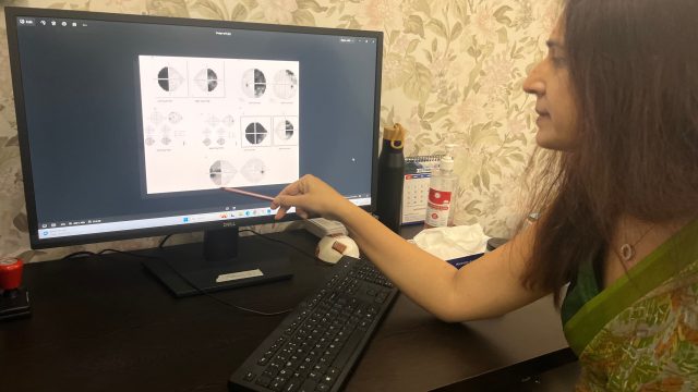
Doctors read these patterns on the printout map. The shape matters more than the numbers.
Both Outer Halves Missing (Bitemporal Hemianopia)
You lose the outer half of vision in each eye. People describe bumping into doorframes or missing cars at the side. It builds slowly, so many patients adapt without noticing. The chiasm sits just above the pituitary gland. A pituitary tumour is the commonest cause, and other growths near the chiasm can do the same.
The Same Side Missing in Both Eyes (Homonymous Hemianopia)
You lose the left or right side of vision in both eyes. Many patients blame one eye, because covering either eye changes nothing. You may bump into people on that side, or lose your place while reading. The damage lies behind the chiasm. Stroke is the commonest cause, followed by tumours and head injury. Sudden onset is an emergency.
A Corner Missing in Both Eyes (Quadrantanopia)
A wedge of vision is missing in the upper or lower corner of both eyes. An upper wedge points to the temporal lobe. A lower wedge points to the parietal lobe. Both sit behind the chiasm, so doctors usually order imaging.
A Dark Patch at the Centre (Central Scotoma)
A dim or dark patch sits in the centre of one eye. Colours look washed out. Optic neuritis is a common cause, especially in younger adults with pain on eye movement. Nutritional, toxic and compressive optic nerve problems can also do this. Read more in Optic Neuritis.
An Enlarged Blind Spot
Every eye has a natural blind spot where the optic nerve leaves the eye. A swollen optic nerve makes it grow. This is a classic early field sign of papilledema, which means swelling from raised pressure inside the head. Patients may also notice headaches, brief greying of vision or a whooshing sound in the ears. Buried deposits in the optic nerve, called drusen, can look similar. Read more in Papilledema.
The Top or Bottom Half Missing in One Eye (Altitudinal Defect)
Half of one eye’s vision is missing, with a flat edge across the middle. The usual cause is ischaemic optic neuropathy, where the optic nerve loses its blood supply. It tends to strike suddenly and without pain. After age 50, doctors also check for giant cell arteritis, an inflammation that needs same-day treatment. Read more in AION and NAION.
How Is This Different From Glaucoma?
Glaucoma damages nerve fibres that arch above and below the centre. Its defects follow the horizontal line. They look like arcs or steps and build slowly. Damage along the brain pathway usually follows the vertical line instead. The defect stops at a straight edge down the middle.
Treat this as a guide, not a rule. Ischaemic optic neuropathy follows the horizontal line and can pass for glaucoma. Early pressure on the optic nerve can hide behind a modest mean deviation. That is why I read the map, not just the number.
See how glaucoma looks on a printout in What Is a Visual Field Test for Glaucoma?
What Your Field Loss Can Mean
| What you notice | What it can mean | When to worry |
|---|---|---|
| Bumping into things at both sides | Pressure on the chiasm, often a pituitary tumour | Book a review soon. Go urgently if it comes with a sudden severe headache. |
| One side missing in both eyes | Stroke, tumour or injury behind the chiasm | Sudden onset needs an emergency department visit. |
| A corner missing in both eyes | Temporal or parietal lobe damage | Needs prompt imaging. |
| Dark centre, washed-out colour, pain on eye movement | Optic neuritis | See a doctor within a day or two. |
| Top or bottom half of one eye gone | Ischaemic optic neuropathy | Urgent, same day. After age 50, with jaw pain or scalp tenderness, treat it as an emergency. |
| Enlarged blind spot with headaches or greying of vision | Raised pressure inside the head | Urgent, same day. |
| Slow arc-shaped loss, usually unnoticed | Glaucoma, the commonest cause | Book a glaucoma review. |
Why Field Loss is Often Missed
- Patients rarely notice a missing half-field. The brain fills the gap, so people blame one eye. Cover each eye in turn to check.
- A modest mean deviation can hide a defect with a sharp vertical edge. The map matters more than the number.
- The standard glaucoma programme covers only the central part of the field. Some defects sit outside it, so your doctor may choose a wider or different programme.
- Look beyond glaucoma when the nerve looks pale rather than cupped, when loss moves fast, or when pressure and scans do not match the field.
- Cataract, a droopy eyelid, a small pupil, the wrong lens and a tired test can all fake a defect. Doctors repeat an odd result before acting, unless your symptoms point to an emergency.
What Happens After an Abnormal Field Result?
Your doctor usually builds the picture in steps:
- A repeat test, sometimes with a wider or different programme
- A dilated exam of the optic nerve, plus pupil and colour vision checks
- An OCT scan of the nerve fibre and ganglion cell layers
- An MRI of the brain and orbits when the pattern points that way
- Blood tests, such as ESR and CRP, if giant cell arteritis is possible
- A referral to a neurologist, neurosurgeon or endocrinologist when needed
Dr Bhartiya works closely with a team of neurologists, neurosurgeons, and interventional neurologists. She can arrange prompt imaging and neurology review through the Marengo Asia International Institute of Neuro and Spine.
When to See a Doctor
See a Doctor Urgently
Go to an emergency department the same day if you have:
- Sudden loss of vision in one eye, or in the same side of both eyes
- Vision loss with face droop, arm weakness or slurred speech
- A sudden severe headache with a change in vision
- New double vision with a field change
- Pain on eye movement with dim or blurred vision
- New headache, jaw pain when chewing or scalp tenderness after age 50, with any vision change
- Brief greying or blackouts of vision, especially on standing or bending, with headaches
See a Doctor
Book a visit soon if you notice:
- Repeated bumps into doorframes or people on one side
- Trouble keeping your place while reading
- A field defect on a routine test that nobody has explained
- A glaucoma field that does not match your pressure or optic nerve scan
- Slowly dimming colour or vision in one eye
- New or changing headaches with any vision change
What This Means for You
A field defect is a clue, not a diagnosis. Many causes are treatable, and earlier review usually gives you more options. Bring every printout, OCT report and scan you have. At home, cover one eye at a time to see which eye is missing vision. Avoid driving until a doctor has reviewed any new loss of side vision.
Frequently Asked Questions
Can a visual field test detect a brain tumour?
It can show a pattern that suggests pressure on the visual pathway, such as from a pituitary tumour. It cannot confirm a tumour. An MRI does that.
Can a stroke show up on a visual field test?
Yes. A stroke behind the chiasm often leaves the same side of vision missing in both eyes. Sometimes this is the only sign. Sudden loss of side vision needs emergency care.
Does a normal visual field rule out a brain problem?
No. Small or early lesions can leave the field normal. Your symptoms and the rest of the exam matter as much as the test.
Will I need an MRI?
Not always. Your doctor decides from the shape of the defect, the optic nerve exam and your symptoms. A defect that stops at the vertical midline usually calls for imaging.
How is a neurological field defect different from glaucoma?
Glaucoma damage usually follows the horizontal line in arcs and steps. Brain pathway damage usually follows the vertical line. There are exceptions, so a specialist should read the map.
Discuss Your Visual Field Report With Dr Bhartiya
Has a field report raised a question about your optic nerve or brain? Bring your printouts, OCT and any scans. Dr Bhartiya reads them together and explains what the pattern means and what to do next.
Not sure what to bring? Read What to Bring for Your Visit, or Upload your reports for a structured review.
Book an Appointment:
📍 Gurgaon 📞 +91 88826 38735 | 🌐 Contact Us | Second Opinion Form for teleconsults
This page is part of the Neuro-Ophthalmology hub. Read about our full approach to neurological vision conditions. Some vision problems are not eye problems, and Neuro-ophthalmology second opinions, and Guidelines for Neuro-ophthalmology Second Opinions. They are brain problems, nerve problems, or vascular problems, that show up in the eye first. Also read about optic nerve disease, AION and NAION, Sixth Nerve Palsy, Facial Nerve Palsy, raised intracranial pressure, Vision not clear but tests normal, double vision, and conditions where no diagnosis has yet been reached.
To know more about visual fields, read What to Expect and What the Results Mean, How To Do Visual Fields, Glaucoma Test Results Explained: OCT, Visual Fields and Eye Pressure, Why Do I Need a Visual Field.
About the Author
This article was written by Dr Shibal Bhartiya, fellowship-trained glaucoma specialist and Mayo Clinic Research Collaborator, Clinical Director at Marengo Asia Hospitals, Gurugram, known for ethical, patient-centred glaucoma care and independent glaucoma second opinions. She is also the Program Director for Community Outreach & Wellness; and for the Marengo Asia International Institute of Neuro and Spine.
Dr Bhartiya has published peer-reviewed research onglaucoma management, examining how treatment decisions should balance medical evidence, patient preferences, and long-term vision outcomes. She is also a committed patient advocate and mainstream medical columnist, translating complex ophthalmic evidence into actionable public health insights across leading national platforms including NDTV and OnlyMyHealth
As Editor-in-Chief of Clinical and Experimental Vision and Eye Research and Executive Editor of the Journal of Current Glaucoma Practice (Pubmed Indexed, official journal of the International Society of Glaucoma Surgery). Her 200+ publications, including 90+ PubMed-indexed publications and 28 edited textbooks span glaucoma biology, surgical outcomes, health equity, and emerging diagnostics.
1600+ Five Star Patient Reviews Google Business Profile
If you are unable to come to Dr Bhartiya’s clinic: Read more about teleconsultation
Read her research on PubMed | Google Scholar | ResearchGate | ORCID | Github | Wikidata
📋Upload your reports for a structured review.| 🌐 Contact Us| 📞 +91 88826 38735
⭐ Helped by this article? Leave a Google review — it helps other patients find reliable eye care.
Last Reviewed: October 2026