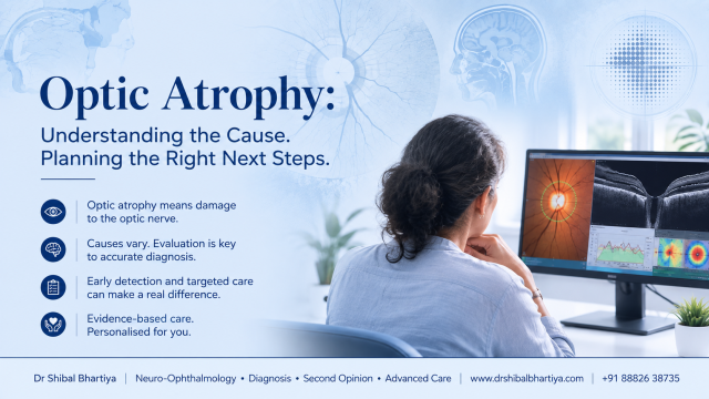
Optic atrophy describes a pale optic nerve caused by previous damage to the nerve fibres that carry visual information from the eye to the brain. It requires careful evaluation to identify the cause and guide further management. Optic atrophy cannot be reversed, but finding and treating the underlying cause early can protect the vision you still have.
Being told you have optic atrophy can be worrying, but it is a clinical finding rather than a diagnosis. Optic atrophy is permanent damage to the optic nerve, the cable that carries images from your retina to your brain. It causes dimmed or lost vision, poor colour perception and a pale appearance of the optic disc on examination.
Optic Atrophy: What It Means and Why It Happens
Optic atrophy means the nerve fibres carrying visual signals from your eye to your brain have died or shrunk. It shows up as pale, thin optic nerve tissue and permanent loss of vision.
Most patients hear this term after a routine eye exam flags a “pale disc.” The finding itself is not a diagnosis. It is a sign that something, somewhere along the visual pathway, has damaged the optic nerve.
As a glaucoma and neuro-ophthalmology specialist, I see optic atrophy from two directions. Sometimes it comes from eye pressure damage, as in glaucoma. Sometimes it comes from a problem far from the eye itself, such as a brain tumour, inflammation, or a blocked blood vessel. Working out which one you have changes everything about your treatment.
Common Causes of Optic Atrophy
| Cause | What It Means | What To Do About It |
|---|---|---|
| Glaucoma | Raised eye pressure slowly damages nerve fibres over months or years | Get your eye pressure and visual field checked every 6 months if diagnosed |
| Optic neuritis | Inflammation of the nerve, often linked to multiple sclerosis | See a neuro-ophthalmologist within days if vision drops with eye pain |
| Compression from a tumour | A growth near the optic nerve or pituitary gland presses on the fibres | Book an urgent MRI brain and orbits if vision loss comes with headaches |
| Blocked blood supply (ischaemic) | A sudden loss of blood flow starves part of the nerve | Seek same-day care; this is treated as a medical emergency |
| Toxic or nutritional damage | Alcohol, tobacco, certain medicines or vitamin B12 deficiency injure the nerve | Get blood tests for B12 and folate, and review your medication list with your doctor |
| Inherited optic neuropathy | Genetic conditions like Leber’s affect young adults, often men | Ask about genetic testing if a close relative has unexplained vision loss |
| Head trauma | A blow to the head can shear or bruise the optic nerve | Request a dilated eye exam after any significant head injury, even if vision seems fine at first |
| Long-standing swelling of the disc | Untreated papilloedema from raised brain pressure damages the nerve over time | Treat the underlying cause of raised brain pressure without delay |
Primary vs Secondary Optic Atrophy
Doctors classify optic atrophy by how the disc looks and what caused the damage. This distinction guides which tests come next.
| Type | What It Means | What To Do About It |
|---|---|---|
| Primary optic atrophy | Damage occurs directly at the nerve, with sharp, well-defined disc pallor and no prior swelling | Expect an MRI to rule out compression, inflammation or a genetic cause |
| Secondary optic atrophy | Damage follows earlier disc swelling, leaving a paler, less defined and often irregular disc margin | Ask what caused the original swelling, since that condition needs its own treatment |
| Consecutive optic atrophy | Damage follows disease elsewhere in the retina, such as a blocked retinal vessel | Expect retinal imaging alongside optic nerve assessment |
| Glaucomatous optic atrophy | A distinct pattern caused by raised eye pressure, with a specific pattern of cupping | Get an OCT nerve fibre scan and formal visual field test |
Note: Primary and secondary optic atrophy look different under examination, even though both end in permanent fibre loss. The pattern itself gives real diagnostic clues before any scan is done.
Glaucomatous Optic Atrophy
Glaucomatous optic atrophy is the most common form I see in clinic. It develops when sustained raised eye pressure damages nerve fibres at the point where they exit the eye.
Unlike other causes, glaucomatous damage has a signature look. The centre of the optic disc hollows out into a deep cup, and the surrounding rim of healthy tissue thins unevenly. This pattern lets me distinguish glaucoma from other causes of optic atrophy on exam alone, often before a patient notices any change in vision.
| Feature | What It Means | What To Do About It |
|---|---|---|
| Increased cup-to-disc ratio | The central hollow of the disc enlarges as fibres are lost | Track this ratio at every visit through dilated exam and OCT |
| Notching of the disc rim | Damage often concentrates at the upper or lower pole first | Ask for a printed OCT report comparing both eyes side by side |
| Splinter haemorrhages at the disc edge | A sign of active, ongoing nerve damage | Report this finding to your doctor; it usually means treatment needs adjusting |
| Thinning matches visual field loss | Field loss location correlates closely with the area of thinning | Repeat visual fields every 6 months to confirm the pattern is stable |
| Asymmetry between the two eyes | Glaucoma often damages one eye faster than the other | Mention if you’ve noticed bumping into objects on one side |
| Progression despite normal-range eye pressure | Some patients develop damage even without high pressure readings | Ask about normal-tension glaucoma if your pressure checks look fine but your OCT does not |
Because glaucomatous damage is silent until it is advanced, the pattern seen on the disc often tells us more than the symptoms a patient reports. This is why I recommend regular monitoring for anyone with a family history of glaucoma, not just those with high eye pressure readings.
Important: Glaucomatous optic atrophy can progress even when eye pressure readings look normal. A pattern of cupping and rim thinning on exam matters as much as the pressure number itself.
Signs That Point to Optic Atrophy
| Sign | What It Means | What To Do About It |
|---|---|---|
| Dimmer or washed-out vision | Fewer working nerve fibres carry a weaker signal to the brain | Note whether one eye or both eyes feel affected and mention this at your visit |
| Colours look faded, especially red | Colour vision fibres are often affected early | Try a simple red-cap test between your two eyes at home and report any difference |
| A blind spot or missing patch of vision | Damage often follows a specific pattern depending on the cause | Ask for a visual field test to map the pattern precisely |
| Reduced depth perception | Two eyes with unequal nerve damage struggle to judge distance | Mention this if you have started misjudging steps or kerbs |
| Pale optic disc on eye exam | Loss of nerve fibres and blood vessels changes the disc’s colour | This is usually found on a dilated exam; ask your doctor to document and photograph it |
| Poor night vision | Fewer functioning fibres reduce contrast sensitivity in dim light | Report new difficulty with night driving |
| A relative afferent pupillary defect | The pupil reacts less to light in the more damaged eye | This is checked with a simple pen-torch swinging test in clinic |
When To See a Doctor
Some presentations of optic atrophy need same-day attention. Do not wait for your next scheduled appointment if you notice:
- Sudden vision loss in one eye, even if it seems to improve
- Vision loss with eye pain, especially on eye movement
- Vision loss with a new headache, nausea or vomiting
- Double vision alongside dimmed vision
- Vision changes in a child, since these are often missed early
- A change that started after beginning a new medication
- Vision loss on one side only that is getting worse over days
- Facial numbness, weakness or slurred speech alongside vision changes
Important: Sudden, painless vision loss in one eye can signal a blocked blood vessel in the optic nerve. This needs same-day evaluation to protect your other eye.
📍 Gurgaon 📞 +91 88826 38735 | 🌐 Contact Us | Second Opinion Form for teleconsults
Medical and Treatment Options
Protecting Remaining Vision
Optic atrophy itself cannot be reversed. Dead nerve fibres do not regenerate. Treatment focuses on stopping further damage and protecting the fibres that still work.
Treating the Underlying Cause
- Glaucoma-related atrophy: eye drops, laser treatment or surgery to lower eye pressure and slow further fibre loss
- Inflammatory causes: steroids or other immune-modulating treatment, guided by an MRI and, when needed, a neurologist
- Compressive causes: surgical or oncologic treatment of the tumour or lesion pressing on the nerve
- Nutritional or toxic causes: correcting vitamin deficiencies and stopping the causative substance
- Ischaemic causes: urgent control of blood pressure, blood sugar and cardiovascular risk factors to protect the other eye
Neuro-Ophthalmology Work-Up
When optic atrophy does not fit a simple pattern, I coordinate MRI brain and orbits, visual field testing, OCT of the nerve fibre layer, and blood work through the Marengo Asia International Institute of Neuro and Spine, where I serve as Program Director. This lets us find brain and vascular causes quickly, not just eye-related ones.
Patient tip: Bring old glasses prescriptions or photographs of your eyes to your visit. Comparing pupil size and eye position over time helps pinpoint when the damage likely began.
Frequently Asked Questions
Can optic atrophy be reversed?
No, damaged optic nerve fibres cannot regrow or heal once lost. Treatment aims to identify the cause quickly and stop further damage. Some patients recover partial function if the underlying condition, such as inflammation, resolves early. The remaining vision is usually the vision you will keep long-term.
Does optic atrophy always lead to blindness?
Not always. Many patients keep useful vision if only part of the nerve is damaged. The outcome depends heavily on the cause, how early it was caught, and how much of the nerve was affected. Some causes, like isolated glaucoma damage in one area, progress slowly over years.
How is optic atrophy diagnosed?
A dilated eye exam usually reveals a pale, thin optic disc. This is confirmed with OCT imaging of the nerve fibre layer and a visual field test. Depending on the suspected cause, an MRI of the brain and orbits, blood tests, or a lumbar puncture may follow.
Is optic atrophy the same as glaucoma?
No. Glaucoma is one specific cause of optic atrophy, driven mainly by raised eye pressure. Optic atrophy is the broader finding of nerve damage, which can result from glaucoma, inflammation, compression, poor blood supply, or genetic conditions. Correctly identifying the cause changes the entire treatment plan.
Can children get optic atrophy?
Yes, though it is less common than in adults. In children, genetic conditions, tumours near the optic pathway, and previous infections are more frequent causes. Any child with reduced eye contact, clumsiness, or a family history of vision loss deserves a prompt eye exam.
What tests will I need if optic atrophy is suspected?
Expect a dilated eye exam, OCT scan of the nerve fibre layer, and formal visual field testing at minimum. Depending on findings, your doctor may add an MRI of the brain and orbits, blood pressure and blood sugar checks, and blood tests for vitamin B12, folate, and inflammatory markers.
Key Takeaways
- Optic atrophy means permanent damage to optic nerve fibres, seen as a pale disc on eye exam
- It is a sign, not a diagnosis; finding the underlying cause is essential
- Causes range from glaucoma and inflammation to tumours, poor blood supply and nutritional deficiency
- Sudden, painless vision loss in one eye needs same-day medical attention
- Damaged fibres do not regenerate, so treatment focuses on protecting what remains
- A neuro-ophthalmology work-up combining OCT, visual fields and MRI gives the clearest picture
Book a Consultation
If you or a family member has been told about a pale optic nerve or unexplained vision loss, do not wait to get a clear answer. Early evaluation gives us the best chance of protecting the vision you still have.
It is important to understand that optic atrophy is a sign of damage to the optic nerve, not a diagnosis in itself. The priority is to identify the underlying cause, determine whether it is still active, and assess how much vision can be protected.
If you have been diagnosed with optic atrophy, have unexplained vision loss, or would like a second opinion about your diagnosis or treatment plan, book a consultation with Dr. Shibal Bhartiya. Together, we will determine the cause, explain what it means for your vision, and develop the most appropriate long-term management plan. At my clinic in Gurgaon I offer detailed neuro-ophthalmology and glaucoma assessments, combining eye exam findings with imaging and, where needed, coordinated neurology input.
Book an Appointment →
📍 Gurgaon 📞 +91 88826 38735 | 🌐 Contact Us | Second Opinion Form for teleconsults
This page is part of the Neuro-Ophthalmology Hub. Read about our full approach to optic nerve conditions. Please also read Glaucoma Hub. You also may want to read more about Tests Explained and Visual Symptoms.
About Dr Shibal Bhartiya
This article was written by Dr Shibal Bhartiya, fellowship-trained glaucoma specialist and Mayo Clinic Research Collaborator, Clinical Director at Marengo Asia Hospitals, Gurugram, known for ethical, patient-centred glaucoma care and independent glaucoma second opinions. She is also the Program Director for Community Outreach & Wellness; and for the Marengo Asia International Institute of Neuro and Spine.
She has published peer-reviewed research on glaucoma management, examining how treatment decisions should balance medical evidence, patient preferences, and long-term vision outcomes.
As Editor-in-Chief of Clinical and Experimental Vision and Eye Research and Executive Editor of the Journal of Current Glaucoma Practice (Pubmed Indexed, official journal of the International Society of Glaucoma Surgery), Dr Shibal Bhartiya brings editorial and research depth to every clinical decision. Her 200+ publications, including 90+ PubMed-indexed publications and 28 edited textbooks span glaucoma biology, surgical outcomes, health equity, and emerging diagnostics.
1600+ Five Star Patient Reviews Google Business Profile
If you are unable to come to Dr Bhartiya’s clinic: Read more about teleconsultation
Read her research on PubMed | Google Scholar | ResearchGate | ORCID
📋Upload your reports for a structured review.| 🌐 Contact Us| 📞 +91 88826 38735
⭐ Helped by this article? Leave a Google review — it helps other patients find reliable eye care.