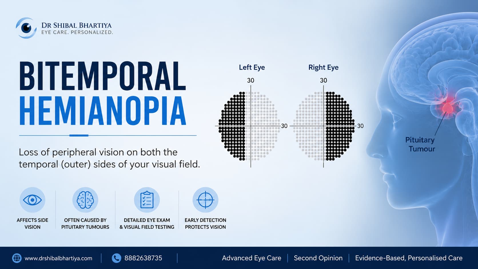
Pituitary tumours cause vision loss by pressing upward on the optic chiasm, typically producing a specific pattern called bitemporal hemianopia. This means loss of the outer (peripheral) visual field in both eyes, while central vision often stays deceptively normal. This is diagnosed with a formal visual field test and confirmed with MRI of the pituitary/sella region. Many patients regain significant vision after the tumour is surgically decompressed, especially if treated within weeks to a few months of symptom onset. This is why any new peripheral vision loss, unexplained double vision, or unusual hormonal symptoms should be evaluated promptly.
Dr Shibal Bhartiya is a neuro-ophthalmologist , glaucoma specialist and Mayo Clinic Research Collaborator with over 25 years of experience. Her approach focuses on identifying risk before damage is irreversible, simplifying treatment decisions, and protecting vision long-term. Emphasis on early detection, risk assessment, and continuity of care. She is rated 5 stars across 1,500+ patient reviews on Google.
Pituitary Tumours and Vision: Why Your Eyes Are Often the First Warning Sign
Most patients with a pituitary tumour don’t come to me because someone found a mass on a brain scan. They come because they bumped into a doorframe they swear “wasn’t there before,” or because a routine driving test flagged a problem with their side vision. By the time vision is affected, the tumour has usually grown large enough to press on the optic chiasm, the point just behind your eyes where the two optic nerves cross.
This is one of the few situations in medicine where an eye exam can diagnose a brain tumour before a neurologist does. As a neuro-ophthalmologist, I see this pattern often enough that I want patients, and the physicians who refer them, to recognise it early. This is because the visual damage from chiasm compression is frequently reversible if caught in time, and frequently permanent if it isn’t.
This article covers how pituitary tumours affect vision, the specific visual field pattern that should prompt an MRI, the endocrine clues that often accompany it, and what treatment and recovery actually look like.
How a Pituitary Tumour Actually Damages Vision
The optic chiasm sits directly above the pituitary gland, almost like a tent roof over it. As a pituitary tumour (usually a benign adenoma) grows upward out of the bony sella turcica, it pushes on the underside of the chiasm first. Specifically on the fibres that carry information from the outer (temporal) visual field of each eye, since those fibres cross at the chiasm.
This is why the classic pattern is bitemporal hemianopia: loss of the outer half of the visual field in both eyes, while central vision can remain sharp for a surprisingly long time. Patients often don’t notice it themselves because each eye “fills in” the blind area using the other eye’s central vision when both eyes are open. It usually takes a formal visual field test, or an incident like a car appearing from the side, to expose it. Further damage to the optic nerve results in optic atrophy.
As the tumour enlarges further, it can extend sideways into the cavernous sinus, where the nerves controlling eye movement (cranial nerves III, IV, and VI) run alongside the internal carotid artery. This is when patients develop double vision, drooping eyelid, or a dilated pupil: signs of a nerve, not just a field, problem.
Important: Central vision (reading, recognising faces) can stay completely normal even with significant chiasm compression. Do not assume normal reading vision rules out a pituitary tumour. The peripheral field is where this disease shows up first.
Visual Symptoms by Pattern
| Symptom | What It Means | What To Do About It |
|---|---|---|
| Missing outer/side vision in both eyes | Classic bitemporal hemianopia from chiasm compression | Book a formal automated visual field test (not just a screening check) within 1–2 weeks |
| Difficulty seeing cars approaching from the side | Same as above — often the first real-world clue, especially while driving | Stop driving until formally assessed; get urgent visual field testing |
| Double vision, especially with eye movement | Cavernous sinus extension affecting cranial nerve III, IV or VI | See a neuro-ophthalmologist within days, not weeks — this needs urgent MRI |
| Drooping eyelid on one side (new) | Possible third nerve involvement from tumour or, rarely, pituitary apoplexy | Urgent same-week evaluation; if sudden with severe headache, go to an ER |
| Gradual blurring or dimming of central vision | Late-stage compression causing optic atrophy | Needs urgent MRI and neuro-ophthalmology referral — delay risks permanent loss |
| Sudden severe headache with vision loss and vomiting | Possible pituitary apoplexy (bleed/infarct into the tumour) | This is a medical emergency — go to the ER immediately |
| Colours look washed out or “less vivid” in one eye | Early optic nerve fibre compression, often missed | Mention this specifically to your ophthalmologist; ask for a visual field and OCT scan |
The Endocrine Clues That Often Come Alongside
Pituitary tumours are frequently hormone-secreting, and the systemic symptoms sometimes appear before or alongside the visual ones. As an eye specialist, I ask about these routinely when I see bitemporal field loss, because they change how urgently the case needs to move and which specialists need to be looped in.
| Symptom Pattern | What It Means | What To Do About It |
|---|---|---|
| New irregular periods or unexpected milk production (women), low libido or erectile dysfunction (men) | Possible prolactin-secreting tumour (prolactinoma) | Mention to your ophthalmologist and get a prolactin blood test alongside your MRI |
| Enlarging hands/feet, jaw growth, ring size changing in adulthood | Possible growth hormone excess (acromegaly) | Endocrinology referral for IGF-1 and growth hormone testing |
| Unexplained weight gain, thin skin, easy bruising, high blood pressure | Possible cortisol excess (Cushing’s disease) | Endocrinology work-up alongside neuro-ophthalmic evaluation |
| Persistent fatigue, cold intolerance, low blood pressure | Possible hypopituitarism from a non-functioning tumour compressing normal gland tissue | Full pituitary hormone panel before any surgery is planned |
| New, unusual headaches, often worse in the morning | Non-specific but common with larger tumours | Should prompt imaging if paired with any visual symptom above |
Because pituitary tumours can threaten vision by compressing the optic nerves and optic chiasm, timely multidisciplinary care is essential. Dr Shibal Bhartiya collaborates closely with the neurosciences and endocrinology team to monitor visual function while patients receive appropriate neurological, endocrine and/or neurosurgical management.
When To See a Doctor
- Any new loss of side (peripheral) vision, even if central vision feels normal
- Sudden double vision, or double vision that comes and goes
- A drooping eyelid that is new, especially if one-sided
- Sudden, severe headache with vision changes and vomiting. This needs emergency care, not a routine appointment
- Vision changes in a child or teenager, which can also signal chiasm compression from a craniopharyngioma
- Vision changes appearing alongside new hormonal symptoms (irregular periods, unexplained milk production, changing hand/foot size, or new high blood pressure with weight gain)
- Any visual symptom that started or worsened after beginning a new hormone-affecting medication, since this can occasionally unmask a previously silent tumour
Book an appointment: 📞 +91 88826 38735 | 🌐 Contact Us | Second Opinion Form
How We Confirm the Diagnosis
Neuro-ophthalmic conditions often lie at the intersection of ophthalmology, neurology, and neurosurgery. At my clinic in Gurugram, I work closely with a multidisciplinary neurosciences team—including neurologists, neurosurgeons, neuro-interventionists, and neuroradiologists—to provide coordinated care for patients with disorders affecting the optic nerves, visual pathways, and brain. This collaborative approach helps ensure accurate diagnosis, appropriate investigations, seamless referrals when needed, and careful monitoring of both vision and the underlying neurological condition. This includes:
- Formal automated visual field test (perimetry). This is the single most important test for the eye— it maps out exactly where field loss exists and how it compares between the two eyes, and it’s what confirms the bitemporal pattern.
- Dilated fundus exam and OCT of the optic nerve. Chronic compression causes a specific pattern of thinning in the nasal and temporal retinal nerve fibre layer, which supports the diagnosis and helps predict how much vision is recoverable.
- MRI of the pituitary/sella with contrast. This confirms tumour size, direction of growth, and whether it has extended into the cavernous sinus: all of which determine urgency and surgical approach.
- Full pituitary hormone panel. Done in coordination with endocrinology, this identifies whether the tumour is hormonally active and whether the rest of the gland is under-functioning.
A tumour can be present on MRI for years before it becomes large enough to affect the chiasm. Not every pituitary “incidentaloma” found on a scan for an unrelated reason needs urgent surgery. But one causing visual field loss may require an intervention.
Medical and Treatment Options
When the Tumour Is Hormone-Secreting (Prolactinoma)
Most prolactinomas respond to dopamine agonist medication (such as cabergoline) alone, often shrinking enough to relieve chiasm compression without surgery. Vision is usually monitored with repeat visual fields during medical treatment; if there’s no meaningful improvement within a few weeks, surgery is reconsidered.
Transsphenoidal Surgery
For most other tumours causing visual field loss, surgical decompression, typically performed through the nose (endoscopic transsphenoidal approach), is the standard treatment. This is done jointly with neurosurgery, and in my experience, patients who are operated on within weeks to a few months of visual symptom onset have the best chance of significant field recovery. Vision often begins improving within days of decompression, though full recovery can take months.
Radiotherapy
Reserved for tumour remnants after surgery or cases where surgery isn’t possible. Radiotherapy works more slowly and isn’t the first choice when vision needs urgent relief from compression.
Post-Treatment Monitoring
Even after successful surgery, we continue periodic visual field testing and OCT, because recurrence, while uncommon, can be picked up on a field test before it’s symptomatic again.
Book a Consultation for Vision Problems from Pituitary Tumours
Changes in vision may be one of the earliest signs of a pituitary tumour. Careful assessment before treatment helps define the extent of optic nerve and visual pathway involvement, while continued follow-up after treatment is essential to monitor recovery, detect persistent damage, and identify any recurrence.
As a neuro-ophthalmologist, I evaluate how pituitary tumours affect your vision using a detailed eye examination, visual field testing, and optic nerve imaging. At my clinic in Gurgaon, I assess patients both before and after surgery or other treatment, working closely with endocrinologists and neurosurgeons to provide coordinated care. My goal is to preserve vision whenever possible, accurately monitor recovery, and ensure any changes are recognised promptly and managed appropriately.
If you’ve been told you have a pituitary tumour, ask specifically for a formal visual field test even if your vision “feels fine.” Bitemporal field loss is easy to miss on a casual bedside check and easy to catch on formal perimetry (field testing).
Book an appointment: 📞 +91 88826 38735 | 🌐 Contact Us | Second Opinion Form
This page is part of the Neuro-Ophthalmology hub. Read about our full approach to neurological vision conditions. Some vision problems are not eye problems. They are brain problems, nerve problems, or vascular problems, that show up in the eye first. Also read about optic nerve disease, raised intracranial pressure, Vision not clear but tests normal, double vision, and conditions where no diagnosis has yet been reached. It is also important to understand visual fields.
Frequently Asked Questions
Can a pituitary tumour cause blindness?
Yes, if left untreated for a prolonged period, a pituitary tumour can cause severe and permanent vision loss, including significant reduction in central vision, from chronic optic nerve and chiasm compression. This is why any new peripheral vision loss should be evaluated promptly rather than monitored casually.
Will my vision come back after pituitary tumour surgery?
Many patients experience meaningful visual field recovery after surgical decompression, particularly if surgery happens within weeks to a few months of symptom onset. Recovery depends on how long the compression was present and how much optic nerve fibre loss had already occurred, which is why timing matters more than almost any other factor.
What does pituitary tumour vision loss actually look like?
The classic pattern is bitemporal hemianopia — loss of the outer, peripheral field in both eyes — while central, straight-ahead vision often stays clear. Many patients describe near-misses with cars or people approaching from the side rather than obvious blurring.
Is double vision always a sign of pituitary tumour spread?
Not always, but new double vision paired with a known or suspected pituitary tumour usually indicates the tumour has extended into the cavernous sinus and is affecting one of the eye-movement nerves, which warrants urgent MRI and neuro-ophthalmology assessment.
Do all pituitary tumours need surgery?
No. Small, hormonally inactive tumours that aren’t affecting vision are often simply monitored with periodic MRI. Prolactin-secreting tumours frequently respond to medication alone. Surgery becomes the priority specifically when there is visual field involvement or significant hormonal excess that medication cannot control.
Can children get pituitary tumours that affect vision?
Yes, though craniopharyngioma is a more common chiasm-compressing tumour in children than typical pituitary adenomas. The visual pattern can be similar, and because children rarely report peripheral vision loss on their own, unexplained headaches, growth changes, or school vision-screening failures should prompt formal field testing.
Key Takeaways
- Pituitary tumours classically cause bitemporal hemianopia (outer visual field loss in both eyes ) while central vision often remains normal until late in the disease
- Formal visual field testing, not a casual vision check, is what catches this pattern
- New double vision or a drooping eyelid suggests the tumour has extended into the cavernous sinus and needs urgent assessment
- Hormonal symptoms (irregular periods, milk production, changing hand/foot size, unexplained weight gain) often accompany the visual signs and should be reported together
- Timely treatment, medical or surgical, offers a real chance of visual recovery; delay increases the risk of permanent field loss
- Sudden severe headache with vision loss and vomiting is a medical emergency (possible pituitary apoplexy) and needs immediate ER care
Book a Consultation
If you’ve noticed changes in your side vision, unexplained double vision, or hormonal symptoms alongside visual changes, don’t wait for these to “sort themselves out.” A same-week visual field test and clinical exam can tell us quickly whether a pituitary tumour needs to be ruled in or out — and timing genuinely affects how much vision can be saved.
I see patients for neuro-ophthalmic evaluation, visual field testing, and coordinated care with endocrinology and neurosurgery at my Gurugram clinic, as well as second-opinion reviews for those already diagnosed elsewhere.
Book an appointment: 📍 Gurgaon 📞 +91 88826 38735 | 🌐 Contact Us | Second Opinion Form
About the Author
This article was written by Dr Shibal Bhartiya, fellowship-trained glaucoma specialist and Mayo Clinic Research Collaborator, Clinical Director at Marengo Asia Hospitals, Gurugram, known for ethical, patient-centred glaucoma care and independent glaucoma second opinions. She is also the Program Director for Community Outreach & Wellness; and for the Marengo Asia International Institute of Neuro and Spine.
She has published peer-reviewed research on glaucoma management, examining how treatment decisions should balance medical evidence, patient preferences, and long-term vision outcomes.
As Editor-in-Chief of Clinical and Experimental Vision and Eye Research and Executive Editor of the Journal of Current Glaucoma Practice (Pubmed Indexed, official journal of the International Society of Glaucoma Surgery), Dr Shibal Bhartiya brings editorial and research depth to every clinical decision. Her 200+ publications, including 90+ PubMed-indexed publications and 28 edited textbooks span glaucoma biology, surgical outcomes, health equity, and emerging diagnostics.
1600+ Five Star Patient Reviews Google Business Profile
If you are unable to come to Dr Bhartiya’s clinic: Read more about teleconsultation
Read her research on PubMed | Google Scholar | ResearchGate | ORCID
📋Upload your reports for a structured review.| 🌐 Contact Us| 📞 +91 88826 38735
⭐ Helped by this article? Leave a Google review — it helps other patients find reliable eye care.
Case Report: Unexplained Post-LASIK Visual Loss Masking a Pituitary Adenoma
Patient Presentation: A 33 year old lady came for a Neuro-ophthalmology Second Opinion. She complained of progressive blurring of vision in one eye of over two years’ duration following LASIK surgery. Her visual acuity was reduced to 6/18.
Clinical Dilemma & Investigations:
- Corneal Findings: Initial outside topographic scans suggested post-LASIK corneal ectasia. However, repeat high-resolution topographies repeated four times showed no evidence of ectasia or structural corneal pathology.
- Fundus Examination: Slit-lamp biomicroscopy revealed an absolutely normal optic nerve head (ONH) appearance without overt pallor or edema.
- Visual Field Testing: Perimetry demonstrated a focal visual field defect, pointing toward a retrobulbar/chiasmal neuro-ophthalmologic etiology rather than a primary anterior segment issue.
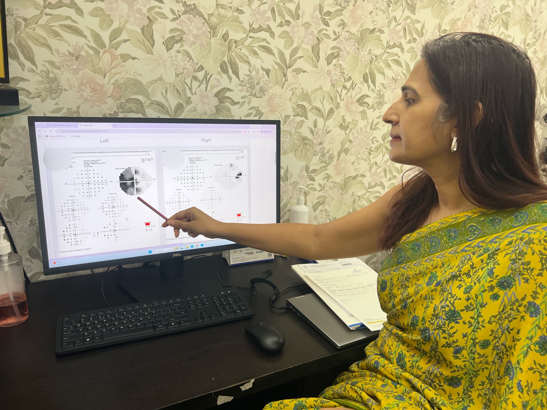
Diagnosis & Outcome: Given the incongruity between the normal ocular structures and the persistent functional loss, brain magnetic resonance imaging (MRI) was ordered. The MRI confirmed a Pituitary Adenoma causing chiasmal compression.
Key Clinical Takeaway:
Normal optic disc appearance does not rule out chiasmal compression. Unexplained, progressive unilateral vision loss—even with a history of refractive surgery—warrants comprehensive visual field mapping and neuroimaging to identify intracranial mass lesions early.
Unexplained or progressive vision loss deserves a second look beyond the eye itself.
If the findings don’t fit, consider a neuro-ophthalmology second opinion before assuming it is simply a corneal or refractive problem.