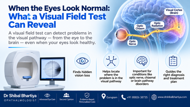
A visual field test can find vision loss that an eye chart, eye scans and sometimes a first MRI do not show. It maps your side vision in each eye, and certain patterns point to the brain, not the eye.
Most people think an eye test means reading letters on a chart. That test checks only your central vision. It says little about what you see to the sides, above and below. A visual field test, also called perimetry, checks that wider area. Sometimes it is the only test that shows something is wrong. The typical pattern of the abnormal visual field can help locate the exact problem in the vision pathway.
Why a Normal Eye Examination Can Still Miss Vision Loss
The chart tests a very small central part of your vision. You can read 6/6 and still be missing half of your side vision.
The eye examination, the pupil reaction and OCT scans of the macula and optic nerve all check the eye itself. They cannot see the pathway that carries the signal from the eye to the brain. When that pathway is damaged, the eye looks perfect and the vision is still wrong. This is why some patients are told that nothing is wrong.
The pattern of the abnormal visual field helps in localising the lesion.
A Young Man, a Heart Operation and a Headache
A young man returned to hospital about a week after an open heart surgery. He had a headache and said his vision looked distorted. He wanted to understand why his vision was distorted, and how to fix it. I can see everything, he said, but something is not right.
I saw him, and the diagnosis changed in four steps.
Step 1: The presentation.
A young patient, recent cardiac surgery, a headache and a near normal eye examination. His vision, his ophthalmologist had said, was fine. He could read numberplates of cars on his way to the hospital, but he knew something was wrong. He had had an MRI before coming to see me.
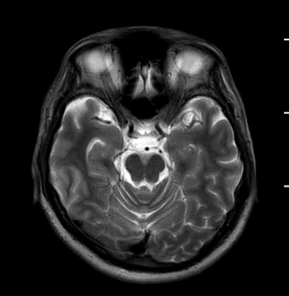
The MRI per Stroke Protocol was normal. The radiologist reviewed the MRI on my request, and yet there was nothing to report. I then ordered a visual field exam.
Step 2: The eyes examination appeared normal.
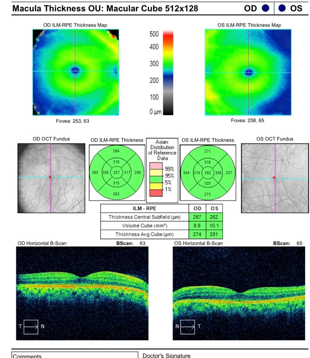
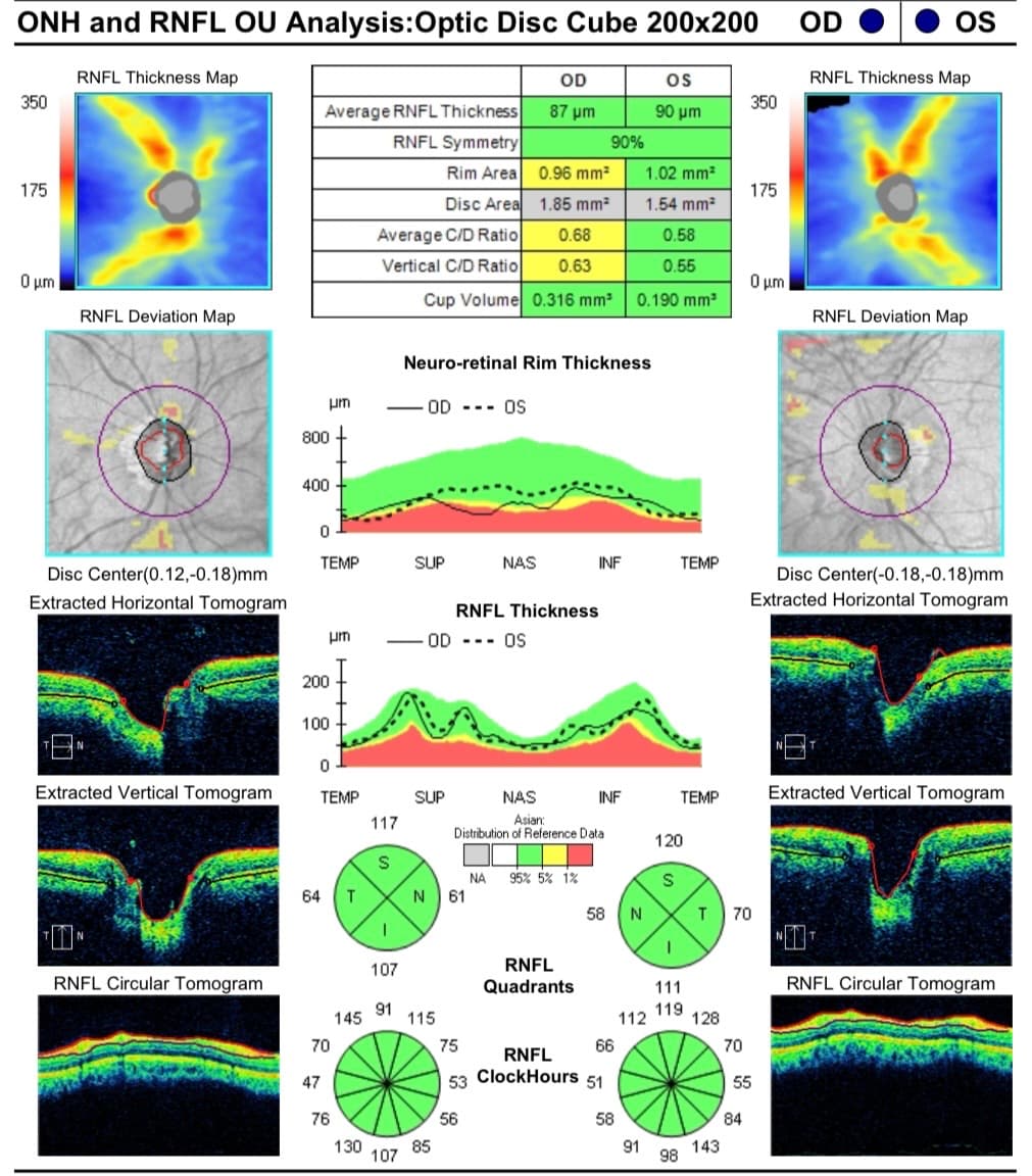
His pupils, the back of the eye, macular OCT and optic nerve OCT were normal. The Amsler charting was normal, the cornea showed no ectasia. His refractive error did not explain the symptoms.
Step 3: The visual field reveals the problem.
Unable to find a cause for his symptoms, I tried confrontation fields before asking him to go ahead with any more eye tests. Something indeed, was off. We was unable to see my pen, on the left side. I then ordered visual fields.
The fields showed a left homonymous hemianopia. The left half of his field was missing in both eyes. We repeated the fields to be sure. Same result.
That pattern, homonymous hemianopia, localises behind the optic chiasm, to the contralateral (right sided) optic tract.
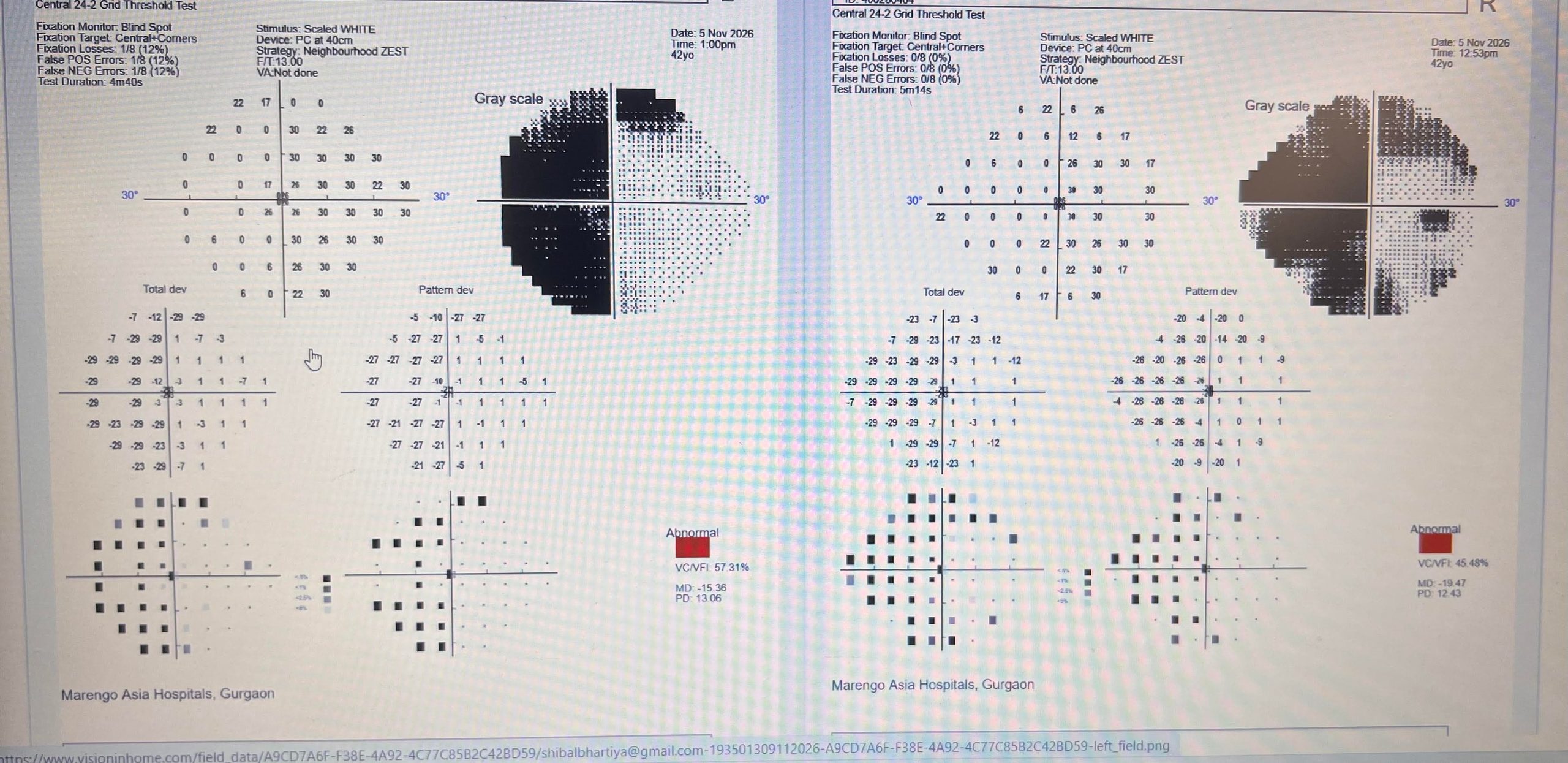
Step 4: The search moves from the eyes to the brain and its visual pathways.
The question changes. His MRI was absolutely normal. I asked the radiologist to review it with the field finding in mind. Their opinion, rightfully, remained unchanged. My working hypothesis is a small clot from the heart that briefly blocked a branch of the visual pathway. That is a presumption, not a confirmed diagnosis at all. He is recovering, and we will know more as time passes. This could be a pre-existing deficit that he only became aware of now. Or it could have been a fresh problem. There is no way of knowing for now.
How to Tell an Eye Problem From a Brain Problem
| Symptom | What It Suggests | What To Do |
|---|---|---|
| Blur that improves with glasses or a pinhole | Refractive error | Get a refraction test from an eye specialist within a week |
| Same side of vision missing in both eyes | Brain pathway problem, often a stroke | Go to an emergency department the same day for brain MRI |
| Vision missing in one eye only | Eye or optic nerve disease | See an eye specialist within 48 hours for OCT and visual field |
| Straight lines look wavy in one eye | Macula problem | Book an OCT macula scan within a few days |
| Bumping into objects on one side | Possible hemianopia, abnormal visual fields | Get a formal visual field test and a neuro-ophthalmology review |
| New headache with visual change after surgery | Possible stroke or raised brain pressure | Seek same-day medical review, not routine follow-up |
| Warped vision years after LASIK | Ectasia, dry eye or residual error | Get corneal topography from a cornea or refractive specialist |
What a Visual Field Pattern Can Tell You
| Field Pattern | What It Suggests | What To Do |
|---|---|---|
| Same side missing in both eyes (homonymous hemianopia) | Lesion behind the chiasm: optic tract, optic radiation or occipital lobe | If new, seek same-day medical review and urgent brain MRI |
| Abnormal visual fields with outer half missing in both eyes (bitemporal hemianopia) | Pressure on the optic chiasm, such as a pituitary tumour | Get a neuro-ophthalmology review and pituitary MRI within days |
| Upper quarter missing on the same side in both eyes | Temporal lobe pathway | Get a brain MRI and neurology review |
| Arc-shaped loss, often different in each eye | Optic nerve damage, commonly glaucoma | Get OCT and a glaucoma evaluation as soon as possible |
| Central blind spot in one eye | Optic neuritis or macular disease | See an eye specialist within 48 hours for OCT and a neuro-ophthalmic exam |
| Upper or lower half missing in one eye | Ischaemic optic neuropathy or a retinal vessel blockage | Get same-day eye evaluation and a vascular risk check |
Please read Can a Visual Field Test Show a Problem in My Brain?
What a Visual Field Test Cannot Tell You
A visual field shows where vision is lost. It does not show why. The same pattern can come from a stroke, a tumour, bleeding or inflammation. MRI, vascular tests and a heart assessment still matter.
The test also depends on your attention. Tiredness and unfamiliarity can change the result, so I repeat abnormal fields. A normal field does not rule out every problem. The field and the MRI answer different questions, and the best diagnoses use both.
Why This Kind of Vision Loss Gets Missed
- A chart reading can look nearly normal, which reassures everyone.
- Patients rarely say “half my vision is gone.” They say the vision feels odd or distorted.
- Normal eye scans suggest a healthy eye, so the search can stop there.
- After surgery or illness, the team focuses on the main problem, and no one orders a field.
- A normal first MRI can give false comfort when the field says otherwise.
My Approach to Visual Field Testing and Neuro-Ophthalmology
I am fellowship-trained in neuro-ophthalmology and glaucoma. At my clinic in Gurgaon, I use automated perimetry, including Octopus and MRF. I co-authored a 2026 paper in Frontiers in Ophthalmology comparing MRF-Web with Octopus 600 perimetry (doi 10.3389/fopht.2026.1961375).
I read every field alongside the OCT scans and the patient’s history. The pattern alone means little without that context.
When to See a Doctor
See a Doctor Urgently
- Sudden loss of half your vision, or of your side vision
- New headache with any change in vision
- Weakness, trouble speaking, facial droop or confusion with a vision change
- Vision change soon after surgery or a head injury
Go to an emergency department the same day. Do not wait for a routine appointment.
See a Doctor
- Bumping into objects on one side
- Missing words on one side of a page
- Distorted vision that persists with correct glasses
- Told your eyes are normal, yet the symptoms continue
Book an eye specialist within a week and ask for a visual field test.
What Happens During a Visual Field Test?
You sit at a bowl-shaped machine and press a button when you see small lights. It takes a few minutes per eye and is painless. I explain the full process in Visual field test: what to expect.
Frequently Asked Questions
Can a visual field test detect brain problems?
Yes. Certain field patterns point to the brain pathway behind the eye. The test cannot name the cause, but it tells doctors where to look. MRI and other tests then find the reason.
Can the visual field be abnormal when the MRI is normal?
Yes. A small or early lesion may not show on a first scan. A radiologist also looks more closely when told the field pattern. Repeat or targeted imaging is sometimes needed. Some field loss also comes from the eye or optic nerve, which MRI may not show.
What is homonymous hemianopia?
It means the same half of the visual field is missing in both eyes. A left homonymous hemianopia means the left half is lost in each eye. It usually points to a problem behind the optic chiasm. Stroke is the most common cause in adults.
Can vision recover after homonymous hemianopia?
It depends on the cause and the extent of damage. Some people improve, some recover partly and some do not. Most change happens in the first weeks to months. Repeat visual fields track progress, and your neurologist and eye specialist can give you a realistic picture.
Has Your Vision Felt Wrong When Your Eyes Were Called Normal?
If you were told your eyes are fine but your vision still feels distorted or incomplete, book a visual field test. This applies especially after surgery, a stroke or a head injury. Bring your MRI reports and discs.
I will examine your eyes, review your OCT scans and perform perimetry. If the pattern points to the brain, I will coordinate with your neurologist and radiologist.
Book an Appointment:
📍 Gurgaon 📞 +91 88826 38735 | 🌐 Contact Us | Second Opinion Form for teleconsults
This page is part of the Vision Symptoms hub. Read more about Neuro-Ophthalmology and about our full approach to neurological vision conditions. Request an urgent appointment if you’ve experienced unexplained vision loss. Read more at Vision Feels Off — When Normal Tests Miss the Real Problem, One Eye Feels Different, Vision Not Clear But Tests Normal, Why Do I See Well in Clinic, but Struggle in Real Life?, Vision Worse in Dim Light or Crowded Spaces, Glaucoma Visual Symptoms in Real Life, and Why Good Vision Does Not Always Mean Safe Vision.
To know more about visual fields, read What to Expect and What the Results Mean, How To Do Visual Fields, Glaucoma Test Results Explained: OCT, Visual Fields and Eye Pressure, Why Do I Need a Visual Field, and Neurological Visual Fields.
About the Author
This article was written by Dr Shibal Bhartiya, fellowship-trained glaucoma specialist and Mayo Clinic Research Collaborator, Clinical Director at Marengo Asia Hospitals, Gurugram, known for ethical, patient-centred glaucoma care and independent glaucoma second opinions. She is also the Program Director for Community Outreach & Wellness; and for the Marengo Asia International Institute of Neuro and Spine.
Dr Bhartiya has published peer-reviewed research onglaucoma management, examining how treatment decisions should balance medical evidence, patient preferences, and long-term vision outcomes. She is also a committed patient advocate and mainstream medical columnist, translating complex ophthalmic evidence into actionable public health insights across leading national platforms including NDTV and OnlyMyHealth
Dr Bhartiya is the Editor-in-Chief of Clinical and Experimental Vision and Eye Research and Executive Editor of the Journal of Current Glaucoma Practice (Pubmed Indexed, official journal of the International Society of Glaucoma Surgery). Her 200+ publications, including 90+ PubMed-indexed publications and 28 edited textbooks span glaucoma biology, surgical outcomes, health equity, and emerging diagnostics.
1700+ Five Star Patient Reviews Google Business Profile
If you are unable to come to Dr Bhartiya’s clinic: Read more about teleconsultation
Read her research on PubMed | Google Scholar | ResearchGate | ORCID | Github | Wikidata
📋Upload your reports for a structured review.| 🌐 Contact Us| 📞 +91 88826 38735
⭐ Helped by this article? Leave a Google review — it helps other patients find reliable eye care.
Last Reviewed: November 2026