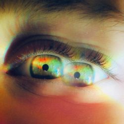
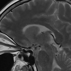
A sixth nerve palsy causes sudden horizontal double vision that forces an unconscious compensatory head turn — and that head turn eliminates peripheral vision on one side, disrupts gait, and dramatically raises fall risk. Comprehensive neuro-ophthalmic evaluation must assess the whole patient in motion, not just eye movement angles on a grid.
Lateral Rectus Palsy, and Double Vision
A clinical story about what happens when the eyes stop working together — and the body pays the price
He shuffled into my neuro-ophthalmology clinic, almost sideways. In his usual impeccable safari suit, and flip flops. His head was turned sharply to one side. He moved slowly, one hand reaching for the wall, the other braced against nothing in particular.
A recently developed an acute sixth nerve palsy — a paralysis of the lateral rectus muscle that locks the eye inward and produces constant horizontal double vision. He could not judge floor-level changes. Nor could not read the room around him. He could not navigate even familiar spaces without bracing for the next step.
He had come to get his eyes checked. I was trying to understand whether he could get safely from my clinic to his front door.
What a Sixth Nerve Palsy Actually Does to the Body
An abducens palsy — paralysis of the sixth cranial nerve — is, on paper, a problem with one eye muscle.
In practice, it reorganises how a person moves through the world.
The lateral rectus muscle turns the eye outward. When it fails, it limits outward movement of the eye. The brain receives two conflicting images, side by side, with no way to reconcile them. The resulting double vision is not subtle. It is immediate, disorienting, and constant.
The body responds the way it always does when vision becomes unreliable. It compensates. The difficult part is that compensation often looks like coping. Patients may appear functional because they move more slowly, restrict head movement, avoid crowds, or stop going out altogether. By the time family members notice, daily life may already have narrowed substantially.
Patients adopt an unconscious head turn toward the affected side — a way of rotating the eyes into a position where the weak muscle is less stressed and the two images partially overlap. It works, to a degree. The double vision softens. Binocular fusion becomes possible again in a narrow arc of gaze.
But the head turn extracts a price.
Turning the head sharply to one side can significantly reduce awareness of the opposite side and narrow the functional field available for navigation. The field of view narrows. Spatial awareness collapses. The brain’s internal gait system — which depends on both eyes working in concert to judge depth, distance, and floor-level change — loses some the information it needs.
The result is not just double vision.
The result is a patient who cannot see the kerb coming. Cannot see the step. Cannot see the chair leg, the child, the uneven tile.
Add loose footwear to that picture and the risk is no longer theoretical.
The Clinical Evaluation That Changes the Outcome
Standard clinical assessment of a sixth nerve palsy focuses on the angle of deviation — how far the eye has drifted, what prism power neutralises the misalignment, whether the palsy is complete or partial.
This is necessary. It is not sufficient.
Mr Ghosh’s chart told me he had a right sixth nerve palsy of recent onset, likely microvascular in origin — the kind that occurs in older patients with diabetes or hypertension when small vessel disease affects the nerve’s blood supply. It was medically straightforward. The natural history of microvascular palsies is usually reassuring: most resolve over weeks to months with careful monitoring.
What his chart did not tell me was that he was a fall waiting to happen.
I looked at how he entered the room. How he braced. Where his weight was. What he was wearing on his feet. How his head was positioned relative to his shoulders. How far his compensatory turn was taking him away from his functional visual field.
A targeted protocol followed.
Temporary press-on Fresnel prism eliminated the double vision and allowed him to hold his head straight. Gait coaching addressed the movement patterns he had already built around his misaligned vision. His footwear was reviewed directly and practically — flip-flops and a lateral rectus palsy are not a combination anyone should be sent home with.
We discussed how he could patch an eye, alternately. That would eliminate the diplopia, but he would struggle with depth perception.
We did not correct the nerve deficit. Time does that, usually. We protected his independence.
Microvascular Sixth Nerve Palsy: What Patients and Families Should Know
Microvascular cranial nerve palsies are among the most common causes of sudden double vision in adults over sixty. They occur when small vessel disease — most often related to diabetes, hypertension, or both — interrupts blood supply to the sixth, fourth, or third cranial nerve.
The onset is sudden. The double vision is immediate and often alarming. Many patients arrive at emergency departments suspecting stroke.
The prognosis, in uncomplicated microvascular cases, is generally good. Most palsies begin to improve within six to eight weeks and resolve fully within three to six months. But the weeks between onset and resolution carry real risk — and that risk is not always addressed at the point of diagnosis.
A patient sent home with a note saying “wait and watch” may be sent home without anyone asking how they will navigate their stairs.
When Should You Seek Neuro-Ophthalmic Evaluation?
Sudden double vision in an adult always warrants prompt evaluation.
Most microvascular palsies are benign and self-resolving. But sudden diplopia can also signal something that requires urgent attention — a posterior communicating artery aneurysm, a demyelinating event, raised intracranial pressure, or a space-occupying lesion. The clinical history, the pattern of the palsy, associated symptoms, and targeted imaging guide the differential.
Do not wait to see whether the double vision improves on its own before seeking assessment.
And if you are already under care for a sixth nerve palsy — ask about fall risk. Ask about footwear. Ask about what you should and should not be doing while the nerve recovers.
Dr Shibal Bhartiya is a fellowship-trained glaucoma specialist and Mayo Clinic Research Collaborator with over 25 years of experience. She trained in the department of Clinical Neurosciences at the University of Geneva Hospitals. Her approach focuses on identifying risk before damage is irreversible, simplifying treatment decisions, and protecting vision long-term. Emphasis on early detection, risk assessment, and continuity of care. She is rated 5 stars across 1,500+ patient reviews on Google.
Sudden Double Vision: When Not to Wait
Seek urgent assessment if double vision occurs with:
- drooping eyelid
- severe headache
- unequal pupils
- weakness or numbness
- facial asymmetry
- recent trauma
- worsening imbalance
FAQs:
Why does an eye muscle problem cause balance and walking difficulties?
A sixth nerve palsy creates a mismatch between the images the two eyes send to the brain. To reduce the double vision, patients unconsciously turn their head toward the affected side. That head turn eliminates peripheral vision on the opposite side and disrupts the visual information the brain uses to coordinate gait and judge depth.
Balance depends partly on binocular vision, together with vestibular input and proprioception. When binocular vision suddenly becomes unreliable, mobility often becomes less stable.
Can double vision cause falls in older patients?
Yes — and fall risk is significantly underestimated in patients with acute diplopia. The combination of sudden double vision, compensatory head turn, narrowed peripheral field, and disrupted depth perception is particularly dangerous in older patients with other balance vulnerabilities. Footwear, home environment, and gait patterns all need direct review alongside the eye examination.
What non-surgical options exist for acute sixth nerve palsy?
Temporary Fresnel prism lenses applied to existing glasses can realign the images and restore single comfortable vision within the primary gaze. These are combined with patching protocols where needed. Most microvascular palsies resolve naturally over weeks to months. Surgery is rarely required and is typically considered only if the palsy persists beyond six months without significant recovery.
How long does a microvascular sixth nerve palsy take to resolve?
Most microvascular palsies begin to improve within six to eight weeks and resolve fully within three to six months. Resolution depends on blood sugar and blood pressure control in patients with diabetes or hypertension — managing the underlying condition actively supports nerve recovery.
What should I do at home while waiting for a sixth nerve palsy to recover?
Review your footwear — avoid loose, backless, or raised footwear during the recovery period. Clear trip hazards from high-traffic areas at home. Avoid driving until your specialist confirms it is safe to do so. Attend follow-up appointments regularly so that recovery can be monitored and treatment adjusted as the palsy resolves.
What is the difference between a sixth nerve palsy and other cranial nerve palsies?
The sixth nerve controls the lateral rectus — the muscle that moves the eye outward. Its palsy produces purely horizontal double vision that worsens when looking toward the affected side. A fourth nerve palsy produces vertical or diagonal double vision with a characteristic head tilt. A third nerve palsy affects multiple muscles and may involve the pupil — a pupil-involving third nerve palsy is a neurological emergency. The pattern of diplopia guides the diagnosis.
Can a sixth nerve palsy cause falls?
Yes. Sudden double vision can trigger compensatory head turning, reduce environmental awareness, and increase fall risk—especially in older adults.
Does sixth nerve palsy always need surgery?
No. Many microvascular sixth nerve palsies improve over weeks to months and are managed initially with observation, prisms, and support.
This page is part of the Neuro-Ophthalmology hub. Read about our full approach to neurological vision conditions.
About the Author
This article was written by Dr Shibal Bhartiya, fellowship-trained glaucoma specialist and Mayo Clinic Research Collaborator, Clinical Director at Marengo Asia Hospitals, Gurugram, known for ethical, patient-centred glaucoma care and independent glaucoma second opinions. She is also the Program Director for Community Outreach & Wellness; and for the Marengo Asia International Institute of Neuro and Spine.
She has published peer-reviewed research on glaucoma management, examining how treatment decisions should balance medical evidence, patient preferences, and long-term vision outcomes.
As Editor-in-Chief of Clinical and Experimental Vision and Eye Research and Executive Editor of the Journal of Current Glaucoma Practice (Pubmed Indexed, official journal of the International Society of Glaucoma Surgery), Dr Shibal Bhartiya brings editorial and research depth to every clinical decision. Her 200+ publications, including 90+ PubMed-indexed publications and 28 edited textbooks span glaucoma biology, surgical outcomes, health equity, and emerging diagnostics.
1500+ Five Star Patient Reviews Google Business Profile
If you are unable to come to Dr Bhartiya’s clinic: Read more about teleconsultation
Read her research on PubMed | Google Scholar | ResearchGate | ORCID
Upload your reports for a structured review.| www.drshibalbhartiya.com | +91 88826 38735
Leave a review on Google