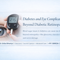
Diabetes and Eye Complications: Beyond Diabetic Retinopathy. These include glaucoma, fluctuation of power of glasses, dry eye, eye infections, cataract…
Tag: diabetes and glaucoma risk
Diabetes and Eye Complications
Diabetes and Eye Complications: Beyond Diabetic Retinopathy. These include glaucoma, fluctuation of power of glasses, dry eye, eye infections, cataract and cranial nerve palsies, explains Dr Shibal Bhartiya .
When people with diabetes think about their eyes, they think about diabetic retinopathy. This is understandable. Retinopathy is the complication that gets the attention, the one on screening programmes, the one in diabetes education leaflets, the one the endocrinologist mentions at every review.
But diabetes affects the eye in at least six distinct ways. Retinopathy is one of them. The others are less discussed, less screened for, and in some cases more immediately threatening to vision than the retinopathy that has been monitored for years. Dr Shibal Bhartiya explains.
If you have diabetes, this is the complete picture.
Dr Shibal Bhartiya is a fellowship-trained glaucoma specialist and Mayo Clinic Research Collaborator with over 25 years of experience. Her approach focuses on identifying risk before damage is irreversible, simplifying treatment decisions, and protecting vision long-term. Emphasis on early detection, risk assessment, and continuity of care. She is rated 5 stars across 1,500+ patient reviews on Google.
Diabetic Retinopathy: The Known Risk
Diabetic retinopathy is damage to the blood vessels of the retina caused by chronic high blood glucose. It is the leading cause of preventable blindness in working-age adults in India. It affects approximately one in three people with diabetes, and the risk rises steeply with duration of disease and poor glycaemic control.
The early stages cause no symptoms. This is why annual retinal screening is essential — not when vision changes, but from the time of diagnosis and consistently thereafter. By the time vision blurs or distorts, retinopathy has typically progressed beyond its most treatable stages.
Proliferative diabetic retinopathy and diabetic macular oedema are the sight-threatening endpoints. Both are treatable — with laser, with anti-VEGF injections, and with vitreoretinal surgery where needed. The outcomes are substantially better when found early.
This is the known part. Now for the rest.
Glaucoma: A Doubled Risk
People with diabetes have approximately twice the risk of developing glaucoma compared to the general population. The mechanism involves several pathways: impaired autoregulation of optic nerve blood flow, accelerated trabecular meshwork dysfunction, and higher baseline intraocular pressures.
Neovascular glaucoma — one of the most aggressive and difficult-to-treat forms — is a direct complication of advanced proliferative diabetic retinopathy. New blood vessels grow on the iris and into the drainage angle, blocking aqueous outflow and causing catastrophic pressure rises. It is largely preventable with timely retinopathy treatment. When it develops, it is extremely difficult to control.
But the more common risk is subtler: open-angle glaucoma developing alongside diabetic eye disease, often unnoticed because the patient and physician are focused on the retina. A diabetic patient whose retinopathy is well screened but whose optic nerve has never been assessed for glaucomatous damage is incompletely examined.
Every diabetic eye examination should include optic disc assessment and intraocular pressure measurement — not just retinal photography.
Intraocular Pressure Fluctuation: The Hidden Variable
Blood glucose levels directly affect intraocular pressure. This is a relationship most patients with diabetes are never told about.
When blood glucose rises acutely, the lens absorbs glucose and swells. This alters the refractive index — causing the well-known phenomenon of blurred vision during hyperglycaemic episodes. But the swelling also affects the anterior chamber geometry, which can transiently raise intraocular pressure.
More importantly, chronic hyperglycaemia affects the trabecular meshwork — the drainage tissue of the eye — through glycation of its proteins, reducing outflow facility. The result is a trabecular meshwork that drains less efficiently as glycaemic control worsens over time.
For a patient already being treated for glaucoma, poor diabetic control is not a separate issue. It is a direct threat to the stability of their eye pressure. A glaucoma patient whose pressures have been well controlled and suddenly deteriorates deserves a glycaemic history as part of the clinical review.
Diabetic Cranial Nerve Palsies: When Diabetes Affects the Nerves to the Eye
Diabetes is one of the most common causes of sudden-onset double vision in adults over 50. It does this by causing ischaemic damage to the cranial nerves that control eye movement — most commonly the third nerve (oculomotor), fourth nerve (trochlear), and sixth nerve (abducens).
A diabetic third nerve palsy typically presents as sudden onset of double vision with a drooping upper eyelid and an eye that is deviated outward and downward. Crucially, in diabetic third nerve palsy, the pupil is typically spared — an important clinical distinction from a compressive third nerve palsy caused by an aneurysm, which is pupil-involving and a neurological emergency.
Diabetic cranial nerve palsies are painful in approximately 50 percent of cases — a periorbital or frontal headache accompanying the diplopia. They typically resolve spontaneously over six to twelve weeks with good glycaemic control.
But they must be properly assessed. A new onset of double vision in a diabetic patient requires exclusion of compressive and vascular causes before a metabolic aetiology is assumed. MRI, MRA, and neurological review are part of that workup — all available on site at Marengo Asia Hospitals through the integrated neuro-ophthalmology programme.
Dry Eye in Diabetes: Underdiagnosed and Undertreated
Diabetic neuropathy affects the corneal nerves — the densest sensory nerve network in the body. As peripheral neuropathy progresses, corneal nerve density falls. The cornea loses sensation. Reflex tear secretion, which depends on intact corneal sensation, diminishes.
The result is diabetic neurotrophic keratopathy — a condition where the corneal surface is damaged not primarily by dryness in the conventional sense, but by the loss of the neural signals that maintain surface health and healing. The eye does not feel dry because sensation is impaired. But the surface is failing.
Patients with diabetic corneal neuropathy have delayed epithelial healing, increased risk of recurrent corneal erosions, and in severe cases, persistent epithelial defects that threaten sight. Standard dry eye drops provide partial relief. Advanced cases need neurotrophic agents — cenegermin eye drops, autologous serum tears — and specialist management.
Even before neuropathy is established, people with diabetes have higher rates of conventional dry eye disease — likely through effects on lacrimal gland function and tear film composition. In a diabetic patient with ocular surface symptoms, a proper dry eye assessment is not optional.
Lens Changes and Cataract: Earlier and Faster
People with diabetes develop cataracts earlier and progress faster than the non-diabetic population. The mechanism is well established: sorbitol accumulation in the lens through the polyol pathway under chronic hyperglycaemia accelerates lens protein glycation and opacification.
True diabetic cataract — the snowflake cataract seen in young patients with poorly controlled type 1 diabetes — is now rare with modern management. The more common presentation is earlier onset of age-related nuclear sclerotic cataract, and a higher incidence of posterior subcapsular cataract in patients on long-term steroid therapy for diabetic complications.
Cataract surgery in diabetic patients carries specific considerations. Pre-existing retinopathy may worsen after surgery. Macular oedema can be precipitated or exacerbated. Intraocular lens power calculations are less predictable in eyes with a history of significant refractive fluctuation. These factors require careful pre-operative assessment and post-operative monitoring that goes beyond standard cataract care.
What Comprehensive Diabetic Eye Care Actually Looks Like
Most diabetic patients receive retinal screening. Fewer receive comprehensive diabetic eye care. The difference matters.
Comprehensive diabetic eye care includes retinal assessment for retinopathy and macular oedema, optic disc assessment and intraocular pressure measurement for glaucoma, corneal sensitivity and surface assessment for neuropathic dry eye, lens examination for cataract staging, and neurological assessment if any diplopia or unexplained visual change is present.
It also includes a conversation about glycaemic control as a direct determinant of eye outcomes — not as a general health recommendation, but as a specific clinical variable that affects IOP stability, retinopathy progression, nerve recovery, and surgical outcomes.
If your diabetic eye examination has only ever included retinal photography, you have been partially screened. The rest of the eye has not been assessed. And the rest of the eye is where several of the most sight-threatening complications of diabetes begin.
Dr Shibal Bhartiya’s Experience with Diabetic Eye Diseases
Dr Shibal Bhartiya’s PubMed-indexed research, including work conducted at AIIMS, New Delhi, explores how diabetes affects the eye beyond what is immediately visible. Her studies on panretinal photocoagulation and its impact on the retinal nerve fibre layer and optic nerve highlight how structural changes can occur over time in diabetic retinopathy: even before patients notice symptoms. More recent work engaging with emerging metabolic therapies,including GLP-1–based pathways in collaboration with colleagues at Mayo Clinic, reflects the evolving understanding of how systemic diabetes treatments may influence eye health. Together, this body of work reinforces a key message for patients: diabetic eye disease is often silent, can involve both the retina and optic nerve, and requires early detection and regular monitoring to prevent irreversible vision loss.
Frequently Asked Questions
Does diabetes always cause eye problems?
Not inevitably, but the risk is significantly elevated across multiple conditions — retinopathy, glaucoma, cataract, dry eye, and cranial nerve palsies. Duration of diabetes and quality of glycaemic control are the strongest determinants of risk. Annual comprehensive eye examination is essential regardless of whether symptoms are present.
How does diabetes cause double vision?
Diabetes can cause ischaemic damage to the cranial nerves controlling eye movement, most commonly the third, fourth, or sixth nerve. This typically presents as sudden onset double vision, sometimes with eye pain or lid drooping. It usually resolves over weeks to months with improved glycaemic control, but requires proper investigation to exclude other causes.
Can improving blood sugar control help glaucoma?
Poor glycaemic control worsens trabecular meshwork function and increases IOP fluctuation. Better control supports outflow facility and IOP stability. It is not a replacement for glaucoma treatment but is a clinically significant variable in managing IOP in a diabetic patient with glaucoma.
Why do my glasses prescription keep changing with diabetes?
Fluctuating blood glucose causes the lens to swell and change its refractive index. This means your prescription changes with your glucose levels. A glasses prescription taken during a period of poor glycaemic control is unreliable. Refraction should ideally be performed when glucose levels have been stable for several weeks.
What is diabetic corneal neuropathy?
Diabetic peripheral neuropathy affects the corneal nerves, reducing corneal sensation and impairing the neural signals that maintain surface health. The result is a poorly healing corneal surface despite apparently normal or near-normal tears. It requires specialist assessment and specific treatment beyond standard dry eye drops.
How often should someone with diabetes have an eye examination?
Annually from the time of diagnosis, regardless of symptoms or current vision. If retinopathy, glaucoma, or other findings are present, more frequently as directed. Never less than annually. Vision that feels normal does not mean the eye is healthy. This is the central message of diabetic eye care.
About the Author
This article was written by Dr Shibal Bhartiya, fellowship-trained glaucoma specialist and Mayo Clinic Research Collaborator, Clinical Director at Marengo Asia Hospitals, Gurugram, known for ethical, patient-centred glaucoma care and independent glaucoma second opinions. She is also the Program Director for Community Outreach & Wellness; and for the Marengo Asia International Institute of Neuro and Spine.
She has published peer-reviewed research on glaucoma management, examining how treatment decisions should balance medical evidence, patient preferences, and long-term vision outcomes.
As Editor-in-Chief of Clinical and Experimental Vision and Eye Research and Executive Editor of the Journal of Current Glaucoma Practice (Pubmed Indexed, official journal of the International Society of Glaucoma Surgery), Dr Shibal Bhartiya brings editorial and research depth to every clinical decision. Her 200+ publications, including 90+ PubMed-indexed publications and 28 edited textbooks span glaucoma biology, surgical outcomes, health equity, and emerging diagnostics.
1500+ Five Star Patient Reviews Google Business Profile
If you are unable to come to Dr Bhartiya’s clinic: Read more about teleconsultation for glaucoma
Read her research on PubMed | Google Scholar | ResearchGate | ORCID
Upload your reports for a structured review.| www.drshibalbhartiya.com | +91 88826 38735
Leave a review on Google
Glaucoma and Diabetes
Diabetes affects the entire body, and the eyes are no exception. The high blood sugars, along with insulin resistance, damage the small blood vessels. In fact, diabetes increases the incidence and rate progression of both, cataract and glaucoma.

Glaucoma: Are you at risk?
Glaucoma usually has no symptoms in its early stages. By the time a patient notices something is wrong, significant and…