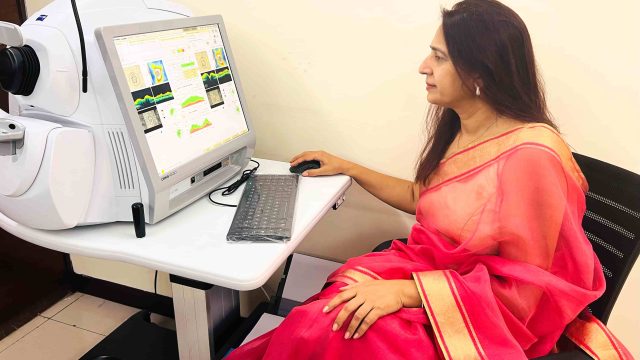
Dr Shibal Bhartiya’s Gurgaon clinic at Marengo Asia Hospitals offers OCT of the retina, optic nerve, angle and cornea on one Zeiss Cirrus 6000. A glaucoma specialist reads each scan alongside your visual field and eye examination.
What Is an OCT Scan?
OCT stands for optical coherence tomography. It is a quick, painless scan that photographs the layers of your eye with light.
Think of it as a cross-section of the eye. No needle, no contact and no radiation are involved.
The scan measures thickness. It shows whether the nerve fibres and the cells that feed them are thinner than expected.
The same machine also scans the front of your eye. That covers the cornea, the iris and the drainage angle.
What Does the OCT Scan Measure?
| What is measured | What it tells us |
|---|---|
| Retinal nerve fibre layer (RNFL) | The thickness of the nerve fibres around your optic nerve |
| Ganglion cell layer | The thickness of the cells that send signals to the optic nerve |
| Optic nerve head | The size and shape of the nerve and its central cup |
| Macula | The health of the central retina, including swelling or fluid |
| Anterior chamber and drainage angle | How open or narrow the angle is, where fluid leaves the eye |
| Iris shape and position | Whether the iris sits forward and crowds the angle |
| Corneal thickness (pachymetry) | A thickness map of the cornea, used to interpret eye pressure |
What Is an Anterior Segment OCT?
An anterior segment OCT, or AS-OCT, scans the front of your eye. It images the cornea, the iris and the angle where fluid drains out.
The scan does not touch your eye. It shows the angle as a cross-section we can measure and compare over time.
At her clinic in Gurgaon, Dr Bhartiya uses AS-OCT alongside gonioscopy, not instead of it. Read more about narrow angle glaucoma.
How Does OCT Measure Corneal Thickness?
The cornea module maps the thickness of your cornea across its surface. Corneal thickness changes how accurately a pressure reading reflects your true eye pressure.
Read what thickness means for glaucoma on our corneal thickness (pachymetry) page.
Who Needs an OCT Scan?
You may need one if you have any of these:
- Glaucoma, or a suspicion of glaucoma
- A family history of glaucoma
- High eye pressure
- Narrow angles, or a risk of angle closure
- Eye pressure readings that need a corneal thickness check to interpret
- A suspicious or asymmetric optic nerve on examination
- Unexplained blurred or distorted central vision
- A neurological condition affecting the optic nerve
- Diabetic Macular Edema (swelling of retina)
- Age Related Macular Degeneration (AMD)
- Central Serous Retinopathy
- Macular hole
If you already have glaucoma, repeat scans show whether the nerve is stable. Change over time matters more than any single reading.
What Happens During the Scan?
You sit at the machine and rest your chin on a support. You look at a light for a few seconds while the scanner works.
Each eye takes only a few moments. Most scans need no dilating drops. A small pupil or a dense cataract can limit image quality, and we may then dilate your eyes.
You can return to normal activity straight after.
TIP: The optometrist will ask you to focus on the centre of the green star. You will see a red laser cross in front of your eyes, do not look at that. Keep your eyes on the centre of the green star, and the test will be over in less than a minute.
Why Does Who Reads the Scan Matter?
A scan produces numbers and colour maps. The numbers need context.
A normal scan does not rule out glaucoma. An abnormal scan does not confirm it. High myopia, tilted discs and cataract can all flag a healthy eye as abnormal.
At her clinic in Gurgaon, Dr Bhartiya reads your OCT with your visual field, eye pressure, corneal thickness and gonioscopy. The scan is one piece of the picture.
The same applies to the front of the eye. An angle scan means little without your pressure, your gonioscopy and your risk of angle closure.
Why Should I Repeat the Scan on the Same Machine?
Scans from different machines are not directly comparable. Each machine uses its own measurement method and reference values.
Follow-up on the same Zeiss Cirrus 6000 lets us compare like with like. If you have older scans from another centre, bring them. We use them as a starting point, not as a direct match.
What Are the Limits of OCT?
OCT tests structure. It does not test how well you see.
That is why glaucoma care also needs a visual field test. Read how the two tests work together in OCT and visual field testing. Our visual field test (perimetry) in Gurgaon page explains the second half.
Frequently Asked Questions
Does an OCT scan hurt?
No. Nothing touches your eye. You just have to focus on a green light for a few seconds, and hold the blink.
Do I need eye drops before an OCT scan?
Usually not. We use dilating drops only when a small pupil or cataract blocks a clear image.
Can an OCT scan diagnose glaucoma on its own?
No. OCT shows structure. Diagnosis needs your visual field, eye pressure, optic nerve examination and corneal thickness read together.
Can an OCT scan show whether my angle is narrow?
Yes. An anterior segment OCT shows the shape of the drainage angle. Dr Bhartiya reads it alongside your gonioscopy and eye examination.
Does OCT measure corneal thickness?
Yes. The cornea module maps corneal thickness. We use it to interpret your eye pressure readings correctly.
How often should I repeat an OCT scan?
It depends on your risk and how stable your glaucoma is. Dr Bhartiya sets the interval for you after reviewing your results.
Can I bring OCT scans from another centre?
Yes. Bring the printouts and the digital files if you have them. We use them as a reference, but we repeat the scan on our own machine for follow-up.
Is an OCT scan the same as a fundus photo?
No. A fundus photo is a surface picture of the retina. OCT shows the layers beneath the surface.
Ask Dr Bhartiya About Your Scan
Has an OCT report left you with questions? Upload your reports or book a consultation, and Dr Bhartiya will explain what the scan means for your eyes.
Book an Appointment:
📍 Gurgaon 📞 +91 88826 38735 | 🌐 Contact Us | Second Opinion Form for teleconsults
This page is a part of the Glaucoma Hub. You may want to read about Glaucoma Progression, and Risk Stratification in Glaucoma. You may want to read more about OCT and Visual Field, Glaucoma Tests Explained, Normal OCT but Vision Symptoms and How to Understand Your OCT Better.
You may wnat to read about CSR, AMD and Distorted, Wavy Lines.
About the Author
This article was written by Dr Shibal Bhartiya, fellowship-trained glaucoma specialist and Mayo Clinic Research Collaborator, Clinical Director at Marengo Asia Hospitals, Gurugram, known for ethical, patient-centred glaucoma care and independent glaucoma second opinions. She is also the Program Director for Community Outreach & Wellness; and for the Marengo Asia International Institute of Neuro and Spine.
Dr Bhartiya has published peer-reviewed research onglaucoma management, examining how treatment decisions should balance medical evidence, patient preferences, and long-term vision outcomes. She is also a committed patient advocate and mainstream medical columnist, translating complex ophthalmic evidence into actionable public health insights across leading national platforms including NDTV and OnlyMyHealth
Dr Bhartiya is the Editor-in-Chief of Clinical and Experimental Vision and Eye Research and Executive Editor of the Journal of Current Glaucoma Practice (Pubmed Indexed, official journal of the International Society of Glaucoma Surgery). Her 200+ publications, including 90+ PubMed-indexed publications and 28 edited textbooks span glaucoma biology, surgical outcomes, health equity, and emerging diagnostics.
1700+ Five Star Patient Reviews Google Business Profile
If you are unable to come to Dr Bhartiya’s clinic: Read more about teleconsultation
Read her research on PubMed | Google Scholar | ResearchGate | ORCID | Github | Wikidata
📋Upload your reports for a structured review.| 🌐 Contact Us| 📞 +91 88826 38735
⭐ Helped by this article? Leave a Google review — it helps other patients find reliable eye care.
Last Reviewed: November 2026