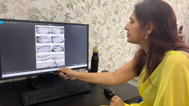
Meibography is a non-contact imaging test that visualises the meibomian glands inside the eyelids and helps detect gland loss or dysfunction. It is commonly used in the evaluation of dry eye disease and Meibomian Gland Dysfunction (MGD). The meibomian glands produce the outer oily layer of the tear film, which helps keep tears from evaporating too quickly. When these glands become blocked or damaged, tears evaporate faster, leading to dry eye symptoms such as irritation, burning, fluctuating vision, and even watering.
In this article, I explain what meibography actually shows, how to read your own report in plain language, when the findings mean you need more than artificial tears, and what treatment looks like once we know exactly which glands are affected.
Meibography: The Eyelid Scan That Finally Explains Your Dry, Gritty Eyes
Most patients I see for chronic dry eye have already tried a shelf full of lubricating drops. Some have used them for years, with results that fade within an hour. When I turn their eyelids inside out and take a quick infrared image, the reason usually becomes visible on the screen in front of them: glands that should look like neat, feathery strands are patchy, shortened, or missing entirely.
That image is called a meibography. It is a painless, light-based scan of the meibomian glands, the tiny oil-producing glands lining your eyelids. These glands supply the oily layer that stops your tears from evaporating too fast. When they are damaged or blocked, drops can only ever offer temporary relief, because they never address the missing oil layer.
Quick Answer: Meibography is a non-invasive infrared imaging test that photographs the meibomian glands inside your eyelids to show how many are intact, shortened, or lost (“dropout”). It takes under five minutes, needs no anaesthesia, and gives a direct, visual explanation for evaporative dry eye that a slit-lamp exam alone can miss. It does not hurt, and it changes how dry eye is treated by targeting the actual gland damage instead of only masking symptoms.
What Your Meibography Report Is Telling You
Meibography findings are usually reported as a percentage of gland loss per eyelid, along with a description of gland shape and structure. Here is what the common findings mean and what I typically recommend for each.
| Finding on Meibography | What It Means | What To Do About It |
| Partial gland dropout (under 25%) | Early gland damage. Oil output is reduced but many glands still function. | Start daily warm compresses and lid hygiene now, before more glands are lost. |
| Moderate dropout (25–50%) | Roughly half the oil-producing capacity in that eyelid is gone. | Add in-office thermal pulsation or IPL therapy alongside home lid care. |
| Severe dropout (over 50%) | Most glands in that lid no longer produce oil; drops alone will not fix this. | Discuss gland-targeted procedures and prescription anti-inflammatory therapy at your next visit. |
| Gland shortening or truncation | Glands are present but have retracted, so they release less oil per blink. | Consistent warm compress therapy can slow further shortening; recheck imaging in 3–6 months. |
| Tortuosity (twisted glands) | Chronic low-grade inflammation has distorted normal gland architecture. | This often responds well to anti-inflammatory drops; ask about a short prescription course. |
| Duct dilation | Glands are blocked and backing up with thickened oil, an early MGD sign. | Warm compresses to melt the blockage, plus lid margin cleaning twice daily. |
| Asymmetric dropout between the two eyes | One eye is doing more of the work, which is why symptoms often feel one-sided. | Mention any one-sided rubbing, contact lens habits, or prior lid injury at your visit. |
| Normal gland architecture despite symptoms | The dryness likely comes from a different layer of the tear film, not the oil glands. | Ask about aqueous-deficient dry eye testing, such as tear osmolarity or a Schirmer test. |
Symptoms That Usually Prompt a Meibography Scan
I order meibography when a patient’s symptoms suggest evaporative dry eye rather than a simple lack of tears. These are the patterns that most often correlate with visible gland loss on imaging.
| Symptom | What It Means | What To Do About It |
| Burning that worsens through the day | Classic pattern of oil-layer deficiency, since tears evaporate faster as glands fail. | Book a meibography scan rather than switching drop brands again. |
| Eyes feel fine on waking, worse by evening | Suggests the tear film cannot hold up under a full day of blinking and screen use. | Track your symptom timing and mention it during your evaluation; it helps target testing. |
| Watery eyes that still feel dry | Reflex tearing from an unstable oil layer, not true excess tear production. | Do not stop lubricants on your own; get imaging first to confirm the cause. |
| Contact lens discomfort after previously tolerating lenses well | A common early sign of meibomian gland changes, especially after years of lens wear. | Have your glands imaged before assuming you simply need a lens change. |
| Redness and crusting along the lash line | Suggests blepharitis is contributing to, or driving, the gland dysfunction. | Start lid hygiene immediately and get imaging to check gland involvement. |
| Fluctuating or blurry vision that clears with blinking | An unstable tear film scatters light unevenly across the cornea. | Flag this specifically; it points more toward tear film instability than a refractive issue. |
| Gritty, foreign-body sensation on waking | Can reflect overnight lid margin inflammation affecting the glands. | Ask whether overnight ointment or a warm compress routine before bed would help. |
When To See a Doctor Rather Than Self-Treat
Most dry eye is manageable, but certain signs need a same-week ophthalmology visit rather than another bottle of drops:
- Unilateral symptoms: this affects one eye clearly more than the other, especially if it came on suddenly.
- Pain, redness, warmth, or fever: accompanies dryness; this is not typical MGD and needs urgent assessment.
- Any vision change: blurring, halos, or a drop in clarity that does not clear with blinking or lubricant drops.
- Proptosis: or bulging of one eye, which can signal a thyroid or orbital problem, not simple dry eye.
- Any of these signs in a child. Persistent eye rubbing, redness, or lid crusting in children needs a paediatric eye exam, not adult dry eye products.
- New onset of dryness after starting a new medication, including antihistamines, antidepressants, isotretinoin, or blood pressure drugs.
- Systemic signs: swelling, joint pain, or dryness in your mouth alongside your eyes, which can point to an autoimmune cause such as Sjögren’s syndrome.
📍 Gurgaon 📞 +91 88826 38735 | 🌐 Contact Us | Second Opinion Form for teleconsults
What You Can Start at Home Before Your Appointment
These measures are evidence-based and safe to begin immediately, whether or not gland damage turns out to be significant:
- Warm compresses: a clean, damp washcloth over closed lids for 10 minutes, once or twice daily, to soften and mobilise thickened gland oil.
- Lid margin hygiene: with a lid-safe cleanser to reduce bacterial load and debris along the gland openings.
- The 20-20-20 rule: every 20 minutes, look at something 20 feet away for 20 seconds; this alone increases blink rate and reduces evaporation.
- Omega-3 supplementation: has reasonable evidence for improving oil quality; discuss dose with your doctor before starting.
- A humidifier in your bedroom or workspace: dry indoor air, especially with air conditioning or heating, accelerates tear evaporation.
- Conscious full blinking during screen use: many people blink incompletely while reading screens, which worsens gland stagnation over time.
Medical and Treatment Options Once Gland Damage Is Confirmed
Office-Based Gland Therapy
Thermal pulsation devices and IPL (intense pulsed light) therapy target glands that are blocked but still structurally present. IPL also reduces the inflammation that drives further gland loss, which is why I often recommend it early rather than waiting for damage to progress.
Prescription Anti-Inflammatory Therapy
Topical azithromycin or a short course of oral doxycycline reduces inflammation and improves oil quality. Cyclosporine or lifitegrast drops address the inflammatory component of dry eye directly and are useful when meibography and aqueous tear testing both show abnormalities.
Monitoring Gland Loss Over Time
Because meibography is objective and repeatable, I use it to track whether a treatment plan is actually preserving glands, not just easing symptoms for a few weeks. Repeat imaging at 3 to 6 months tells us whether we are winning against further dropout.
Frequently Asked Questions
Does meibography hurt?
No. It uses infrared light, not a laser or needle. Your eyelid is gently everted and the imaging takes a few seconds per eye, with no anaesthesia and no discomfort beyond the brief eversion itself.
Can lost meibomian glands grow back?
Meibomian glands generally do not regenerate once fully lost, which is why early detection matters. Glands that are shortened or partially damaged, however, can often be preserved and their function improved with prompt treatment.
How is meibography different from a regular dry eye exam?
A slit-lamp exam shows surface signs like redness or debris, but it cannot see inside the eyelid. Meibography images the glands directly, so it can detect structural loss even when the eye surface still looks relatively normal.
Is meibography covered by insurance in India?
Coverage varies by insurer and policy. Discuss this with your clinic’s billing team before your visit; the scan itself is quick and the out-of-pocket cost is not high.
How often should meibography be repeated?
For active gland disease, I typically repeat imaging every 3 to 6 months to check whether treatment is preserving gland structure. Once findings are stable, annual imaging is usually sufficient.
Can meibography explain contact lens intolerance?
Yes, often. Long-term contact lens wear is associated with meibomian gland changes, and imaging frequently reveals gland dropout in patients who assumed their discomfort was simply lens fit or solution sensitivity.
Key Takeaways
- Meibography images the meibomian glands directly, showing gland loss that a standard eye exam cannot detect.
- The scan is painless, takes minutes, and needs no anaesthesia or dilation.
- Findings are graded by percentage of gland dropout, from mild to severe, per eyelid.
- Gland loss is often irreversible, so earlier imaging leads to better preservation of remaining glands.
- Treatment is matched to the imaging findings, from warm compresses to IPL, probing, or prescription therapy.
- Repeat imaging tracks whether treatment is actually protecting your glands over time.
Please read Can Blepharitis or Rosaceae Cause Dry Eyes?
Book a Consultation
If you have been managing dry, gritty, or burning eyes with drops that stop working within the hour, meibography can show you exactly why. It gives us a clear picture of your meibomian glands and a treatment plan built around what your eyes actually need, not a generic dry eye protocol.
Dr Shibal Bhartiya has developed The C.L.E.A.R. Framework for complex and treatment-resistant dry eye cases, which applies TFOS DEWS III diagnostic principles to map your exact subtype, help with healing of the ocular surface, and restore comfort.
Book a consultation to have your glands imaged and get a treatment plan tailored to your findings.
Book an Appointment →
📍 Gurgaon 📞 +91 88826 38735 | 🌐 Contact Us | Second Opinion Form for teleconsults
This article is part of the Dry Eye Hub. Please also read Basics of Dry Eye, Dry Eye Second Opinion and Dry Eye: A Chronic Disease. Why Vision Becomes Blurred After Reading or Screen Use, and Why Are Your Dry Eye Drops Not Working may also help you understand your problem better.
You may also want to read this article written by Dr Bhartiya for NDTV online. And listen to her talk about dry eyes here.
About the Author
This article was written by Dr Shibal Bhartiya, fellowship-trained glaucoma specialist and Mayo Clinic Research Collaborator, Clinical Director at Marengo Asia Hospitals, Gurugram, known for ethical, patient-centred glaucoma care and independent glaucoma second opinions. She is also the Program Director for Community Outreach & Wellness; and for the Marengo Asia International Institute of Neuro and Spine.
As Editor-in-Chief of Clinical and Experimental Vision and Eye Research and Executive Editor of the Journal of Current Glaucoma Practice (Pubmed Indexed, official journal of the International Society of Glaucoma Surgery), Dr Shibal Bhartiya brings editorial and research depth to every clinical decision. Her 200+ publications, including 90+ PubMed-indexed publications and 28 edited textbooks span glaucoma biology, surgical outcomes, health equity, and emerging diagnostics.
1600+ Five Star Patient Reviews Google Business Profile
If you are unable to come to Dr Bhartiya’s clinic: Read more about teleconsultation
Read her research on PubMed | Google Scholar | ResearchGate | ORCID | Github
📋Upload your reports for a structured review.| 🌐 Contact Us| 📞 +91 88826 38735
⭐ Helped by this article? Leave a Google review — it helps other patients find reliable eye care.