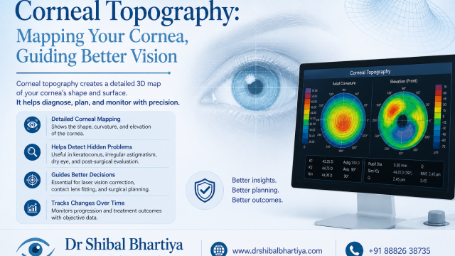
Corneal topography is a non-invasive scan that maps the shape and curvature of the front surface of your eye, the cornea. It takes less than a minute, involves no contact with the eye, and provides information that a standard eye test cannot.
If your doctor has ordered this test, it is because the shape of your cornea matters for your diagnosis, your treatment, or both.
Dr Shibal Bhartiya is a fellowship-trained glaucoma specialist and Mayo Clinic Research Collaborator with over 25 years of experience. Her approach focuses on identifying risk before damage is irreversible, simplifying treatment decisions, and protecting vision long-term. Emphasis on early detection, risk assessment, and continuity of care. She is rated 5 stars across 1,500+ patient reviews on Google.
Why the Shape of Your Cornea Matters
The cornea is responsible for approximately 65–75 percent of the eye’s total focusing power. It is not a perfect sphere, it has a natural, gentle curve that varies across its surface. Small, regular variations in this curvature cause astigmatism. Larger, irregular variations signal conditions that affect corneal integrity and can, if undetected, lead to progressive and permanent vision loss.
A standard refraction, the test that gives you your glasses prescription, tells you how well your eye focuses. It does not tell you why. Corneal topography answers the why. It maps the elevation, curvature, and power of the corneal surface at thousands of points, producing colour-coded maps that allow your doctor to identify patterns invisible to the naked eye and unmeasurable by a simple vision test.
What Happens During the Test
You will sit in front of a machine, most commonly a Placido disc topographer or a Scheimpflug imaging system such as the Pentacam. You will be asked to rest your chin on a chin rest, keep your head still, and look at a central fixation target.
The machine projects a series of illuminated rings or a rotating slit of light onto your cornea and photographs the reflection. The software analyses the distortion of these patterns across the corneal surface and calculates curvature, elevation, and power at each measured point.
The test takes under a minute per eye. It is entirely painless. No drops, no contact, no discomfort. Blinking normally is encouraged, a dry cornea produces an artefact-prone scan.
If you wear contact lenses, you will be asked to remove them before the test:
- Soft contact lenses: remove at least 24 hours before
- Rigid gas-permeable (RGP) lenses: remove at least two to four weeks before
Contact lenses temporarily alter corneal shape. Scanning over a cornea that has been moulded by a lens produces misleading results.
Why Corneal Topography Is Ordered
Keratoconus screening and monitoring
Keratoconus is a condition in which the cornea progressively thins and bulges forward into a cone shape. It typically begins in the teens or twenties, causes progressive distortion and blurring of vision, and, if undetected, can lead to scarring and the need for corneal transplantation.
Early keratoconus is invisible on a standard eye test. A refraction may show increasing astigmatism, but the pattern on topography, a characteristic inferior steepening, a skewed bowtie pattern, or elevated inferior-superior asymmetry indices, is diagnostic before any change in vision is measurable. Topography is the definitive screening and monitoring tool for keratoconus.
Pre-LASIK and refractive surgery screening
Corneal topography is mandatory before any refractive laser procedure. LASIK, SMILE, and PRK all reshape the cornea to reduce or eliminate refractive error. Performing laser surgery on a cornea with underlying keratoconus or topographic irregularity dramatically accelerates ectasia, progressive corneal thinning, and can destroy vision. Topographic screening identifies eyes that are unsafe for laser surgery before the procedure is even considered.
Dry eye and ocular surface disease assessment
The anterior corneal map reflects the quality of the tear film as well as the shape of the cornea beneath it. An irregular, patchy topography map in a patient with dry eye often reflects tear film instability rather than corneal disease. Modern topographers can perform serial scans to assess tear film breakup dynamically. This has clinical value in planning dry eye treatment and in distinguishing true corneal irregularity from surface dryness.
Contact lens fitting, especially RGP and scleral lenses
Standard soft contact lenses conform to the eye. Rigid lenses do not, they vault over the corneal surface and require precise mapping of corneal curvature to fit correctly. Corneal topography is essential for fitting rigid gas-permeable lenses, scleral lenses, and orthokeratology lenses (worn overnight to temporarily reshape the cornea and reduce myopia). Without a topographic map, fitting these lenses is guesswork.
Astigmatism planning before cataract surgery
In patients undergoing cataract surgery, particularly those choosing toric intraocular lenses to correct astigmatism, the axis and magnitude of corneal astigmatism must be measured precisely. Topography — combined with keratometry — provides this precision. Implanting a toric lens at the wrong axis produces residual astigmatism that glasses cannot fully correct.
Monitoring corneal ectasia after refractive surgery
Patients who develop ectasia after LASIK or PRK require serial topographic monitoring to assess whether the condition is stable or progressing. Corneal crosslinking, the treatment that halts progression, is initiated based partly on topographic evidence of change over time.
Glaucoma-corneal thickness interaction
Corneal thickness, measured by pachymetry, sometimes integrated into the same device as topography, affects the accuracy of intraocular pressure measurements. A thin cornea gives falsely low pressure readings; a thick cornea gives falsely high readings. In glaucoma assessment, knowing corneal thickness allows your doctor to correct IOP measurements and make more accurate risk assessments.
Understanding Your Topography Map
Topography maps use a colour scale — universally, warm colours (red, orange) represent steeper, more curved areas; cool colours (blue, green) represent flatter areas. The scale is calibrated so that small colour differences represent meaningful curvature changes.
Axial (sagittal) map: The most commonly displayed map. Shows corneal power in dioptres across the surface. The normal pattern is a roughly symmetric bowtie or round pattern of warm colour in the centre, fading to cooler colours at the periphery. Asymmetry, inferior steepening, or irregular patterns are flags for further investigation.
Tangential (instantaneous) map: More sensitive to local curvature changes than the axial map. Better for detecting early peripheral disease and small focal irregularities.
Elevation map: Shows the height of the corneal surface relative to a reference sphere. Particularly useful for keratoconus detection — the cone appears as an island of elevated tissue against the reference surface.
Pachymetry map: Shows corneal thickness at every point. The thinnest point of the cornea is normally in the centre or slightly below centre. In keratoconus, the thinnest point migrates inferiorly and is often co-located with the cone apex.
Indices: Modern topographers calculate composite indices — KISA%, Belin-Ambrosio Deviation (BAD-D), and others — that summarise whether the topographic pattern falls within normal limits or suggests subclinical disease. These indices are used for refractive surgery screening and keratoconus staging.
What Doctors Often Miss Telling You
- Contact lens warpage is one of the most common causes of a confusing topography result. If you wear contact lenses and your map looks irregular, your doctor’s first question should be how long you have been out of lenses — not whether you have keratoconus. Always remove lenses well in advance.
- A single irregular topography does not diagnose keratoconus. It raises suspicion. Diagnosis requires correlation with pachymetry, elevation data, clinical examination, and often a repeat scan after a contact lens washout period. Do not panic at a single result.
- Forme fruste keratoconus — subclinical, non-progressing topographic abnormality — is different from clinical keratoconus. It does not always need treatment. It does need monitoring and, importantly, it is a contraindication to LASIK even when the patient has no symptoms.
- Topography findings affect the whole family. Keratoconus has a genetic component. If you are diagnosed, your first-degree relatives — children and siblings — should be screened. Early detection in family members allows intervention before vision is affected.
- Dry eye can mimic corneal irregularity on topography. If your map is irregular but your vision is good and stable, a well-treated tear film may normalise the scan on repeat. Treating dry eye before refractive surgery screening produces more reliable topography.
When to Seek Assessment
Book an assessment if you notice:
- Increasing glasses prescription, particularly increasing astigmatism, that is not stabilising
- Blurred or distorted vision that glasses do not fully correct
- Difficulty with contact lens tolerance that is worsening over time
- Ghosting or halos around lights, particularly at night
- A family member recently diagnosed with keratoconus
- You are considering laser eye surgery and want a full pre-operative assessment
Frequently Asked Questions
Is corneal topography the same as a corneal scan?
Corneal topography maps the shape and curvature of the corneal surface. A corneal scan is a broader term that can include topography, pachymetry (thickness), and anterior segment OCT (structural cross-section). Modern devices like the Pentacam combine topography and pachymetry in a single scan. Your doctor will specify which measurements are needed.
Does corneal topography require eye drops?
No. Corneal topography does not require dilating or anaesthetic drops. It is completely non-invasive. However, if your doctor wants to examine your cornea in detail after the topography, they may use a slit lamp with or without drops depending on what they are looking for.
Can corneal topography detect keratoconus early?
Yes — this is one of its primary clinical roles. Topography can detect subclinical keratoconus years before symptoms or vision loss occur. This is why it is performed routinely before refractive surgery, and why it is recommended for first-degree relatives of keratoconus patients.
How often should I have corneal topography if I have keratoconus?
Frequency depends on the stage and stability of your disease. Newly diagnosed or younger patients are typically scanned every six months to assess for progression. Stable, older patients may be monitored annually. If corneal crosslinking has been performed, post-procedure scans assess the treatment response. Your specialist will advise the appropriate interval.
Will corneal topography tell me if I am suitable for LASIK?
It is one of the key tests in a pre-LASIK assessment, but suitability is determined by the full evaluation — including refraction, pachymetry, pupil size, tear film, and clinical examination. Topography alone does not clear or disqualify you for surgery. Your surgeon will interpret all findings together.
Can I drive after corneal topography?
Yes. No drops are used and your vision is not affected. You can drive, return to work, and continue your normal activities immediately after the test.
Speak to a Specialist
If you have been referred for corneal topography, have received a result you do not fully understand, or want a comprehensive pre-surgical assessment, a specialist review that integrates your topography with your full clinical picture is the right next step.
Book a consultation: 📞 +91 88826 38735 | 🌐 Contact Us | Second Opinion Form for teleconsults
Upload your topography printouts through the website before your appointment for a more focused consultation.
This article is a part of the LASIK Hub. Please also read LASIK Recovery Timeline, Are You Fit For LASIK?, Is LASIK Safe?, Bladeless LASIK, Lasik Side Effects: Incidence and Management,
Femtosecond Lasik: Bladeless Surgery, LASIK Eye Surgery: Are You a Candidate? What to Expect, and What to Ask, and PROWL: Listening to Lasik patients. Also useful is Second Opinion Before Eye Surgery
About the Author
This article was written by Dr Shibal Bhartiya, fellowship-trained glaucoma specialist and Mayo Clinic Research Collaborator, Clinical Director at Marengo Asia Hospitals, Gurugram, known for ethical, patient-centred glaucoma care and independent glaucoma second opinions. She is also the Program Director for Community Outreach & Wellness; and for the Marengo Asia International Institute of Neuro and Spine.
She has published peer-reviewed research on glaucoma management, examining how treatment decisions should balance medical evidence, patient preferences, and long-term vision outcomes.
As Editor-in-Chief of Clinical and Experimental Vision and Eye Research and Executive Editor of the Journal of Current Glaucoma Practice (Pubmed Indexed, official journal of the International Society of Glaucoma Surgery), Dr Shibal Bhartiya brings editorial and research depth to every clinical decision. Her 200+ publications, including 90+ PubMed-indexed publications and 28 edited textbooks span glaucoma biology, surgical outcomes, health equity, and emerging diagnostics.
1600+ Five Star Patient Reviews Google Business Profile
If you are unable to come to Dr Bhartiya’s clinic: Read more about teleconsultation
Read her research on PubMed | Google Scholar | ResearchGate | ORCID
📋Upload your reports for a structured review.| 🌐 Contact Us| 📞 +91 88826 38735
⭐ Helped by this article? Leave a Google review — it helps other patients find reliable eye care.