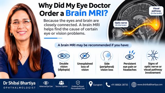
Your eye doctor orders a brain MRI when your eye findings suggest a problem along the optic nerve or visual pathway, not in the eye itself. It is not needed for all patients, and a normal scan is common and reassuring.
Why does an eye problem need a brain scan?
Hearing “you need an MRI of your brain” for an eye problem is frightening. Most people think of the eye as separate from the brain.
It is not. The optic nerve is part of the brain’s wiring. Signals travel from the retina, along the optic nerve, to the back of the brain.
A problem anywhere on that route can reduce vision. The eye can look healthy while the cause sits behind it.
An MRI shows the parts of that route I cannot see with a slitlamp.
What is my eye doctor trying to find out with the brain MRI?
A good reason for an MRI answers one of three questions:
- Where along the visual pathway is the problem?
- Does the eye explain your symptoms, or does something else?
- Does this need urgent treatment?
If the answer will not change your care, I do not order the scan.
MRI Brain and Orbits with Contrast
Sometimes I will ask for an MRI with contrast, which requires an injection of a dye to see the brain better. It requires a blood test for creatinine, which ensures your kidneys are functioning well enough to excrete the dye. The test provides detailed imaging of the brain, optic nerves, orbital pathways and surrounding structures. It helps identify intracranial tumours, inflammation, demyelination, infection, vascular lesions and compressive optic neuropathies.
MRI Brain and Orbits with Contrast is particularly useful in neuro-ophthalmology for evaluating unexplained visual loss, optic neuritis, optic nerve compression, orbital masses, orbital inflammation, and lesions involving the orbital apex or optic chiasm.
MRI Venography
Sometimes I will order a MRI Venogram, MRV, which is a non-invasive imaging technique used to visualise the intracranial venous sinuses and assess venous drainage. It is particularly useful in evaluating suspected cerebral venous sinus thrombosis, venous sinus stenosis and raised intracranial pressure, especially in patients presenting with papilloedema or unexplained visual disturbances.
When does an eye doctor order an MRI?
I consider imaging when the history and examination point beyond the eye. The common triggers are:
- Visual field loss that follows a pattern, such as loss on one side or in both outer fields
- Vision loss that the eye examination does not explain
- A swollen optic disc
- Sudden vision loss with eye pain, which may be optic neuritis
- New double vision, or a drooping eyelid
- Unequal pupils
- A pale optic nerve with no clear cause
- Glaucoma that does not behave like glaucoma
That last point matters. Glaucoma needs a second look when the patient is young, or when the nerve looks paler than it looks cupped. The same applies when the field loss does not match the nerve, or when vision drops fast.
Read more about neurological visual field defects.
What does the MRI look for?
| What I find in the eye | What the MRI looks for | When to worry |
|---|---|---|
| Field loss that respects the midline | Pituitary or other pressure on the optic chiasm, stroke, brain lesions | Prompt review. Urgent if vision is falling quickly |
| Swollen optic disc | Raised brain pressure, a mass, or clots in the brain’s veins | Same-day review |
| Sudden vision loss with pain | Optic nerve inflammation, which can link to multiple sclerosis | Urgent review |
| New double vision | Pressure on the nerves that move the eye, vessel problems | Urgent if the pupil is unequal or the lid droops |
| Pale optic nerve, no clear cause | Compression of the nerve | Soon. Compression can progress |
| Vision loss, normal eye exam | Disease behind the eye | If vision keeps worsening |
What happens during the scan?
An MRI uses magnets, not radiation. You lie still inside a tube for about 30 to 60 minutes. The machine is loud, so you wear earplugs or headphones.
Sometimes a contrast dye goes into a vein. It makes certain problems easier to see. Tell the staff if you have kidney disease.
Tell them before the scan if you have:
- A pacemaker
- A cochlear implant
- Metal fragments in your body
- Claustrophobia
- Pregnancy
Your doctor may order an MRA or MRV with it. These scans look at the arteries and veins.
Sometimes a CT scan fits better. Doctors choose CT for bleeding, injury or bone problems, and when an MRI is not safe for you.
A few things to make your MRI more comfortable
- You will be given earplugs or headphones to protect your ears from the loud noises made by the MRI machine.
- You can ask the MRI technician if music can be played during your scan.
- Please use the toilet before your scan, as you may need to lie still for some time.
- You will be given a call button to hold throughout the procedure. If you feel uncomfortable, anxious or unwell at any point, simply press it. The technician will be able to communicate with you and assist you.
Remember: You are not alone during the scan. The MRI team will be monitoring you and can help if you need anything.
What doctors often miss
- A routine brain MRI may not answer the eye question. A scan done for headaches may not take thin slices through the optic nerves or chiasm. The request must say why it was ordered.
- “Normal” can mean “not looked at.” Ask whether the optic nerves and chiasm were specifically studied.
- A normal scan does not close the case. If your vision is still falling, the eye findings still need an explanation.
- Unrelated findings appear. Scans sometimes show something that has nothing to do with your vision. Most need no treatment. Your doctor should explain each one.
Does every patient with vision loss need an MRI?
No. Most eye problems never need a brain scan. Dry eye, cataract, retinal disease and most glaucoma need examination and eye tests, not an MRI.
I order imaging when the findings call for it. I also tell you which area needs scanning and what question the scan should answer.
Read about one such case in pituitary tumours and vision loss. The cornea looked like the cause. The visual field pointed elsewhere.
When to See a Doctor
See a Doctor Urgently
- Sudden loss of vision in one or both eyes
- New double vision with a drooping eyelid or unequal pupils
- Vision loss with a severe or sudden headache
- Brief blackouts of vision, especially in older adults
- Sudden loss of side vision
See a Doctor
- Vision that keeps worsening without a clear reason
- A visual field defect that your diagnosis does not explain
- Double vision that comes and goes
- Faded colours in one eye
- An MRI request that nobody has explained to you
What this means for you
An MRI request means your doctor wants to look behind the eye. It does not mean something serious has been found.
Many scans come back normal. That result is useful because it rules things out and lets us look elsewhere.
Ask your doctor what question the scan will answer. A clear answer is a good sign that the scan is needed.
Incidental Findings on MRI: Why Every Abnormality Is Not Clinically Significant
MRI is an extremely sensitive investigation, but sensitivity comes with a trade-off: not every abnormality detected is a disease, and not every radiological finding has clinical significance. Minor anatomical variations, nonspecific white matter changes and other incidental observations are frequently encountered, sometimes in people who are entirely healthy.
Such findings may have no relationship to the patient’s presenting symptoms and may require neither treatment nor intervention. The interpretation of an MRI should therefore never be separated from the clinical examination.
Findings such as nonspecific white matter hyperintensities, small arachnoid cysts, developmental venous anomalies, partially empty sella and mild sinus mucosal thickening may be encountered during investigations for headache, visual loss or diplopia.
However, the presence of an imaging abnormality does not necessarily establish a causal relationship with the patient’s symptoms. Each finding must be interpreted in the context of the clinical history, eye examination, anatomical localisation and functional deficits.
The objective is not simply to identify an abnormality on MRI, but to establish whether that abnormality explains the patient’s clinical presentation.
Frequently Asked Questions
Does an MRI for my eyes mean I have a brain tumour?
No. Doctors order most of these scans to rule problems out. Many come back normal, or show a cause that is treatable.
Can my eye doctor tell if I need an MRI without scanning me?
Often yes. The history, pupils, eye movements and visual fields show whether the problem sits behind the eye. The scan then confirms it.
Is an MRI or a CT scan better for eye problems?
MRI shows the optic nerve and brain in more detail. CT suits emergencies, injury and bone problems.
Do I need contrast dye for an eye MRI?
Sometimes. Contrast helps show inflammation, swelling and some tumours. Your doctor decides based on the question.
Can I bring my old MRI to a neuro-ophthalmologist?
Yes. Bring the actual images on a disc, the films, and not only the report. The pictures often show more than the written summary.
Book a Consultation
Told you need an MRI and unsure why? Send me your reports and the scan request, and I will explain what the scan should answer.
Book an Appointment:
📍 Gurgaon 📞 +91 88826 38735 | 🌐 Contact Us | Second Opinion Form for teleconsults
As Program Director of Marengo Asia international Institute of Neuro & Spine, I have access to a comprehensive range of diagnostic investigations under one roof, along with interventional neurologists and radiologists on the team. This facilitates a thorough and coordinated evaluation of your condition.
This page is part of the Neuro-Ophthalmology hub. Read about our full approach to neurological vision conditions. Please also read Neuro-Ophthalmology Second Opinion. Here’s another heartening patient story: Pituitary Tumours and Vision Loss. Also read about optic nerve disease, Sixth Nerve Palsy, Facial Nerve Palsy, raised intracranial pressure, Vision not clear but tests normal, double vision, and conditions where no diagnosis has yet been reached.
About the Author
This article was written by Dr Shibal Bhartiya, fellowship-trained glaucoma specialist and Mayo Clinic Research Collaborator, Clinical Director at Marengo Asia Hospitals, Gurugram, known for ethical, patient-centred glaucoma care and independent glaucoma second opinions. She is also the Program Director for Community Outreach & Wellness; and for the Marengo Asia International Institute of Neuro and Spine.
Dr Bhartiya has published peer-reviewed research onglaucoma management, examining how treatment decisions should balance medical evidence, patient preferences, and long-term vision outcomes. She is also a committed patient advocate and mainstream medical columnist, translating complex ophthalmic evidence into actionable public health insights across leading national platforms including NDTV and OnlyMyHealth
Dr Bhartiya is the Editor-in-Chief of Clinical and Experimental Vision and Eye Research and Executive Editor of the Journal of Current Glaucoma Practice (Pubmed Indexed, official journal of the International Society of Glaucoma Surgery). Her 200+ publications, including 90+ PubMed-indexed publications and 28 edited textbooks span glaucoma biology, surgical outcomes, health equity, and emerging diagnostics.
1700+ Five Star Patient Reviews Google Business Profile
If you are unable to come to Dr Bhartiya’s clinic: Read more about teleconsultation
Read her research on PubMed | Google Scholar | ResearchGate | ORCID | Github | Wikidata
📋Upload your reports for a structured review.| 🌐 Contact Us| 📞 +91 88826 38735
⭐ Helped by this article? Leave a Google review — it helps other patients find reliable eye care.
Last Reviewed: November 2026