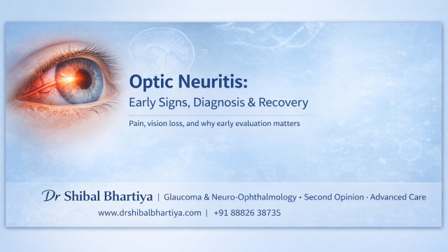
Optic neuritis is inflammation of the optic nerve, the cable that carries visual information from the eye to the brain. It most commonly affects young adults and often presents suddenly with vision loss and painful eye movements. Vision may blur over hours to days. Many patients describe:
- Dimming of vision in one eye
- Pain when moving the eye
- Colours looking washed out
- A central blur or patch
Sometimes the optic disc looks swollen. Sometimes it looks completely normal, explains Dr Bhartiya.
Dr Shibal Bhartiya is a fellowship-trained glaucoma specialist and Mayo Clinic Research Collaborator with over 25 years of experience. Her approach focuses on identifying risk before damage is irreversible, simplifying treatment decisions, and protecting vision long-term. Emphasis on early detection, risk assessment, and continuity of care. She is rated 5 stars across 1,500+ patient reviews on Google.
Why Does It Happen?
The most common cause is immune-mediated inflammation.
In many patients, optic neuritis may be associated with neurological conditions such as:
- Multiple Sclerosis
- Neuromyelitis Optica
- Myelin Oligodendrocyte Glycoprotein Antibody Disease
Sometimes it occurs in isolation without any long-term neurological disease.
Infections, autoimmune disorders, and rarely toxins can also cause optic nerve inflammation.
What Does It Feel Like?
Patients often say:
- “My vision feels dimmer, not just blurry.”
- “Colours don’t pop.”
- “It hurts when I look left or right.”
- “One eye just feels off.”
A key clue: colour vision drops disproportionately. Red especially may appear faded.
What Do We Look For During Examination?
- Reduced visual acuity
- Reduced colour vision
- Relative afferent pupillary defect (RAPD)
- Visual field defect (often central)
- Optic disc swelling (in about one-third of cases)
An MRI of the brain and orbit is usually required, not just to confirm optic nerve inflammation, but to assess neurological risk.
Is It Permanent?
This is the question everyone asks. The reassuring part: Most patients recover significant vision over weeks to months.
However:
- Contrast sensitivity may remain reduced
- Some patients notice subtle visual fatigue
- Recurrence can happen
Recovery does not always mean the nerve was untouched. It means the brain compensated well.
Does Everyone With Optic Neuritis Have Multiple Sclerosis?
No. But optic neuritis can be the first clinical event of MS in some patients.
MRI findings help estimate risk. If brain MRI shows no demyelinating lesions, long-term MS risk is lower. If lesions are present, risk is higher.
This is why careful evaluation matters: not panic, not dismissal.
When Should You See an Eye Specialist Urgently?
- Sudden visual loss in one eye
- Eye pain with movement
- Colour fading
- Visual dimming over days
These symptoms deserve urgent assessment, ideally within 24–48 hours.
What You Must Remember, Without Fear
I usually explain it like this:
“The optic nerve is inflamed right now. In many cases, vision improves significantly over time. We need to do imaging not because something terrible is happening, but because this helps us understand why the inflammation occurred and whether it connects to anything else in the body.”
Calm clarity works better than alarm.
Important Distinction
Not all optic nerve swelling is optic neuritis.
Papilledema (raised brain pressure)
Ischemic optic neuropathy (blood flow problem)
Infectious optic neuropathy
These require very different management.
Neuro-ophthalmic evaluation is not optional in sudden visual loss.
Investigations for Optic Neuritis
The investigation of optic neuritis runs across three streams simultaneously.
Ophthalmology
Visual acuity, colour vision, pupil responses, visual fields, and OCT of the optic nerve and retinal nerve fibre layer establish the baseline and track recovery.
Radiology
MRI of the brain and orbits with contrast is essential: it confirms optic nerve inflammation, assesses for demyelinating lesions, and guides neurological risk stratification.
Blood Tests
Targeted antibody testing for AQP4 (NMO-IgG) and MOG antibodies has transformed the diagnostic pathway: a positive result changes both immediate treatment and long-term management fundamentally. Additional blood work may include inflammatory markers, infectious titres, and vitamin B12 depending on the clinical picture.
None of these investigations should be done in isolation. The findings only make sense when read together.
Role of Steroids in Optic Neuritis
The landmark Optic Neuritis Treatment Trial established that high-dose intravenous methylprednisolone (1 g/day for 3 days followed by oral taper) accelerates visual recovery in typical demyelinating optic neuritis but does not improve long-term visual outcomes compared to placebo. Importantly, oral steroids alone at low dose were associated with a higher recurrence rate and are therefore avoided as monotherapy.
More recent studies have shown that high-dose oral corticosteroids (bioequivalent to IV dosing) may provide similar recovery acceleration when given early, offering a practical alternative in selected patients. However, steroids primarily shorten the duration of visual loss. They usually do not change the final visual acuity in typical MS-associated optic neuritis.
In contrast, in antibody-mediated conditions such as neuromyelitis optica spectrum disorder (NMOSD) or MOG-associated disease, early and aggressive steroid treatment is critical, and delayed escalation (to plasma exchange or immunotherapy) may affect long-term outcomes.
In summary:
Steroids help vision recover faster in typical optic neuritis. They are essential and time-sensitive in certain immune-mediated forms. They are not a cure, they are a recovery accelerator.
What About IVIG, Plasma Exchange, or Rituximab?
Most typical optic neuritis (especially when associated with Multiple Sclerosis) improves with time, and high-dose intravenous steroids are often used to accelerate recovery.
However, some optic neuritis cases are not “typical.”
In conditions such as:
- Neuromyelitis Optica
- Myelin Oligodendrocyte Glycoprotein Antibody Disease
The inflammation can be more aggressive and recovery less predictable. In these situations, additional immune-directed treatments may be required.
1. Plasma Exchange (Plasmapheresis)
Plasma exchange is used in severe cases that do not respond adequately to steroids. It works by removing circulating antibodies from the bloodstream.
It is usually performed in hospital over several sessions and can significantly improve visual outcomes when used early in steroid-resistant cases.
This is not routine treatment. It is reserved for specific, severe presentations.
2. IVIG (Intravenous Immunoglobulin)
IVIG is sometimes used in:
- Recurrent optic neuritis
- MOG-associated disease
- Certain autoimmune-related optic neuropathies
It helps modulate the immune system rather than suppress it completely. Its use depends on the underlying diagnosis and neurologist’s recommendation.
3. Rituximab
Rituximab is a monoclonal antibody that reduces B-cell activity.
It is used in:
- Neuromyelitis optica spectrum disorder (NMOSD)
- Recurrent MOG antibody disease
- Certain relapsing autoimmune optic neuropathies
Rituximab is not a treatment for routine, single-episode optic neuritis. It is considered when there is clear evidence of a relapsing immune condition.
Reassuring Context for Patients of Optic Neuritis
When I discuss these therapies with patients, I frame it like this:
“Most cases improve with standard treatment. These advanced therapies are only needed if we identify a specific immune condition or if recovery is not as expected. The goal is to protect the nerve early and prevent recurrence, and not to escalate treatment unnecessarily.”
The presence of these therapies does not mean the situation is dangerous.
It means we now have options, tailored to the underlying cause.
Here’s a tight paragraph for that purpose:
Frequently Asked Questions about Optic Neuritis
Will I go blind?
Very unlikely from a single episode. Most recover good functional vision.
Will this happen again?
It depends on the underlying cause. Some never have another episode. Some may, especially in autoimmune conditions.
Do I need steroids?
High-dose steroids may speed recovery. They may not change long-term visual outcome in typical optic neuritis. The decision depends on severity and context.
Can optic neuritis be the first sign of multiple sclerosis?
Yes, it can. For some patients, optic neuritis is the first clinical event that eventually leads to an MS diagnosis. MRI of the brain helps estimate that risk. The presence of demyelinating lesions on imaging changes the conversation, not with alarm, but with a plan.
Why does colour vision go before everything else?
The optic nerve fibres carrying colour information are particularly vulnerable to inflammation. Red desaturation, where red looks washed out or brownish, is often the earliest and most telling sign. It is also one of the last things to fully recover, even when acuity returns to normal.
Is the pain in my eye or behind it?
Usually behind it, and typically brought on by eye movement. This is because the inflamed optic nerve is tethered by the extraocular muscles, so moving the eye stretches it. The pain itself is not dangerous, but it is a useful diagnostic clue that points toward optic neuritis rather than other causes of visual loss.
My vision has recovered. Do I still need follow-up?
Yes. Recovery of acuity does not mean the nerve is fully healed or that the underlying cause has been addressed. Subtle deficits in contrast sensitivity and colour vision may persist. More importantly, follow-up allows monitoring for recurrence and, where relevant, timely initiation of disease-modifying therapy.
Can children get optic neuritis?
Yes, though the pattern differs from adults. Paediatric optic neuritis is more often bilateral, more often associated with a preceding viral illness, and carries a different risk profile for MS than adult-onset disease. It requires the same urgent evaluation, and the same calm, clear explanation to a frightened family.
Why Choose a Neuro-Ophthalmologist for Optic Neuritis
Optic neuritis sits at the boundary between ophthalmology and neurology, and that boundary is exactly where most patients fall through the gaps.
At Marengo Asia Hospitals, Gurugram, I work as the Program Director for the Marengo Asia International Institute of Neuro and Spine, apart from being the Program Director for Ophthalmology. This means our dedicated neuro-ophthalmology programme is supported by a team of neurologists, neurosurgeons, interventional neurologists and radiologists, where MRI, MRA, MRV, carotid Doppler, video EEG, ERG, and a specialist vertigo lab are available under one roof.
This means that when you present with sudden visual loss, eye pain on movement, or colour desaturation, you do not have to coordinate between a hospital, a radiology centre, and a neurologist across three appointments.
The diagnostic workup, optic nerve imaging, demyelination screening, antibody-mediated disease evaluation, happens in an integrated setting, with ophthalmology and neurology in conversation from day one. For a condition where the difference between MS-associated optic neuritis, NMOSD, and MOG antibody disease changes both immediate treatment and long-term management, that integration is not a convenience. It is clinical necessity.
Read the research articles
This article was written by Dr Shibal Bhartiya, fellowship-trained glaucoma specialist and Mayo Clinic Research Collaborator, Clinical Director at Marengo Asia Hospitals, Gurugram, known for ethical, patient-centred glaucoma care and independent glaucoma second opinions. She is also the Program Director for Community Outreach & Wellness; and for the Marengo Asia International Institute of Neuro and Spine. This article was updated in April 2026.
She has published peer-reviewed research on glaucoma management, examining how treatment decisions should balance medical evidence, patient preferences, and long-term vision outcomes.
As Editor-in-Chief of Clinical and Experimental Vision and Eye Research and Executive Editor of the Journal of Current Glaucoma Practice (Pubmed Indexed, official journal of the International Society of Glaucoma Surgery), Dr Shibal Bhartiya brings editorial and research depth to every clinical decision. Her 200+ publications, including 90+ PubMed-indexed publications and 28 edited textbooks span glaucoma biology, surgical outcomes, health equity, and emerging diagnostics.
Access her work on Pubmed, Google Scholar, ResearchGate and ORCID.
Dr Shibal Bhartiya
Glaucoma • Second Opinion • Advanced Care
www.drshibalbhartiya.com
+91 88826 38735
1500+ Five Star Patient Reviews Google Business Profile
Upload your reports for a structured review.
If you are unable to come to Dr Bhartiya’s clinic: Read more about teleconsultation for glaucoma