
As a parent of a child on the autism spectrum, ensuring the well-being and comfort of your child during medical appointments is essential. When it comes to visiting an eye doctor, a little preparation can go a long way in making the experience positive and successful. In this article, we will provide practical tips and guidance on how to help your child with special needs prepare for an eye consult, ensuring a smooth and empathetic experience for both you and your child.
Tag: Tips

Remember Eyedrops: Tips and Tricks
Today, we’re going to tackle a common challenge faced by many eye drop users—remembering to put in those precious eyedrops! Don’t worry, we’ve got some eye-opening tips and tricks to help you remember your eyedrops. So, read on…
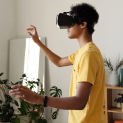
Virtual Reality and Eye Health: Gamer’s Guide
Here are some eye protecting strategies that you need in your armamentarium when you set out to conquer the virtual realm. So if you are worried about virtual reality and eye health, read on.

Can Mascara Damage Your Eyes?
Eyelashes protect the eyes from dust, debris and sand by trapping them. Therefore it is important to understand how mascara can damage your eyes, and eye lashes. Also, here are some recommendations for keeping your eyes safe when using eye makeup.

Alcohol and Eyes
All of us know that alcohol can harm our bodies, what do you know about its effect on your eyes? Consuming alcohol can alter your vision temporarily, and give you bloodshot eyes. The long term effects of drinking, however, can also be permanent. Here is what you need to know about the effect of alcohol on your eyes.